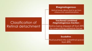
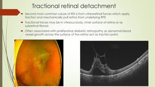
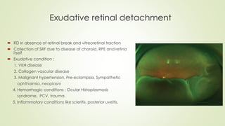
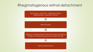
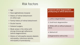
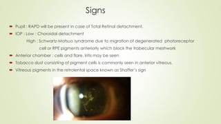
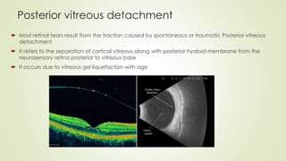
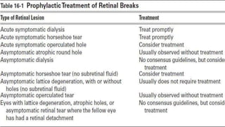
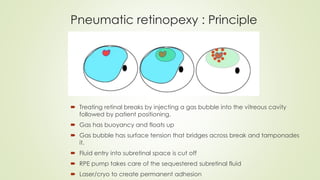
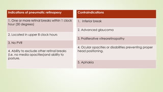
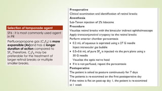
Retinal Detachment
Presented by : Dr Shagun Gaur
JR Ophthalmology
Moderator : Dr Neha Singh
Associate professor
Introduction
 Separation of inner nine layers of the retina from retinal pigment epithelium.
Classification of
Retinal detachment
Rhegmatogenous :
Retinal break allows fluid to go from
vitreous into subretinal space
Tractional/combined
rhegmatogenous-traction :
Retina being stripped off from RPE
by overcoming adhesive forces.
Exudative :
Fluid pushed into subretinal space
from RPE.
Tractional retinal detachment
 Second most common cause of RD is from vitreoretinal forces which apply
traction and mechanically pull retina from underlying RPE
 Tractional forces may be in vitreous body, inner surface of retina or as
subretinal fibrosis
 Often associated with proliferative diabetic retinopathy as abnormal blood
vessel growth across the surface of the retina act as traction point.
Exudative retinal detachment
 RD in absence of retinal break and vitreoretinal traction
 Collection of SRF due to disease of choroid, RPE and retina
itself
 Exudative condition :
1. VKH disease
2. Collagen vascular disease
3. Malignant hypertension, Pre-eclampsia, Sympathetic
ophthalmia, neoplasm
4. Hemorrhagic conditions : Ocular Histoplasmosis
syndrome, PCV, trauma.
5. Inflammatory conditions like scleritis, posterior uveitis.
Rhegmatogenous retinal detachment
Risk factors, senile PVD , Peripheral retinal
degenerations, aphakia, trauma
Retinal break
Seeping of degenerated vitreous fluid through retinal
break into subretinal space and collects as SRF
Retinal detachment
Factors keeping retina attached
Tamponading effect of intact vitreous gel
Active RPE pumping of subretinal fluid to choroid
Interdigitation between the RPE and photoreceptor outer segment.
Interphotoreceptor matrix plays a role of glue between RPE and photoreceptors.
Dehydration of subretinal space enhances its binding properties.
Risk factors
Peripheral retinal degenerations
predisposing to retinal detachment
1. Lattice degeneration
2. Snail track degeneration
3. White with or without pressure
areas
4. Retinal tuft
5.Degenerative retinochisis
Symptoms of retinal detachment
Signs
 Pupil : RAPD will be present in case of Total Retinal detachment.
 IOP : Low : Choroidal detachment
High : Schwartz-Matsuo syndrome due to migration of degenerated photoreceptor
cell or RPE pigments anteriorly which block the trabecular meshwork
 Anterior chamber : cells and flare. Iritis may be seen
 Tobacco dust consisting of pigment cells is commonly seen in anterior vitreous.
 Vitreous pigments in the retrolental space known as Shaffer’s sign
Prerequisite for
retinal detachment
Liquefaction of
vitreous
Vitreoretinal
traction
Retinal break
Posterior vitreous detachment
 Most retinal tears result from the traction caused by spontaneous or traumatic Posterior vitreous
detachment
 It refers to the separation of cortical vitreous along with posterior hyaloid membrane from the
neurosensory retina posterior to vitreous base
 It occurs due to vitreous gel liquefaction with age
Vitreoretinal traction
Atrophy/degeneration
Trauma
Retinal break
• Primary break : Responsible for RD
• Secondary break : not responsible for RD
Types of retinal breaks
Atrophic
retinal holes
Operculated
retinal holes
Horse shoe
shaped tears
/ flap tears
Giant retinal
tears
And giant
retinal
dialysis
Atrophic retinal hole
• Break in the retina not associated with vitreoretinal traction and occur secondary to focal
degeneration of neurosensory retina
• Associated commonly with lattice degeneration
• May have a cuff of fluid surrounding them leading to chronic rhegmatogenous retinal detachment.
Horse shoe shaped tears
 Full thickness breaks of neurosensory retina
 Occurs as a result of vitreous traction or PVD
 Most common in superotemporal quadrant (greater
chances of macula off RD)
 Flap tears can be associated with reactive RPE
hyperplasia of the retina ( long standing tears)
 Goal of treatment is to create chorioretinal
adhesion.
Horns
Base
Apex
Operculated Retinal hole
 There is overlying free floating plug of
retinal tissue (operculum) that adheres
to the posterior hyaloid of the vitreous.
 In areas of focal vitreoretinal
abnormalities.
Giant retinal tear
 Extends 90 degree or 3 clock hours or more
and usually occurs along the posterior edge
of vitreous base.
 It has strong vitreous adhesion to the anterior
margin of the break.
 Posterior margin is free from vitreous
attachment.
 Due to gravity and retinal elasticity, posterior
flap can invert over itself.
 Progress very fast to RD.
Giant retinal dialysis
 Tear of the retina that results in its disinsertion from
the ora serrata
 Vitreous remains attached to the posterior margin
of the break
 PVD is absent preventing the posterior margin from
inverting
Prophylaxis for retinal detachment
Lesions
1. Lattice
2. Holes
3. Holes with operculum
4.Horse shoe tear
When is prophylaxis always indicated
1. Symptomatic patients (Flashes or acute
onset floaters
2. Fellow eye of RD
3. Family history of RD
Added risk factors where prophylaxis may be
considered
1. Myopia of 6-10D
2. Definite presence of traction : Horse shoe tear or hole
with surrounding rim of subretinal fluid
3. Equatorial or more posterior location of lesion
4. Perivascular lesion
Fresh retinal detachment
1. Grayish white, opaque raised detached
retina (convex corrugated appearance with
loss of choroidal pattern)
2. Dark retinal vessels with no central light reflex
3. Shaffer sign and oscillation of multiple retinal
folds with movement of eye
4. Macular pseudohole and presence of retinal
tear
5. B scan : good mobility of retina and vitreous
Old retinal detachment
1. Retinal thinning (due to atrophy)
2. Fixed retinal folds
3. Secondary intraretinal cysts
4. Subretinal demarcation lines ,aka high
water or tide marks due to proliferation of RPE
cells at junction of flat and detached retina.
Fresh RD
Old RD
Proliferative vitreoretinopathy
 In some retinal detachments, cells proliferate on either or both surfaces of
the retina and on vitreous strands and then contract, the process known as
PVR
 This leads to tangential retinal traction and fixed retinal fold formation.
 Main features are retinal folds and rigidity so that retinal mobility induced
by eye movements is decreased
 Presence of PVR indicates poor prognosis and often requires vitrectomy.
Proliferative vitreoretinopathy
Look for retinal break : Lincoff’s rule
Principles of management
 Identification of all retinal breaks and areas of vitreous traction
 Induction of aseptic chorioretinal inflammation around breaks to seal them
 Release of vitreoretinal traction
 Drainage of subretinal fluid
Surgical
approaches
Tamponade
principle
Pneumatic
retinopexy
Shifiting position
of RPE
Scleral buckling
with drainage
or non drainage
Removal of
traction
Primary
vitreoretinal
surgery
Pneumatic retinopexy : Principle
 Treating retinal breaks by injecting a gas bubble into the vitreous cavity
followed by patient positioning.
 Gas has buoyancy and floats up
 Gas bubble has surface tension that bridges across break and tamponades
it.
 Fluid entry into subretinal space is cut off
 RPE pump takes care of the sequestered subretinal fluid
 Laser/cryo to create permanent adhesion
Indications of pneumatic retinopexy
1. One or more retinal breaks within 1 clock
hour (30 degrees)
2. Located in upper 8 clock hours
3. No PVR
4. Ability to exclude other retinal breaks
(i.e. no media opacities)and ability to
posture.
Contraindications
1. Inferior break
2. Advanced glaucoma
3. Proliferative vitreoretinopathy
4. Ocular opacities or disabilities preventing proper
head positioning.
5. Aphakia
Selection of tamponade agent
SF6 : it is most commonly used agent
in PR
Perfluoropropane gas (C3F8) is more
expansible (4x)and has a longer
duration of action compared to
SF6.Therefore, C3F8 may be
preferable for the treatment of
larger retinal breaks or multiple
smaller breaks.
Complications of pneumatic retinopexy
 Gas in the wrong place
1. Trapped in Petit’s space
2. Leaked out of the eye into subconjunctival
space
3. Subretinal gas due to fish eggs
 Secondary glaucoma
 Macular detachment in eyes with attached
macula ( for this steam roller technique is
performed.
Post op management
 Immediate positioning – for at least 5 days
 Follow up until macula is attached
 Post op examination – day 1 – day3/4- every2/3 weeks till 3 months
 What to see : Positioning
Bubble size
New breaks
Delayed absorption of SRF
Advantages Disadvantages
1. Office procedure 1. Patient should not air travel
2. Minimally invasive 2. Positioning is crucial
3. Reduced recovery time
4. Better post op visual acuity
5. Used in failed buckle surgery
Scleral buckling
Localisation
of break
Retinopexy
Placement of
buckle
Management
of subretinal
fluid
Indications of scleral buckling
Fresh RRD due to retinal break anterior to equator
Young phakic patients without PVD
PVR <C1
Minimal SRF
To counteract anterior PVR
RD with dialysis
360 degree limbal peritomy or
4mm posterior to limbus performed
and tenon conjunctival adhesions
are lysed.
Muscle tagging is done i.e. all four
recti are tagged with silk sutures
preventing hooking of vortex veins.
Localisation of break with the help
of indirect ophthalmoscope. A
diathermy, cautery or marking pen
is used to mark the area of retinal
break in sclera.
Retinopexy
It creates a chorioretinal scar thereby sealing the retinal break. This prevents the liquid vitreous
from entering subretinal space. It can be performed using :
Cryopexy
• Performed under IO
visualisation of fundus.
• A row of contiguous
cryo burns are applied
around retinal break or
lattice degeneration
preventing bare RPE
beneath the break
Laser photocoagulation
• Intraoperatively done
with transscleral
diopexy probe or laser
IO.
• It is used only if there is
shallow fluid
separating the layers
or after drainage of
subretinal fluid
Diathermy
• Provides strongest
adhesion
• It can be applied in
staggered rows of
burns 1.5mm apart
that need not surround
break
• Results in shrinkage of
sclera so needs
lamellar dissection of
sclera so seldom used .
Scleral buckling
• Explants are the elements that is sutured to the surface of sclera to indent it.
• It ma be radial or circumferential ( segmental or encircling)
• It is made up of silicone.
• Construction of scleral
pocket with a crescent
blade
• Scleral suture to secure
encircling silicone band
• Encircling band secured
within scleral pockets and
passed beneath the recti
• Ends of encircling band
secure with Watzke sleeve.
Potential errors in localisation
 Highly elevated RD : Parallax issue
 Localising under muscles
 Confusion between indentation caused by shaft vs tip of
the instrument
Encirclage indications
 Evidence of PVR grade B or more
 Aphakic/pseudophakic
 High myopia
 Multiple breaksin different quadrant
 Retinal dialysis due to trauma
 When no break found
 Extensive lattice
Drainage of subretinal fluid
Indications Preferable site for
drainage
Highly elevated (bullous)
RD
Beneath the area of
maximum SRF
Chronic inferior
detachment
Away from the break
PVR grade B or above Either side of horizontal
recti
Glaucomatous eye Preferably beneath scleral
buckle
Aphakic detachments
DACE- Drainage Air Cryo Encirclage
 In cases with bullous retinal detachments, localization of the break
would be false due to the parallax or breaks that can be hidden in
between the retinal folds
 The application of the cryo in a detached retina may be difficult.
 Drainage of the subretinal fluid helps to better identify the breaks
and avoid false localization.
 The SRF can be replaced by either air or fluid injection in the
vitreous cavity and cryo application is done and the scleral
buckling procedure is completed.
Complications of scleral buckling
Intra-op Early post-op Late post op
Scleral perforation Failure of retinal
reattachment
Strabismus
Rise in IOP Buckle infection Refractive error
Fish mouthing Anterior segment ischemia Extrusion/intrusion of buckle
Accidental damage to
muscle, vortex veins
Misplaced buckle Late detachment
During SRF drainage
1. Choroidal bleeding
2. Suprachoroidal
hemorrhage
3. Vitreous loss and
hemorrhage
4. Retinal perforation and
incarceration
Choroidal detachment Reopening of original break
Vitrectomy
Indications in RD
PVR more than C2
GRT
Multiple or posterior break
Failed scleral buckling
Tractional RD
Vitreous hemorrhage
Absence of apparent break in
pseudophakic patients
• Abnormal vitreous
traction increases the
vitreous mobility by
causing PVD
• Removal of preretinal
structures releases
tractional force causing
break and detachment
• To stabilize and flatten the
detached retina a heavy
liquid is applied.
Thank you

RETINAL DETACHMENTfulll 2-compressed.pdf

  • 1.
    Retinal Detachment Presented by: Dr Shagun Gaur JR Ophthalmology Moderator : Dr Neha Singh Associate professor
  • 2.
    Introduction  Separation ofinner nine layers of the retina from retinal pigment epithelium.
  • 3.
    Classification of Retinal detachment Rhegmatogenous: Retinal break allows fluid to go from vitreous into subretinal space Tractional/combined rhegmatogenous-traction : Retina being stripped off from RPE by overcoming adhesive forces. Exudative : Fluid pushed into subretinal space from RPE.
  • 4.
    Tractional retinal detachment Second most common cause of RD is from vitreoretinal forces which apply traction and mechanically pull retina from underlying RPE  Tractional forces may be in vitreous body, inner surface of retina or as subretinal fibrosis  Often associated with proliferative diabetic retinopathy as abnormal blood vessel growth across the surface of the retina act as traction point.
  • 5.
    Exudative retinal detachment RD in absence of retinal break and vitreoretinal traction  Collection of SRF due to disease of choroid, RPE and retina itself  Exudative condition : 1. VKH disease 2. Collagen vascular disease 3. Malignant hypertension, Pre-eclampsia, Sympathetic ophthalmia, neoplasm 4. Hemorrhagic conditions : Ocular Histoplasmosis syndrome, PCV, trauma. 5. Inflammatory conditions like scleritis, posterior uveitis.
  • 6.
    Rhegmatogenous retinal detachment Riskfactors, senile PVD , Peripheral retinal degenerations, aphakia, trauma Retinal break Seeping of degenerated vitreous fluid through retinal break into subretinal space and collects as SRF Retinal detachment
  • 7.
    Factors keeping retinaattached Tamponading effect of intact vitreous gel Active RPE pumping of subretinal fluid to choroid Interdigitation between the RPE and photoreceptor outer segment. Interphotoreceptor matrix plays a role of glue between RPE and photoreceptors. Dehydration of subretinal space enhances its binding properties.
  • 8.
    Risk factors Peripheral retinaldegenerations predisposing to retinal detachment 1. Lattice degeneration 2. Snail track degeneration 3. White with or without pressure areas 4. Retinal tuft 5.Degenerative retinochisis
  • 9.
  • 10.
    Signs  Pupil :RAPD will be present in case of Total Retinal detachment.  IOP : Low : Choroidal detachment High : Schwartz-Matsuo syndrome due to migration of degenerated photoreceptor cell or RPE pigments anteriorly which block the trabecular meshwork  Anterior chamber : cells and flare. Iritis may be seen  Tobacco dust consisting of pigment cells is commonly seen in anterior vitreous.  Vitreous pigments in the retrolental space known as Shaffer’s sign
  • 11.
    Prerequisite for retinal detachment Liquefactionof vitreous Vitreoretinal traction Retinal break
  • 12.
    Posterior vitreous detachment Most retinal tears result from the traction caused by spontaneous or traumatic Posterior vitreous detachment  It refers to the separation of cortical vitreous along with posterior hyaloid membrane from the neurosensory retina posterior to vitreous base  It occurs due to vitreous gel liquefaction with age
  • 13.
    Vitreoretinal traction Atrophy/degeneration Trauma Retinal break •Primary break : Responsible for RD • Secondary break : not responsible for RD
  • 14.
    Types of retinalbreaks Atrophic retinal holes Operculated retinal holes Horse shoe shaped tears / flap tears Giant retinal tears And giant retinal dialysis
  • 15.
    Atrophic retinal hole •Break in the retina not associated with vitreoretinal traction and occur secondary to focal degeneration of neurosensory retina • Associated commonly with lattice degeneration • May have a cuff of fluid surrounding them leading to chronic rhegmatogenous retinal detachment.
  • 17.
    Horse shoe shapedtears  Full thickness breaks of neurosensory retina  Occurs as a result of vitreous traction or PVD  Most common in superotemporal quadrant (greater chances of macula off RD)  Flap tears can be associated with reactive RPE hyperplasia of the retina ( long standing tears)  Goal of treatment is to create chorioretinal adhesion. Horns Base Apex
  • 18.
    Operculated Retinal hole There is overlying free floating plug of retinal tissue (operculum) that adheres to the posterior hyaloid of the vitreous.  In areas of focal vitreoretinal abnormalities.
  • 19.
    Giant retinal tear Extends 90 degree or 3 clock hours or more and usually occurs along the posterior edge of vitreous base.  It has strong vitreous adhesion to the anterior margin of the break.  Posterior margin is free from vitreous attachment.  Due to gravity and retinal elasticity, posterior flap can invert over itself.  Progress very fast to RD.
  • 20.
    Giant retinal dialysis Tear of the retina that results in its disinsertion from the ora serrata  Vitreous remains attached to the posterior margin of the break  PVD is absent preventing the posterior margin from inverting
  • 21.
    Prophylaxis for retinaldetachment Lesions 1. Lattice 2. Holes 3. Holes with operculum 4.Horse shoe tear When is prophylaxis always indicated 1. Symptomatic patients (Flashes or acute onset floaters 2. Fellow eye of RD 3. Family history of RD Added risk factors where prophylaxis may be considered 1. Myopia of 6-10D 2. Definite presence of traction : Horse shoe tear or hole with surrounding rim of subretinal fluid 3. Equatorial or more posterior location of lesion 4. Perivascular lesion
  • 22.
    Fresh retinal detachment 1.Grayish white, opaque raised detached retina (convex corrugated appearance with loss of choroidal pattern) 2. Dark retinal vessels with no central light reflex 3. Shaffer sign and oscillation of multiple retinal folds with movement of eye 4. Macular pseudohole and presence of retinal tear 5. B scan : good mobility of retina and vitreous Old retinal detachment 1. Retinal thinning (due to atrophy) 2. Fixed retinal folds 3. Secondary intraretinal cysts 4. Subretinal demarcation lines ,aka high water or tide marks due to proliferation of RPE cells at junction of flat and detached retina.
  • 23.
  • 24.
    Proliferative vitreoretinopathy  Insome retinal detachments, cells proliferate on either or both surfaces of the retina and on vitreous strands and then contract, the process known as PVR  This leads to tangential retinal traction and fixed retinal fold formation.  Main features are retinal folds and rigidity so that retinal mobility induced by eye movements is decreased  Presence of PVR indicates poor prognosis and often requires vitrectomy.
  • 25.
  • 29.
    Look for retinalbreak : Lincoff’s rule
  • 30.
    Principles of management Identification of all retinal breaks and areas of vitreous traction  Induction of aseptic chorioretinal inflammation around breaks to seal them  Release of vitreoretinal traction  Drainage of subretinal fluid
  • 31.
    Surgical approaches Tamponade principle Pneumatic retinopexy Shifiting position of RPE Scleralbuckling with drainage or non drainage Removal of traction Primary vitreoretinal surgery
  • 32.
    Pneumatic retinopexy :Principle  Treating retinal breaks by injecting a gas bubble into the vitreous cavity followed by patient positioning.  Gas has buoyancy and floats up  Gas bubble has surface tension that bridges across break and tamponades it.  Fluid entry into subretinal space is cut off  RPE pump takes care of the sequestered subretinal fluid  Laser/cryo to create permanent adhesion
  • 33.
    Indications of pneumaticretinopexy 1. One or more retinal breaks within 1 clock hour (30 degrees) 2. Located in upper 8 clock hours 3. No PVR 4. Ability to exclude other retinal breaks (i.e. no media opacities)and ability to posture. Contraindications 1. Inferior break 2. Advanced glaucoma 3. Proliferative vitreoretinopathy 4. Ocular opacities or disabilities preventing proper head positioning. 5. Aphakia
  • 34.
    Selection of tamponadeagent SF6 : it is most commonly used agent in PR Perfluoropropane gas (C3F8) is more expansible (4x)and has a longer duration of action compared to SF6.Therefore, C3F8 may be preferable for the treatment of larger retinal breaks or multiple smaller breaks.
  • 36.
    Complications of pneumaticretinopexy  Gas in the wrong place 1. Trapped in Petit’s space 2. Leaked out of the eye into subconjunctival space 3. Subretinal gas due to fish eggs  Secondary glaucoma  Macular detachment in eyes with attached macula ( for this steam roller technique is performed.
  • 38.
    Post op management Immediate positioning – for at least 5 days  Follow up until macula is attached  Post op examination – day 1 – day3/4- every2/3 weeks till 3 months  What to see : Positioning Bubble size New breaks Delayed absorption of SRF
  • 39.
    Advantages Disadvantages 1. Officeprocedure 1. Patient should not air travel 2. Minimally invasive 2. Positioning is crucial 3. Reduced recovery time 4. Better post op visual acuity 5. Used in failed buckle surgery
  • 40.
    Scleral buckling Localisation of break Retinopexy Placementof buckle Management of subretinal fluid
  • 41.
    Indications of scleralbuckling Fresh RRD due to retinal break anterior to equator Young phakic patients without PVD PVR <C1 Minimal SRF To counteract anterior PVR RD with dialysis
  • 42.
    360 degree limbalperitomy or 4mm posterior to limbus performed and tenon conjunctival adhesions are lysed. Muscle tagging is done i.e. all four recti are tagged with silk sutures preventing hooking of vortex veins. Localisation of break with the help of indirect ophthalmoscope. A diathermy, cautery or marking pen is used to mark the area of retinal break in sclera.
  • 44.
    Retinopexy It creates achorioretinal scar thereby sealing the retinal break. This prevents the liquid vitreous from entering subretinal space. It can be performed using : Cryopexy • Performed under IO visualisation of fundus. • A row of contiguous cryo burns are applied around retinal break or lattice degeneration preventing bare RPE beneath the break Laser photocoagulation • Intraoperatively done with transscleral diopexy probe or laser IO. • It is used only if there is shallow fluid separating the layers or after drainage of subretinal fluid Diathermy • Provides strongest adhesion • It can be applied in staggered rows of burns 1.5mm apart that need not surround break • Results in shrinkage of sclera so needs lamellar dissection of sclera so seldom used .
  • 45.
    Scleral buckling • Explantsare the elements that is sutured to the surface of sclera to indent it. • It ma be radial or circumferential ( segmental or encircling) • It is made up of silicone.
  • 46.
    • Construction ofscleral pocket with a crescent blade • Scleral suture to secure encircling silicone band • Encircling band secured within scleral pockets and passed beneath the recti • Ends of encircling band secure with Watzke sleeve.
  • 47.
    Potential errors inlocalisation  Highly elevated RD : Parallax issue  Localising under muscles  Confusion between indentation caused by shaft vs tip of the instrument Encirclage indications  Evidence of PVR grade B or more  Aphakic/pseudophakic  High myopia  Multiple breaksin different quadrant  Retinal dialysis due to trauma  When no break found  Extensive lattice
  • 48.
    Drainage of subretinalfluid Indications Preferable site for drainage Highly elevated (bullous) RD Beneath the area of maximum SRF Chronic inferior detachment Away from the break PVR grade B or above Either side of horizontal recti Glaucomatous eye Preferably beneath scleral buckle Aphakic detachments
  • 49.
    DACE- Drainage AirCryo Encirclage  In cases with bullous retinal detachments, localization of the break would be false due to the parallax or breaks that can be hidden in between the retinal folds  The application of the cryo in a detached retina may be difficult.  Drainage of the subretinal fluid helps to better identify the breaks and avoid false localization.  The SRF can be replaced by either air or fluid injection in the vitreous cavity and cryo application is done and the scleral buckling procedure is completed.
  • 50.
    Complications of scleralbuckling Intra-op Early post-op Late post op Scleral perforation Failure of retinal reattachment Strabismus Rise in IOP Buckle infection Refractive error Fish mouthing Anterior segment ischemia Extrusion/intrusion of buckle Accidental damage to muscle, vortex veins Misplaced buckle Late detachment During SRF drainage 1. Choroidal bleeding 2. Suprachoroidal hemorrhage 3. Vitreous loss and hemorrhage 4. Retinal perforation and incarceration Choroidal detachment Reopening of original break
  • 52.
    Vitrectomy Indications in RD PVRmore than C2 GRT Multiple or posterior break Failed scleral buckling Tractional RD Vitreous hemorrhage Absence of apparent break in pseudophakic patients • Abnormal vitreous traction increases the vitreous mobility by causing PVD • Removal of preretinal structures releases tractional force causing break and detachment • To stabilize and flatten the detached retina a heavy liquid is applied.
  • 53.