SlideShare a Scribd company logo
1 of 100
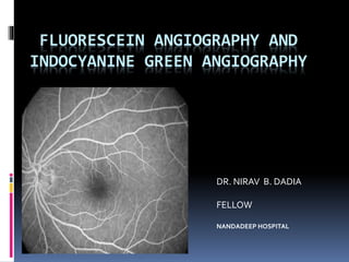
FLUORESCEIN ANGIOGRAPHY AND
INDOCYANINE GREEN ANGIOGRAPHY
DR. NIRAV B. DADIA
FELLOW
NANDADEEP HOSPITAL
INTRODUCTION
 The word Angiography - Greek angeion,
"vessel" and graphien, "to write or record".
 Imaging of vessels, and the resulting pictures
are angiograms.
 In-vivo study of the retinal circulation
Basic principle
 luminescence- emission of light by
excitation of atoms or molecules to higher
energy levels
 fluorescence- luminescence that is
maintained by continuous excitation.
 The emitted energy is often less than the
absorbed energy, though of longer
wavelength (stoke’s law)
 Fluorescein absorbs blue light and emits
yellow green light
 Exciter filter (blue) and barrier filter (green)
 Properties of Flourescein Na
Chemical properties
Fluorescein sodium –
synthesized from the petroleum derivatives resorcinol and
phthalic anhydride
• Low molecular weight
• High solubility
• Rapid diffusion through body fluids but not large enough to
pass through tight junctions of retinal vessels, RPE and large
choroidal vessels.
•This is the basis for the diagnostic value of the test.
Optical properties
• It absorbs blue light, with peak absorption
and excitation occurring at wavelengths
between 465-490nm.
• Fluorescence occurs at the yellow-green
wavelengths of 520 to 530nm
Pharmacological properties
• 80% bound to serum proteins
• also bound to blood cells
• remaining i.e. Unbound dye is seen during
angiography
pharmacokinetics
• Metabolized by kidney, excreted from the body
within 24 to 36 hrs
• Small amounts are lost in bile.
• Skin - a yellowish tinge for a few hours
• Urine - yellow-orange color
• Dye is a biologically inert substance
 Requirement
Relevant anatomy
Outer blood
retinal barrier
Inner blood
retinal Barrier
Fluorescein cannot diffuse through tight
cellular junctions present at two sites within
the fundus
 Angiography is composed of the superimposition of
two separate circulations
 – Choroidal circulation
the fluorescein freely leaks out of the fenestrated
choroidal capillaries, and from there through Bruch's
membrane.
– Retinal circulation
the retinal blood vessel endothelial cells are joined
by tight junctions which prevent leakage of
fluorescein into the retina.
Phases of normal angiogram
• Arm to retina time: Normally 10-15 seconds
elapse between dye injection and arrival of
dye in the eye.
• Retinal ciculation time: Transit of dye
through the retinal circulation takes 15 to 20
seconds.
PHASES OF ANGIOGRAM
 1. PREARTERIAL [ CHOROIDAL FLUSH ] – 10 sec
 2. ARTERIAL – 12sec
 3. ARTERIO-VENOUS
- EARLYTRANSIT – 13 sec
- MIDTRANSIT – 16sec
- LATETRANSIT – 20 sec
3a. Peak phase – 25 sec
4. RECIRCULATION – 30sec
5. LATE FLURESCEINTRANSIT – after 10 min
 Choroidal phase
- initial patching filing of lobules, - followed by a
diffuse (flush) as dye leaks out of the
choroidocapillaris.
 visualisation of choroid depends on retinal
pigmentation - Cilioretinal vessels and
prelaminar optic disc capillaries fill during this
phase.
Arterial phase
• the central retinal artery fills about 1 second
later than choroidal filling
Venous phase
• Early venous phase: filling of the veins is from
tributaries joining their margins, resulting in a
tramline effect (lamellar flow)
Late phase
• after 10 to 15 minutes little dye remains within
the blood circulation.
• Dye which has left the blood to ocular
structures is particularly visible.
• it shows abnormal dye accumulations
indicative of leakage or staining.
DARK APPEARANCE OF FOVEA
The dark appearance of the fovea is caused by three
factors:
Absence of blood vessels in the FAZ.
Blockage of background choroidal fluorescence due
to the high density of xanthophyll at the fovea.
Blockage of background choroidal fluorescence by
the RPE cells at the fovea, which are larger and
contain more melanin and lipofuscin than else
where in the retina
SELECTION OF PATIENT
• Not recommended :
- History of allergy, severe urticaria or bronchial
asthma
- Patient with renal failure and poor general
condition.
• In pregnant women - it may be avoided.
• Safe: In diabetics, hypertensives and history of
previous cardiovascular disorders.
CONTRAINDICATIONS
ABSOLUTE
 1) Known allergy to iodine containing compounds.
 2) H/O adverse reaction to FFA in the past.
RELATIVE
 1) Asthma
 2) Hay fever
 3) Renal failure
 4) Hepatic failure
 5) Cardiac disease – cardiac failure, Myocardial infarction
 6) Previous mild reaction to dye.
 7)Tonic-clonic seizures
 8) Pregnancy ( especially 1st trimester
preparation
• make sure the patient is well dilated
• Log the patient information
• Injectable fluorescein dye comes in 5%(10cc) ,
10% (5cc), and 20% (2cc or 3cc) solutions.
• 20% solution is preferred because this larger
bolus produces better photographic contrast and
detail in the initial phases of the angiogram
analysis
– Sequential analysis
- frame by frame. useful in analysing vascular
disorders of the retinal and choroidal.
– Anatomic analysis
- observes each of the major layers of the posterior
pole of the eye - the choroidal, RPE and
neurosensory retina.
– Morphologic analysis
- considers overall patterns. (hyperfluorescent) or
lighter (hypofluorescent)
reporting
• Start with any striking abnormality and describe
this in detail:
- phase of angiogram
– Hypo/hyperfluorescent components
– Intensity of fluorescence and changes with time
– Area of fluorescence and changes with time
Common abnormalities
•Timing
-arm to eye time and retinal circulation may
be prolonged if the cardiac output is low or
the carotid perfusion is reduced.
• Abnormal dye distribution:
hypofluorescence/
hyperfluorescence
Hypofluorescence
Transmission defect
-blood,
-pigment,
-hard exudates
-abnormal material (eg, the yellow flecks in
patient with Stargardt's disease etc)
 pre-retinal opaque structures superficial to
the retinal circulation will mask both the
retina and choroidal circulation eg. -
Preretinal hemorrhage, -myelinated nerve
fibres
 prechoroidal opaque structures deep to the
retinal circulation but superficial to the
choroidal circulation will mask only the
choroidal circulation for example:
blood
 - retinal haemorrhages
 - subretinal blood from choroidal new vessels
Filling defect due to abnormal
circulation
• arterial non-perfusion is seen in occlusion of
the central retinal artery and its branches
• capillary non-perfusion is an important sign of
retinal ischaemia.
Leakage of dye
 • occurs when there is breakdown of the
tight junction of the RPE or the retinal
endothelium.
Autofluorescence
• Presence of hyperfluorescence in the fundus
seen in pre-injection photographs.
• optic disc drusen is the classic example.
• Others:
astrocytic hamartoma
large deposits of lipofuscin
and exudates
FFA patterns of some common
diseases
ARMD
• FA is not indicated in each and every case and
in every visit
• Indications
- possibility of finding CNVM
metamorphopsia
recent decrease in vision
central or paracentral scotoma
- undergone laser treatment
Wet ARMD
• FA helps in determining the extent and the
type of neo-vascularization.
• Classified into classic and occult variety into
extrafoveal
juxtafoveal
subfoveal
BRAO
• Artery occlusion
• Purtschners retinopathy-
blocked fluoroscence partly due to ischaemia
and intracellular edema
-opacified edematous retina
CHOROIDITIS
Oral FA
Indication:
Psychologically or technically unsuitable for i/v
injection especially children, obese pts.
Dose:
1 gm Na fluorescein (5ml of 20% dye)
(mixed in 200 ml of orange juice – Body weight
40kg.
1.5 gm in pts with a body wt 60 kg
while 2.0 gm is given to pts over 60 kg
 Post-administration photographs taken after
15, 30, 45 and 60 minutes.
 Reserved - lesions resulting in late dye
leakage and pooling like CSR, disciform disc
degeneration etc.
 Not recommended when early circulation
dynamics are to be studied
Adverse events
severity Adverse events percentage
 Mild Nausia, vomiting, 1-10
extravasation
 moderate Urticaria, 1-6
pyrexia,
local tissue necrosis,
nerve palsy
 severe Bronchospasm, 0.05
anaphylaxis,
shock
death 1/222,000
EMERGENCY in FA Case
• Allergic reaction: Local / Generalised
Manifest: redness, itching, oedema & urticaria.
• Stop dye injection.
• Monitor Pulse , BP & Resp.
• Inj. Avil 2ml IV
• Inj Efcorlin 100mg IV
• Normal Saline – wash the local site
Fluorescein Angioscopy
• Indirect Ophthalmoscope along with its blue
filter attachment, is used for viewing of the
fundus periphery.
• Pathology including Eales disease,
sarcoidosis, retrolental fibroplasia and
peripheral vasculitides, both in active
inflammatory stage and later stage, are
effectively visualised by F-scopy
Limitations of FFA
1) Does not permit study of choroidal circulation details due to
a) melanin in RPE
b) b) low mol wt of fluorescein
how to overcome ---- ICG
2) More adverse reaction
3) Inability to obtain angiogram in patient with excess hemoglobin or serum
protein.e.g.
polycythemia weldenstrom macroglobulenaemia
binding of fluorescein with excess Hb or protein
Lack of freely circulating molecule
INDOCYANINE GREEN ANGIOGRAPHY
• Indocyanine green (ICG) angiography (ICGA) is fast emerging as a
popular and useful adjunct to the traditional fundus fluorescein
angiography (FFA) in the diagnosis of macular, choroidal and outer
retinal disorders.
•This technique was introduced in ophthalmology in 1973 by Flower and
Hochheimer.
• FDA approved the ophthalmic use of ICG dye in 1975.
• It remained largely unpopular owing mainly to technical difficulties
With the advent of videoangiogram recordings and the recognition of its
potential in delineating occult choroidal neovascular membranes, the
clinical use of ICGA has increased tremendously .
PRINCIPLES OF ICG
ICG fluorescence is only 1/25th that of fluorescein.
So modern digital ICGA uses high-sensitivity videoangiographic
image capture by means of an appropriately adapted camera.
Both the excitation (805 nm) and emission (835 nm) filters are set
at infrared wavelengths.
Alternatively, scanning laser ophthalmoscopy (SLO) systems
provide high contrast images, with less scattering of light and
fast image acquisition rates facilitating high quality ICG video.
The technique is similar to that of FA, but with an increased
emphasis on the acquisition of later images (up to about 45
minutes) than with FA. A dose of 25–50 mg in 1–2 ml water for
injection is used.
INDOCYANINE GREEN
The indocyanine green (ICG) is a tricarbocyanine dye that comes packaged as a
sterile lyophilized powder and is supplied with an aqueous solvent.
Molecular weight :774.97
It contains less than 5% sodium iodide (in order to increase its solubility).
It has a pH of 5.5 to 6.5 in the dissolved state, has limited stability, and hence
must be used within 10 hours after reconstitution.
98% of the injected dye is bound to plasma proteins, with 80% being bound
to globulins, especially alpha- 1 lipoproteins.
CLEARANCE
The dye is secreted unchanged by the liver into
the bile.
There is no renal excretion of the dye
It does not cross the placenta.
The dye also has a high affinity for vascular
endothelium, and hence persists in the large
choroidal veins, long after injection.
ADVERSE EFFECTS
Nausea, vomiting are uncommon.
Anaphylaxis, approximately equal incidence to FA.
Serious reactions are exceptionally rare.
ICG contains iodide and so should not be given to
patients allergic to iodine or possibly shellfish.
iodine-free preparations such as infracyanine green
are available.
CONTRAINDICATIONS
ICGA is relatively contraindicated in liver disease
(excretion is hepatic)
In patients with a history of a severe reaction to
any allergen.
moderate or severe asthma
significant cardiac disease.
Its safety in pregnancy has not been established
PHASES OF ICGA
Early – up to 60 seconds post-injection
Early mid-phase – 1–3 minutes
Late mid-phase –3–15 minutes
Late phase – 15–45 minute
HYPERFLOURESCENE
A window defect similar to those seen with FA.
Leakage from retinal or choroidal vessels the optic nerve head or the RPE
gives rise to tissue staining or to pooling.
Abnormal retinal or choroidal vessels with an anomalous morphology
exhibiting greater fluorescence than normal
HYPOFLOURECENCE
 Blockage (masking) of fluorescence.
A particular phenomenon to note is that in contrast to its
FA appearance, a pigment epithelial detachment
appears predominantly hypofluorescent on ICGA.
Pigment and blood are self-evident causes, but fibrosis,
infiltrate, exudate and serous fluid also block
fluorescence
Filling defect due to obstruction or loss of choroidal or
retinal circulation.
INDICATIONS OF ICGA
 Polypoidal choroidal vasculopathy (PCV)
 Exudative age-related macular degeneration (AMD)
 Chronic central serous chorioretinopathy.
 Posterior uveitis.
 Choroidal tumors may be imaged effectively
 Breaks in Bruch membrane
 If FA is contraindicated.
ADVANTAGES OF ICGA OVER FFA
FA is an excellent method of studying the retinal circulation, it is of limited
use in delineating the choroidal vasculature, due to masking by the RPE.
ICGA can be used even when the ocular media are too hazy for FFA.This is
due to the phenomenon of Rayleigh scatter.
In contrast, the near-infrared light utilized in indocyanine green
angiography (ICGA) penetrates ocular pigments such as melanin and
xanthophyll, as well as exudate and thin layers of subretinal blood, making
this technique eminently suitable.
ICG fluorescence can be imaged even in the presence of considerable
blood, due to the phenomenon of Mie or forward scatter.
The peak absorption of ICG coincides with the emission spectrum of
diode laser, which allows the selective ablation of chorioretinal lesions
using ICG dye-enhanced laser photocoagulation wherein a target tissue
containing ICG is exposed to the diode laser beam.
Photophobic patients tolerate ICGA better than FFA.
ICGA accurately measures the size of an occult choroidal neovascular
membrane(CNVM).
LIMITATIONS OF ICGA
The
choriocapillaris
cannot be imaged
separately with
ICGA since their
average cross-
sectional diameter
(21 μm) is much
smaller than that
of their feeding
and draining
vessels, and hence
the fluorescence of
the former cannot
be differentiated
from that arising
from the latter
Although superior
to FFA in the
imaging of occult
CNVM, ICGA may
underestimate the
size of the CNVM.
Bright areas do not
necessarily signify
dye leakage due to
the phenomenon
of additive
fluorescence
The phenomenon
of Mie scatter also
masks the unfilled
retinal vessels that
cannot be
visualized well in
low speed
angiography
systems.
ICGA is poorer
than FFA in the
imaging of classic
CNVM since the
early hyper
fluorescence of the
CNVM is
overwhelmed by
the intense
background
choroidal filling
RECENT ADVANCES IN INDOCYANINE GREEN
ANGIOGRAPHY
 Wide-angle angiography:This is carried out by performing ICGA with
the aid of wide angle contact lenses, such asVolk SuperQuad and a
traditionalTopcon fundus camera.This allows real-time imaging of a
wide field of the choroidal circulation up to 160 degrees of field of view.
 Overlay technique:This technique allows lesion on one image to be
traced on to another color or red-free image.
 Digital stereo imaging: Elevated lesions such as PEDs can be better
imaged in this way. ICG as a photo sensitizer: It is considered to be a
cheaper alternative to vertoporfin in photodynamic therapy of
neovascular AMD& other disorders .
 Digital subtraction ICGA: It uses digital subtraction of sequentially
acquired ICG images along with pseudo color imaging. It shows occult
CNVM in greater detail and within a shorter time than conventional
ICGA.
SUMMARY
Fundus fluorescein angiography ICG angiography
For retinal circulation For choroidal circulation
Dye used – sodium fluorescein Dye used – indocyanin green
80% plasma protein bound 98% plasma protein bound
low MW high MW
Light of visible spectrum used Infrared spectrum of light used
Blue green filters used Infrared filters used
More side effects Less side effects
FFA
•Valuable in DR
– Neovascularisation
– CNP areas
- Maculopathy
• CNVM activity
• CSR
•Venous occlusion
• Patient education
FUTURE APPLICATIONS OF INDOCYANINE
GREEN ANGIOGRAPHY
In the future, ICGA is expected to play a more important
and wider role especially in the management of macular
disorders.
Identifying subclinical neovascular lesions in the other eye
of patients with AMD.There are several reports that
mention that 10% of such eyes with no clinical or
fluorescein angiographic evidence of an exudative process
harbor plaques of neovascularization evident on ICGA.
ICG-guided feeder vessel photocoagulation: SLO high-
speed ICGA can adequately image the feeding vessels of
the CNVM which are 0.5 to 3 mm in length and are believed
to lie in the Sattler’s layer of the choroid
 THANK YOU

More Related Content

What's hot

Transpupillary Thermotherapy (TTT)
Transpupillary Thermotherapy (TTT)Transpupillary Thermotherapy (TTT)
Transpupillary Thermotherapy (TTT)Pushkar Dhir
 
Managing bleb failure - A brief guide
Managing bleb failure - A brief guideManaging bleb failure - A brief guide
Managing bleb failure - A brief guidedrindeevarmishra
 
Choroidal detachment
Choroidal detachmentChoroidal detachment
Choroidal detachmentSSSIHMS-PG
 
INDOCYANINE GREEN ANGIOGRAPHY
INDOCYANINE GREEN ANGIOGRAPHYINDOCYANINE GREEN ANGIOGRAPHY
INDOCYANINE GREEN ANGIOGRAPHYPooja Kandula
 
OPTICAL COHERENCE TOMOGRAPHY ANGIOGRAPHY
OPTICAL COHERENCE TOMOGRAPHY  ANGIOGRAPHY  OPTICAL COHERENCE TOMOGRAPHY  ANGIOGRAPHY
OPTICAL COHERENCE TOMOGRAPHY ANGIOGRAPHY Shruti Laddha
 
AS OCT & UBM - Dr Shylesh B Dabke
AS OCT & UBM - Dr Shylesh B DabkeAS OCT & UBM - Dr Shylesh B Dabke
AS OCT & UBM - Dr Shylesh B DabkeShylesh Dabke
 
Inferior Oblique Overaction (IOOA)
Inferior Oblique Overaction (IOOA)Inferior Oblique Overaction (IOOA)
Inferior Oblique Overaction (IOOA)Meironi Waimir
 
Retinal dystrophy
Retinal dystrophyRetinal dystrophy
Retinal dystrophydipusarkar2
 
Polypoidal choroidal vasculopathy
Polypoidal choroidal vasculopathyPolypoidal choroidal vasculopathy
Polypoidal choroidal vasculopathySujay Chauhan
 
Ophthalmic Viscoelastic devices
Ophthalmic Viscoelastic devicesOphthalmic Viscoelastic devices
Ophthalmic Viscoelastic devicesBinny Tyagi
 
Iol power calculation in pediatric patients
Iol power calculation in pediatric patientsIol power calculation in pediatric patients
Iol power calculation in pediatric patientsAnisha Rathod
 
Fundus Fluorescence Angiography
Fundus Fluorescence Angiography Fundus Fluorescence Angiography
Fundus Fluorescence Angiography abhishek ghelani
 
Complications of trabeculectomy
Complications of trabeculectomyComplications of trabeculectomy
Complications of trabeculectomySumeet Agrawal
 

What's hot (20)

Transpupillary Thermotherapy (TTT)
Transpupillary Thermotherapy (TTT)Transpupillary Thermotherapy (TTT)
Transpupillary Thermotherapy (TTT)
 
Managing bleb failure - A brief guide
Managing bleb failure - A brief guideManaging bleb failure - A brief guide
Managing bleb failure - A brief guide
 
FFA and ICG
FFA and ICGFFA and ICG
FFA and ICG
 
Choroidal detachment
Choroidal detachmentChoroidal detachment
Choroidal detachment
 
INDOCYANINE GREEN ANGIOGRAPHY
INDOCYANINE GREEN ANGIOGRAPHYINDOCYANINE GREEN ANGIOGRAPHY
INDOCYANINE GREEN ANGIOGRAPHY
 
OPTICAL COHERENCE TOMOGRAPHY ANGIOGRAPHY
OPTICAL COHERENCE TOMOGRAPHY  ANGIOGRAPHY  OPTICAL COHERENCE TOMOGRAPHY  ANGIOGRAPHY
OPTICAL COHERENCE TOMOGRAPHY ANGIOGRAPHY
 
AS OCT & UBM - Dr Shylesh B Dabke
AS OCT & UBM - Dr Shylesh B DabkeAS OCT & UBM - Dr Shylesh B Dabke
AS OCT & UBM - Dr Shylesh B Dabke
 
Ffa
FfaFfa
Ffa
 
Inferior Oblique Overaction (IOOA)
Inferior Oblique Overaction (IOOA)Inferior Oblique Overaction (IOOA)
Inferior Oblique Overaction (IOOA)
 
Retinal dystrophy
Retinal dystrophyRetinal dystrophy
Retinal dystrophy
 
Oct in glaucoma
Oct in glaucomaOct in glaucoma
Oct in glaucoma
 
Polypoidal choroidal vasculopathy
Polypoidal choroidal vasculopathyPolypoidal choroidal vasculopathy
Polypoidal choroidal vasculopathy
 
Accommodating IOLs
Accommodating IOLsAccommodating IOLs
Accommodating IOLs
 
Dalk
DalkDalk
Dalk
 
Autofluorescence
AutofluorescenceAutofluorescence
Autofluorescence
 
Ophthalmic Viscoelastic devices
Ophthalmic Viscoelastic devicesOphthalmic Viscoelastic devices
Ophthalmic Viscoelastic devices
 
Iol power calculation in pediatric patients
Iol power calculation in pediatric patientsIol power calculation in pediatric patients
Iol power calculation in pediatric patients
 
Fundus Fluorescence Angiography
Fundus Fluorescence Angiography Fundus Fluorescence Angiography
Fundus Fluorescence Angiography
 
Complications of trabeculectomy
Complications of trabeculectomyComplications of trabeculectomy
Complications of trabeculectomy
 
Gonioscopy
GonioscopyGonioscopy
Gonioscopy
 

Similar to Ffa and icg

98967f8c6ec465e03a7f80b29928ece1copy-230926184118-2578187b.pdf
98967f8c6ec465e03a7f80b29928ece1copy-230926184118-2578187b.pdf98967f8c6ec465e03a7f80b29928ece1copy-230926184118-2578187b.pdf
98967f8c6ec465e03a7f80b29928ece1copy-230926184118-2578187b.pdfMainaBidiyasar
 
Ffa by suraj chhetri
Ffa by suraj chhetriFfa by suraj chhetri
Ffa by suraj chhetriSuraj Chhetri
 
fundus fluorescein and indocyanine green angiography
fundus fluorescein and indocyanine green angiographyfundus fluorescein and indocyanine green angiography
fundus fluorescein and indocyanine green angiographyHaitham Al Mahrouqi
 
Fundus fluroscein angiography
Fundus fluroscein angiographyFundus fluroscein angiography
Fundus fluroscein angiographyShruti Laddha
 
Fundus Fluorescein Angiography
   Fundus Fluorescein Angiography   Fundus Fluorescein Angiography
Fundus Fluorescein Angiographykopila kafle
 
fundus flourescien angiography
fundus flourescien angiographyfundus flourescien angiography
fundus flourescien angiographyVASIUR RAHMAN
 
centralretinalarteryocclusion-150821150708-lva1-app6891.pdf
centralretinalarteryocclusion-150821150708-lva1-app6891.pdfcentralretinalarteryocclusion-150821150708-lva1-app6891.pdf
centralretinalarteryocclusion-150821150708-lva1-app6891.pdfManjunathN95
 
Central retinal artery occlusion
Central retinal artery occlusionCentral retinal artery occlusion
Central retinal artery occlusionSSSIHMS-PG
 
FUNDUS FLUORESCEIN ANGIOGRAPHY
FUNDUS FLUORESCEIN ANGIOGRAPHYFUNDUS FLUORESCEIN ANGIOGRAPHY
FUNDUS FLUORESCEIN ANGIOGRAPHYAnuraag Singh
 
Fundus Flourescein Angiography( FFA ) by optometry fans.pptx
Fundus Flourescein Angiography( FFA ) by optometry fans.pptxFundus Flourescein Angiography( FFA ) by optometry fans.pptx
Fundus Flourescein Angiography( FFA ) by optometry fans.pptxoptometry fans
 
centralretinalarteryocclusion-150821150708-lva1-app6891.pptx
centralretinalarteryocclusion-150821150708-lva1-app6891.pptxcentralretinalarteryocclusion-150821150708-lva1-app6891.pptx
centralretinalarteryocclusion-150821150708-lva1-app6891.pptxMukhtarJamac3
 

Similar to Ffa and icg (20)

Fluorescein Angiography
Fluorescein AngiographyFluorescein Angiography
Fluorescein Angiography
 
98967f8c6ec465e03a7f80b29928ece1copy-230926184118-2578187b.pdf
98967f8c6ec465e03a7f80b29928ece1copy-230926184118-2578187b.pdf98967f8c6ec465e03a7f80b29928ece1copy-230926184118-2578187b.pdf
98967f8c6ec465e03a7f80b29928ece1copy-230926184118-2578187b.pdf
 
FFA AND ICG
FFA AND ICGFFA AND ICG
FFA AND ICG
 
Ffa by suraj chhetri
Ffa by suraj chhetriFfa by suraj chhetri
Ffa by suraj chhetri
 
fundus fluorescein and indocyanine green angiography
fundus fluorescein and indocyanine green angiographyfundus fluorescein and indocyanine green angiography
fundus fluorescein and indocyanine green angiography
 
Fundus fluroscein angiography
Fundus fluroscein angiographyFundus fluroscein angiography
Fundus fluroscein angiography
 
CRAO
CRAOCRAO
CRAO
 
FFA ppt.pptx
FFA ppt.pptxFFA ppt.pptx
FFA ppt.pptx
 
Fundus fl. angio
Fundus fl. angioFundus fl. angio
Fundus fl. angio
 
Fundus Fluorescein Angiography
   Fundus Fluorescein Angiography   Fundus Fluorescein Angiography
Fundus Fluorescein Angiography
 
FFA and ICG
FFA and ICGFFA and ICG
FFA and ICG
 
Presentation.pptx
Presentation.pptxPresentation.pptx
Presentation.pptx
 
fundus flourescien angiography
fundus flourescien angiographyfundus flourescien angiography
fundus flourescien angiography
 
centralretinalarteryocclusion-150821150708-lva1-app6891.pdf
centralretinalarteryocclusion-150821150708-lva1-app6891.pdfcentralretinalarteryocclusion-150821150708-lva1-app6891.pdf
centralretinalarteryocclusion-150821150708-lva1-app6891.pdf
 
Central retinal artery occlusion
Central retinal artery occlusionCentral retinal artery occlusion
Central retinal artery occlusion
 
ffa-180529050618.pdf
ffa-180529050618.pdfffa-180529050618.pdf
ffa-180529050618.pdf
 
ffa-180529050618.pdf
ffa-180529050618.pdfffa-180529050618.pdf
ffa-180529050618.pdf
 
FUNDUS FLUORESCEIN ANGIOGRAPHY
FUNDUS FLUORESCEIN ANGIOGRAPHYFUNDUS FLUORESCEIN ANGIOGRAPHY
FUNDUS FLUORESCEIN ANGIOGRAPHY
 
Fundus Flourescein Angiography( FFA ) by optometry fans.pptx
Fundus Flourescein Angiography( FFA ) by optometry fans.pptxFundus Flourescein Angiography( FFA ) by optometry fans.pptx
Fundus Flourescein Angiography( FFA ) by optometry fans.pptx
 
centralretinalarteryocclusion-150821150708-lva1-app6891.pptx
centralretinalarteryocclusion-150821150708-lva1-app6891.pptxcentralretinalarteryocclusion-150821150708-lva1-app6891.pptx
centralretinalarteryocclusion-150821150708-lva1-app6891.pptx
 

Recently uploaded

Russian Call Girls Hyderabad Indira 9907093804 Independent Escort Service Hyd...
Russian Call Girls Hyderabad Indira 9907093804 Independent Escort Service Hyd...Russian Call Girls Hyderabad Indira 9907093804 Independent Escort Service Hyd...
Russian Call Girls Hyderabad Indira 9907093804 Independent Escort Service Hyd...delhimodelshub1
 
Chandigarh Call Girls 👙 7001035870 👙 Genuine WhatsApp Number for Real Meet
Chandigarh Call Girls 👙 7001035870 👙 Genuine WhatsApp Number for Real MeetChandigarh Call Girls 👙 7001035870 👙 Genuine WhatsApp Number for Real Meet
Chandigarh Call Girls 👙 7001035870 👙 Genuine WhatsApp Number for Real Meetpriyashah722354
 
Call Girl Hyderabad Madhuri 9907093804 Independent Escort Service Hyderabad
Call Girl Hyderabad Madhuri 9907093804 Independent Escort Service HyderabadCall Girl Hyderabad Madhuri 9907093804 Independent Escort Service Hyderabad
Call Girl Hyderabad Madhuri 9907093804 Independent Escort Service Hyderabaddelhimodelshub1
 
Call Girls LB Nagar 7001305949 all area service COD available Any Time
Call Girls LB Nagar 7001305949 all area service COD available Any TimeCall Girls LB Nagar 7001305949 all area service COD available Any Time
Call Girls LB Nagar 7001305949 all area service COD available Any Timedelhimodelshub1
 
Dehradun Call Girls Service ❤️🍑 9675010100 👄🫦Independent Escort Service Dehradun
Dehradun Call Girls Service ❤️🍑 9675010100 👄🫦Independent Escort Service DehradunDehradun Call Girls Service ❤️🍑 9675010100 👄🫦Independent Escort Service Dehradun
Dehradun Call Girls Service ❤️🍑 9675010100 👄🫦Independent Escort Service DehradunNiamh verma
 
Vip sexy Call Girls Service In Sector 137,9999965857 Young Female Escorts Ser...
Vip sexy Call Girls Service In Sector 137,9999965857 Young Female Escorts Ser...Vip sexy Call Girls Service In Sector 137,9999965857 Young Female Escorts Ser...
Vip sexy Call Girls Service In Sector 137,9999965857 Young Female Escorts Ser...Call Girls Noida
 
Gurgaon iffco chowk 🔝 Call Girls Service 🔝 ( 8264348440 ) unlimited hard sex ...
Gurgaon iffco chowk 🔝 Call Girls Service 🔝 ( 8264348440 ) unlimited hard sex ...Gurgaon iffco chowk 🔝 Call Girls Service 🔝 ( 8264348440 ) unlimited hard sex ...
Gurgaon iffco chowk 🔝 Call Girls Service 🔝 ( 8264348440 ) unlimited hard sex ...soniya singh
 
VIP Call Girls Hyderabad Megha 9907093804 Independent Escort Service Hyderabad
VIP Call Girls Hyderabad Megha 9907093804 Independent Escort Service HyderabadVIP Call Girls Hyderabad Megha 9907093804 Independent Escort Service Hyderabad
VIP Call Girls Hyderabad Megha 9907093804 Independent Escort Service Hyderabaddelhimodelshub1
 
VIP Call Girl Sector 25 Gurgaon Just Call Me 9899900591
VIP Call Girl Sector 25 Gurgaon Just Call Me 9899900591VIP Call Girl Sector 25 Gurgaon Just Call Me 9899900591
VIP Call Girl Sector 25 Gurgaon Just Call Me 9899900591adityaroy0215
 
Dehradun Call Girls Service 7017441440 Real Russian Girls Looking Models
Dehradun Call Girls Service 7017441440 Real Russian Girls Looking ModelsDehradun Call Girls Service 7017441440 Real Russian Girls Looking Models
Dehradun Call Girls Service 7017441440 Real Russian Girls Looking Modelsindiancallgirl4rent
 
Call Girl Raipur 📲 9999965857 ヅ10k NiGhT Call Girls In Raipur
Call Girl Raipur 📲 9999965857 ヅ10k NiGhT Call Girls In RaipurCall Girl Raipur 📲 9999965857 ヅ10k NiGhT Call Girls In Raipur
Call Girl Raipur 📲 9999965857 ヅ10k NiGhT Call Girls In Raipurgragmanisha42
 
Call Girls in Mohali Surbhi ❤️🍑 9907093804 👄🫦 Independent Escort Service Mohali
Call Girls in Mohali Surbhi ❤️🍑 9907093804 👄🫦 Independent Escort Service MohaliCall Girls in Mohali Surbhi ❤️🍑 9907093804 👄🫦 Independent Escort Service Mohali
Call Girls in Mohali Surbhi ❤️🍑 9907093804 👄🫦 Independent Escort Service MohaliHigh Profile Call Girls Chandigarh Aarushi
 
Vip Kolkata Call Girls Cossipore 👉 8250192130 ❣️💯 Available With Room 24×7
Vip Kolkata Call Girls Cossipore 👉 8250192130 ❣️💯 Available With Room 24×7Vip Kolkata Call Girls Cossipore 👉 8250192130 ❣️💯 Available With Room 24×7
Vip Kolkata Call Girls Cossipore 👉 8250192130 ❣️💯 Available With Room 24×7Miss joya
 
No Advance 9053900678 Chandigarh Call Girls , Indian Call Girls For Full Ni...
No Advance 9053900678 Chandigarh  Call Girls , Indian Call Girls  For Full Ni...No Advance 9053900678 Chandigarh  Call Girls , Indian Call Girls  For Full Ni...
No Advance 9053900678 Chandigarh Call Girls , Indian Call Girls For Full Ni...Vip call girls In Chandigarh
 

Recently uploaded (20)

Russian Call Girls Hyderabad Indira 9907093804 Independent Escort Service Hyd...
Russian Call Girls Hyderabad Indira 9907093804 Independent Escort Service Hyd...Russian Call Girls Hyderabad Indira 9907093804 Independent Escort Service Hyd...
Russian Call Girls Hyderabad Indira 9907093804 Independent Escort Service Hyd...
 
Chandigarh Call Girls 👙 7001035870 👙 Genuine WhatsApp Number for Real Meet
Chandigarh Call Girls 👙 7001035870 👙 Genuine WhatsApp Number for Real MeetChandigarh Call Girls 👙 7001035870 👙 Genuine WhatsApp Number for Real Meet
Chandigarh Call Girls 👙 7001035870 👙 Genuine WhatsApp Number for Real Meet
 
Call Girl Hyderabad Madhuri 9907093804 Independent Escort Service Hyderabad
Call Girl Hyderabad Madhuri 9907093804 Independent Escort Service HyderabadCall Girl Hyderabad Madhuri 9907093804 Independent Escort Service Hyderabad
Call Girl Hyderabad Madhuri 9907093804 Independent Escort Service Hyderabad
 
Call Girls in Lucknow Esha 🔝 8923113531 🔝 🎶 Independent Escort Service Lucknow
Call Girls in Lucknow Esha 🔝 8923113531  🔝 🎶 Independent Escort Service LucknowCall Girls in Lucknow Esha 🔝 8923113531  🔝 🎶 Independent Escort Service Lucknow
Call Girls in Lucknow Esha 🔝 8923113531 🔝 🎶 Independent Escort Service Lucknow
 
Call Girls LB Nagar 7001305949 all area service COD available Any Time
Call Girls LB Nagar 7001305949 all area service COD available Any TimeCall Girls LB Nagar 7001305949 all area service COD available Any Time
Call Girls LB Nagar 7001305949 all area service COD available Any Time
 
Dehradun Call Girls Service ❤️🍑 9675010100 👄🫦Independent Escort Service Dehradun
Dehradun Call Girls Service ❤️🍑 9675010100 👄🫦Independent Escort Service DehradunDehradun Call Girls Service ❤️🍑 9675010100 👄🫦Independent Escort Service Dehradun
Dehradun Call Girls Service ❤️🍑 9675010100 👄🫦Independent Escort Service Dehradun
 
College Call Girls Dehradun Kavya 🔝 7001305949 🔝 📍 Independent Escort Service...
College Call Girls Dehradun Kavya 🔝 7001305949 🔝 📍 Independent Escort Service...College Call Girls Dehradun Kavya 🔝 7001305949 🔝 📍 Independent Escort Service...
College Call Girls Dehradun Kavya 🔝 7001305949 🔝 📍 Independent Escort Service...
 
Vip sexy Call Girls Service In Sector 137,9999965857 Young Female Escorts Ser...
Vip sexy Call Girls Service In Sector 137,9999965857 Young Female Escorts Ser...Vip sexy Call Girls Service In Sector 137,9999965857 Young Female Escorts Ser...
Vip sexy Call Girls Service In Sector 137,9999965857 Young Female Escorts Ser...
 
Gurgaon iffco chowk 🔝 Call Girls Service 🔝 ( 8264348440 ) unlimited hard sex ...
Gurgaon iffco chowk 🔝 Call Girls Service 🔝 ( 8264348440 ) unlimited hard sex ...Gurgaon iffco chowk 🔝 Call Girls Service 🔝 ( 8264348440 ) unlimited hard sex ...
Gurgaon iffco chowk 🔝 Call Girls Service 🔝 ( 8264348440 ) unlimited hard sex ...
 
Call Girl Lucknow Gauri 🔝 8923113531 🔝 🎶 Independent Escort Service Lucknow
Call Girl Lucknow Gauri 🔝 8923113531  🔝 🎶 Independent Escort Service LucknowCall Girl Lucknow Gauri 🔝 8923113531  🔝 🎶 Independent Escort Service Lucknow
Call Girl Lucknow Gauri 🔝 8923113531 🔝 🎶 Independent Escort Service Lucknow
 
VIP Call Girls Hyderabad Megha 9907093804 Independent Escort Service Hyderabad
VIP Call Girls Hyderabad Megha 9907093804 Independent Escort Service HyderabadVIP Call Girls Hyderabad Megha 9907093804 Independent Escort Service Hyderabad
VIP Call Girls Hyderabad Megha 9907093804 Independent Escort Service Hyderabad
 
VIP Call Girl Sector 25 Gurgaon Just Call Me 9899900591
VIP Call Girl Sector 25 Gurgaon Just Call Me 9899900591VIP Call Girl Sector 25 Gurgaon Just Call Me 9899900591
VIP Call Girl Sector 25 Gurgaon Just Call Me 9899900591
 
Call Girl Guwahati Aashi 👉 7001305949 👈 🔝 Independent Escort Service Guwahati
Call Girl Guwahati Aashi 👉 7001305949 👈 🔝 Independent Escort Service GuwahatiCall Girl Guwahati Aashi 👉 7001305949 👈 🔝 Independent Escort Service Guwahati
Call Girl Guwahati Aashi 👉 7001305949 👈 🔝 Independent Escort Service Guwahati
 
Dehradun Call Girls Service 7017441440 Real Russian Girls Looking Models
Dehradun Call Girls Service 7017441440 Real Russian Girls Looking ModelsDehradun Call Girls Service 7017441440 Real Russian Girls Looking Models
Dehradun Call Girls Service 7017441440 Real Russian Girls Looking Models
 
Call Girl Raipur 📲 9999965857 ヅ10k NiGhT Call Girls In Raipur
Call Girl Raipur 📲 9999965857 ヅ10k NiGhT Call Girls In RaipurCall Girl Raipur 📲 9999965857 ヅ10k NiGhT Call Girls In Raipur
Call Girl Raipur 📲 9999965857 ヅ10k NiGhT Call Girls In Raipur
 
Call Girls in Mohali Surbhi ❤️🍑 9907093804 👄🫦 Independent Escort Service Mohali
Call Girls in Mohali Surbhi ❤️🍑 9907093804 👄🫦 Independent Escort Service MohaliCall Girls in Mohali Surbhi ❤️🍑 9907093804 👄🫦 Independent Escort Service Mohali
Call Girls in Mohali Surbhi ❤️🍑 9907093804 👄🫦 Independent Escort Service Mohali
 
VIP Call Girls Lucknow Isha 🔝 9719455033 🔝 🎶 Independent Escort Service Lucknow
VIP Call Girls Lucknow Isha 🔝 9719455033 🔝 🎶 Independent Escort Service LucknowVIP Call Girls Lucknow Isha 🔝 9719455033 🔝 🎶 Independent Escort Service Lucknow
VIP Call Girls Lucknow Isha 🔝 9719455033 🔝 🎶 Independent Escort Service Lucknow
 
Vip Kolkata Call Girls Cossipore 👉 8250192130 ❣️💯 Available With Room 24×7
Vip Kolkata Call Girls Cossipore 👉 8250192130 ❣️💯 Available With Room 24×7Vip Kolkata Call Girls Cossipore 👉 8250192130 ❣️💯 Available With Room 24×7
Vip Kolkata Call Girls Cossipore 👉 8250192130 ❣️💯 Available With Room 24×7
 
#9711199012# African Student Escorts in Delhi 😘 Call Girls Delhi
#9711199012# African Student Escorts in Delhi 😘 Call Girls Delhi#9711199012# African Student Escorts in Delhi 😘 Call Girls Delhi
#9711199012# African Student Escorts in Delhi 😘 Call Girls Delhi
 
No Advance 9053900678 Chandigarh Call Girls , Indian Call Girls For Full Ni...
No Advance 9053900678 Chandigarh  Call Girls , Indian Call Girls  For Full Ni...No Advance 9053900678 Chandigarh  Call Girls , Indian Call Girls  For Full Ni...
No Advance 9053900678 Chandigarh Call Girls , Indian Call Girls For Full Ni...
 

Ffa and icg

  • 1. FLUORESCEIN ANGIOGRAPHY AND INDOCYANINE GREEN ANGIOGRAPHY DR. NIRAV B. DADIA FELLOW NANDADEEP HOSPITAL
  • 2. INTRODUCTION  The word Angiography - Greek angeion, "vessel" and graphien, "to write or record".  Imaging of vessels, and the resulting pictures are angiograms.  In-vivo study of the retinal circulation
  • 3. Basic principle  luminescence- emission of light by excitation of atoms or molecules to higher energy levels  fluorescence- luminescence that is maintained by continuous excitation.
  • 4.
  • 5.  The emitted energy is often less than the absorbed energy, though of longer wavelength (stoke’s law)  Fluorescein absorbs blue light and emits yellow green light  Exciter filter (blue) and barrier filter (green)
  • 6.
  • 7.  Properties of Flourescein Na
  • 8. Chemical properties Fluorescein sodium – synthesized from the petroleum derivatives resorcinol and phthalic anhydride • Low molecular weight • High solubility • Rapid diffusion through body fluids but not large enough to pass through tight junctions of retinal vessels, RPE and large choroidal vessels. •This is the basis for the diagnostic value of the test.
  • 9.
  • 10. Optical properties • It absorbs blue light, with peak absorption and excitation occurring at wavelengths between 465-490nm. • Fluorescence occurs at the yellow-green wavelengths of 520 to 530nm
  • 11.
  • 12. Pharmacological properties • 80% bound to serum proteins • also bound to blood cells • remaining i.e. Unbound dye is seen during angiography
  • 13. pharmacokinetics • Metabolized by kidney, excreted from the body within 24 to 36 hrs • Small amounts are lost in bile. • Skin - a yellowish tinge for a few hours • Urine - yellow-orange color • Dye is a biologically inert substance
  • 15.
  • 16. Relevant anatomy Outer blood retinal barrier Inner blood retinal Barrier Fluorescein cannot diffuse through tight cellular junctions present at two sites within the fundus
  • 17.
  • 18.
  • 19.  Angiography is composed of the superimposition of two separate circulations  – Choroidal circulation the fluorescein freely leaks out of the fenestrated choroidal capillaries, and from there through Bruch's membrane. – Retinal circulation the retinal blood vessel endothelial cells are joined by tight junctions which prevent leakage of fluorescein into the retina.
  • 20. Phases of normal angiogram • Arm to retina time: Normally 10-15 seconds elapse between dye injection and arrival of dye in the eye. • Retinal ciculation time: Transit of dye through the retinal circulation takes 15 to 20 seconds.
  • 21. PHASES OF ANGIOGRAM  1. PREARTERIAL [ CHOROIDAL FLUSH ] – 10 sec  2. ARTERIAL – 12sec  3. ARTERIO-VENOUS - EARLYTRANSIT – 13 sec - MIDTRANSIT – 16sec - LATETRANSIT – 20 sec 3a. Peak phase – 25 sec 4. RECIRCULATION – 30sec 5. LATE FLURESCEINTRANSIT – after 10 min
  • 22.  Choroidal phase - initial patching filing of lobules, - followed by a diffuse (flush) as dye leaks out of the choroidocapillaris.
  • 23.  visualisation of choroid depends on retinal pigmentation - Cilioretinal vessels and prelaminar optic disc capillaries fill during this phase.
  • 24. Arterial phase • the central retinal artery fills about 1 second later than choroidal filling
  • 25.
  • 26. Venous phase • Early venous phase: filling of the veins is from tributaries joining their margins, resulting in a tramline effect (lamellar flow)
  • 27. Late phase • after 10 to 15 minutes little dye remains within the blood circulation. • Dye which has left the blood to ocular structures is particularly visible. • it shows abnormal dye accumulations indicative of leakage or staining.
  • 28.
  • 29. DARK APPEARANCE OF FOVEA The dark appearance of the fovea is caused by three factors: Absence of blood vessels in the FAZ. Blockage of background choroidal fluorescence due to the high density of xanthophyll at the fovea. Blockage of background choroidal fluorescence by the RPE cells at the fovea, which are larger and contain more melanin and lipofuscin than else where in the retina
  • 30.
  • 31. SELECTION OF PATIENT • Not recommended : - History of allergy, severe urticaria or bronchial asthma - Patient with renal failure and poor general condition. • In pregnant women - it may be avoided. • Safe: In diabetics, hypertensives and history of previous cardiovascular disorders.
  • 32. CONTRAINDICATIONS ABSOLUTE  1) Known allergy to iodine containing compounds.  2) H/O adverse reaction to FFA in the past. RELATIVE  1) Asthma  2) Hay fever  3) Renal failure  4) Hepatic failure  5) Cardiac disease – cardiac failure, Myocardial infarction  6) Previous mild reaction to dye.  7)Tonic-clonic seizures  8) Pregnancy ( especially 1st trimester
  • 33. preparation • make sure the patient is well dilated • Log the patient information • Injectable fluorescein dye comes in 5%(10cc) , 10% (5cc), and 20% (2cc or 3cc) solutions. • 20% solution is preferred because this larger bolus produces better photographic contrast and detail in the initial phases of the angiogram
  • 34. analysis – Sequential analysis - frame by frame. useful in analysing vascular disorders of the retinal and choroidal. – Anatomic analysis - observes each of the major layers of the posterior pole of the eye - the choroidal, RPE and neurosensory retina. – Morphologic analysis - considers overall patterns. (hyperfluorescent) or lighter (hypofluorescent)
  • 35. reporting • Start with any striking abnormality and describe this in detail: - phase of angiogram – Hypo/hyperfluorescent components – Intensity of fluorescence and changes with time – Area of fluorescence and changes with time
  • 36. Common abnormalities •Timing -arm to eye time and retinal circulation may be prolonged if the cardiac output is low or the carotid perfusion is reduced. • Abnormal dye distribution: hypofluorescence/ hyperfluorescence
  • 37.
  • 38.
  • 39. Hypofluorescence Transmission defect -blood, -pigment, -hard exudates -abnormal material (eg, the yellow flecks in patient with Stargardt's disease etc)
  • 40.
  • 41.  pre-retinal opaque structures superficial to the retinal circulation will mask both the retina and choroidal circulation eg. - Preretinal hemorrhage, -myelinated nerve fibres
  • 42.  prechoroidal opaque structures deep to the retinal circulation but superficial to the choroidal circulation will mask only the choroidal circulation for example:
  • 43. blood  - retinal haemorrhages  - subretinal blood from choroidal new vessels
  • 44. Filling defect due to abnormal circulation • arterial non-perfusion is seen in occlusion of the central retinal artery and its branches • capillary non-perfusion is an important sign of retinal ischaemia.
  • 45.
  • 46.
  • 47.
  • 48.
  • 49. Leakage of dye  • occurs when there is breakdown of the tight junction of the RPE or the retinal endothelium.
  • 50.
  • 51.
  • 52.
  • 53.
  • 54. Autofluorescence • Presence of hyperfluorescence in the fundus seen in pre-injection photographs. • optic disc drusen is the classic example. • Others: astrocytic hamartoma large deposits of lipofuscin and exudates
  • 55.
  • 56. FFA patterns of some common diseases
  • 57.
  • 58. ARMD • FA is not indicated in each and every case and in every visit • Indications - possibility of finding CNVM metamorphopsia recent decrease in vision central or paracentral scotoma - undergone laser treatment
  • 59.
  • 60. Wet ARMD • FA helps in determining the extent and the type of neo-vascularization. • Classified into classic and occult variety into extrafoveal juxtafoveal subfoveal
  • 61.
  • 62.
  • 63.
  • 64.
  • 65. BRAO • Artery occlusion • Purtschners retinopathy- blocked fluoroscence partly due to ischaemia and intracellular edema -opacified edematous retina
  • 66.
  • 67.
  • 68.
  • 69.
  • 70.
  • 72. Oral FA Indication: Psychologically or technically unsuitable for i/v injection especially children, obese pts. Dose: 1 gm Na fluorescein (5ml of 20% dye) (mixed in 200 ml of orange juice – Body weight 40kg. 1.5 gm in pts with a body wt 60 kg while 2.0 gm is given to pts over 60 kg
  • 73.  Post-administration photographs taken after 15, 30, 45 and 60 minutes.  Reserved - lesions resulting in late dye leakage and pooling like CSR, disciform disc degeneration etc.  Not recommended when early circulation dynamics are to be studied
  • 74. Adverse events severity Adverse events percentage  Mild Nausia, vomiting, 1-10 extravasation  moderate Urticaria, 1-6 pyrexia, local tissue necrosis, nerve palsy  severe Bronchospasm, 0.05 anaphylaxis, shock death 1/222,000
  • 75. EMERGENCY in FA Case • Allergic reaction: Local / Generalised Manifest: redness, itching, oedema & urticaria. • Stop dye injection. • Monitor Pulse , BP & Resp. • Inj. Avil 2ml IV • Inj Efcorlin 100mg IV • Normal Saline – wash the local site
  • 76. Fluorescein Angioscopy • Indirect Ophthalmoscope along with its blue filter attachment, is used for viewing of the fundus periphery. • Pathology including Eales disease, sarcoidosis, retrolental fibroplasia and peripheral vasculitides, both in active inflammatory stage and later stage, are effectively visualised by F-scopy
  • 77. Limitations of FFA 1) Does not permit study of choroidal circulation details due to a) melanin in RPE b) b) low mol wt of fluorescein how to overcome ---- ICG 2) More adverse reaction 3) Inability to obtain angiogram in patient with excess hemoglobin or serum protein.e.g. polycythemia weldenstrom macroglobulenaemia binding of fluorescein with excess Hb or protein Lack of freely circulating molecule
  • 78.
  • 79. INDOCYANINE GREEN ANGIOGRAPHY • Indocyanine green (ICG) angiography (ICGA) is fast emerging as a popular and useful adjunct to the traditional fundus fluorescein angiography (FFA) in the diagnosis of macular, choroidal and outer retinal disorders. •This technique was introduced in ophthalmology in 1973 by Flower and Hochheimer. • FDA approved the ophthalmic use of ICG dye in 1975. • It remained largely unpopular owing mainly to technical difficulties With the advent of videoangiogram recordings and the recognition of its potential in delineating occult choroidal neovascular membranes, the clinical use of ICGA has increased tremendously .
  • 80. PRINCIPLES OF ICG ICG fluorescence is only 1/25th that of fluorescein. So modern digital ICGA uses high-sensitivity videoangiographic image capture by means of an appropriately adapted camera. Both the excitation (805 nm) and emission (835 nm) filters are set at infrared wavelengths. Alternatively, scanning laser ophthalmoscopy (SLO) systems provide high contrast images, with less scattering of light and fast image acquisition rates facilitating high quality ICG video. The technique is similar to that of FA, but with an increased emphasis on the acquisition of later images (up to about 45 minutes) than with FA. A dose of 25–50 mg in 1–2 ml water for injection is used.
  • 81.
  • 82. INDOCYANINE GREEN The indocyanine green (ICG) is a tricarbocyanine dye that comes packaged as a sterile lyophilized powder and is supplied with an aqueous solvent. Molecular weight :774.97 It contains less than 5% sodium iodide (in order to increase its solubility). It has a pH of 5.5 to 6.5 in the dissolved state, has limited stability, and hence must be used within 10 hours after reconstitution. 98% of the injected dye is bound to plasma proteins, with 80% being bound to globulins, especially alpha- 1 lipoproteins.
  • 83. CLEARANCE The dye is secreted unchanged by the liver into the bile. There is no renal excretion of the dye It does not cross the placenta. The dye also has a high affinity for vascular endothelium, and hence persists in the large choroidal veins, long after injection.
  • 84. ADVERSE EFFECTS Nausea, vomiting are uncommon. Anaphylaxis, approximately equal incidence to FA. Serious reactions are exceptionally rare. ICG contains iodide and so should not be given to patients allergic to iodine or possibly shellfish. iodine-free preparations such as infracyanine green are available.
  • 85. CONTRAINDICATIONS ICGA is relatively contraindicated in liver disease (excretion is hepatic) In patients with a history of a severe reaction to any allergen. moderate or severe asthma significant cardiac disease. Its safety in pregnancy has not been established
  • 86.
  • 87. PHASES OF ICGA Early – up to 60 seconds post-injection Early mid-phase – 1–3 minutes Late mid-phase –3–15 minutes Late phase – 15–45 minute
  • 88.
  • 89. HYPERFLOURESCENE A window defect similar to those seen with FA. Leakage from retinal or choroidal vessels the optic nerve head or the RPE gives rise to tissue staining or to pooling. Abnormal retinal or choroidal vessels with an anomalous morphology exhibiting greater fluorescence than normal
  • 90. HYPOFLOURECENCE  Blockage (masking) of fluorescence. A particular phenomenon to note is that in contrast to its FA appearance, a pigment epithelial detachment appears predominantly hypofluorescent on ICGA. Pigment and blood are self-evident causes, but fibrosis, infiltrate, exudate and serous fluid also block fluorescence Filling defect due to obstruction or loss of choroidal or retinal circulation.
  • 91. INDICATIONS OF ICGA  Polypoidal choroidal vasculopathy (PCV)  Exudative age-related macular degeneration (AMD)  Chronic central serous chorioretinopathy.  Posterior uveitis.  Choroidal tumors may be imaged effectively  Breaks in Bruch membrane  If FA is contraindicated.
  • 92. ADVANTAGES OF ICGA OVER FFA FA is an excellent method of studying the retinal circulation, it is of limited use in delineating the choroidal vasculature, due to masking by the RPE. ICGA can be used even when the ocular media are too hazy for FFA.This is due to the phenomenon of Rayleigh scatter. In contrast, the near-infrared light utilized in indocyanine green angiography (ICGA) penetrates ocular pigments such as melanin and xanthophyll, as well as exudate and thin layers of subretinal blood, making this technique eminently suitable. ICG fluorescence can be imaged even in the presence of considerable blood, due to the phenomenon of Mie or forward scatter.
  • 93. The peak absorption of ICG coincides with the emission spectrum of diode laser, which allows the selective ablation of chorioretinal lesions using ICG dye-enhanced laser photocoagulation wherein a target tissue containing ICG is exposed to the diode laser beam. Photophobic patients tolerate ICGA better than FFA. ICGA accurately measures the size of an occult choroidal neovascular membrane(CNVM).
  • 94. LIMITATIONS OF ICGA The choriocapillaris cannot be imaged separately with ICGA since their average cross- sectional diameter (21 μm) is much smaller than that of their feeding and draining vessels, and hence the fluorescence of the former cannot be differentiated from that arising from the latter Although superior to FFA in the imaging of occult CNVM, ICGA may underestimate the size of the CNVM. Bright areas do not necessarily signify dye leakage due to the phenomenon of additive fluorescence The phenomenon of Mie scatter also masks the unfilled retinal vessels that cannot be visualized well in low speed angiography systems. ICGA is poorer than FFA in the imaging of classic CNVM since the early hyper fluorescence of the CNVM is overwhelmed by the intense background choroidal filling
  • 95. RECENT ADVANCES IN INDOCYANINE GREEN ANGIOGRAPHY  Wide-angle angiography:This is carried out by performing ICGA with the aid of wide angle contact lenses, such asVolk SuperQuad and a traditionalTopcon fundus camera.This allows real-time imaging of a wide field of the choroidal circulation up to 160 degrees of field of view.  Overlay technique:This technique allows lesion on one image to be traced on to another color or red-free image.  Digital stereo imaging: Elevated lesions such as PEDs can be better imaged in this way. ICG as a photo sensitizer: It is considered to be a cheaper alternative to vertoporfin in photodynamic therapy of neovascular AMD& other disorders .  Digital subtraction ICGA: It uses digital subtraction of sequentially acquired ICG images along with pseudo color imaging. It shows occult CNVM in greater detail and within a shorter time than conventional ICGA.
  • 97. Fundus fluorescein angiography ICG angiography For retinal circulation For choroidal circulation Dye used – sodium fluorescein Dye used – indocyanin green 80% plasma protein bound 98% plasma protein bound low MW high MW Light of visible spectrum used Infrared spectrum of light used Blue green filters used Infrared filters used More side effects Less side effects
  • 98. FFA •Valuable in DR – Neovascularisation – CNP areas - Maculopathy • CNVM activity • CSR •Venous occlusion • Patient education
  • 99. FUTURE APPLICATIONS OF INDOCYANINE GREEN ANGIOGRAPHY In the future, ICGA is expected to play a more important and wider role especially in the management of macular disorders. Identifying subclinical neovascular lesions in the other eye of patients with AMD.There are several reports that mention that 10% of such eyes with no clinical or fluorescein angiographic evidence of an exudative process harbor plaques of neovascularization evident on ICGA. ICG-guided feeder vessel photocoagulation: SLO high- speed ICGA can adequately image the feeding vessels of the CNVM which are 0.5 to 3 mm in length and are believed to lie in the Sattler’s layer of the choroid