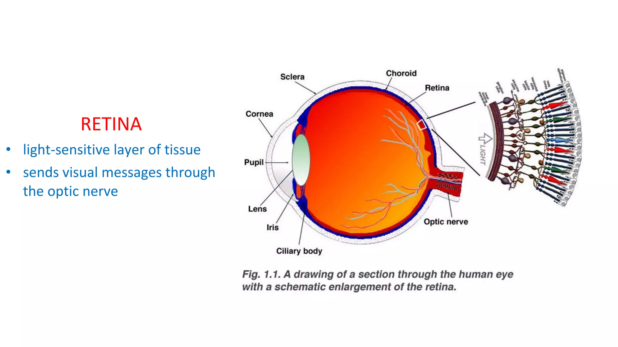
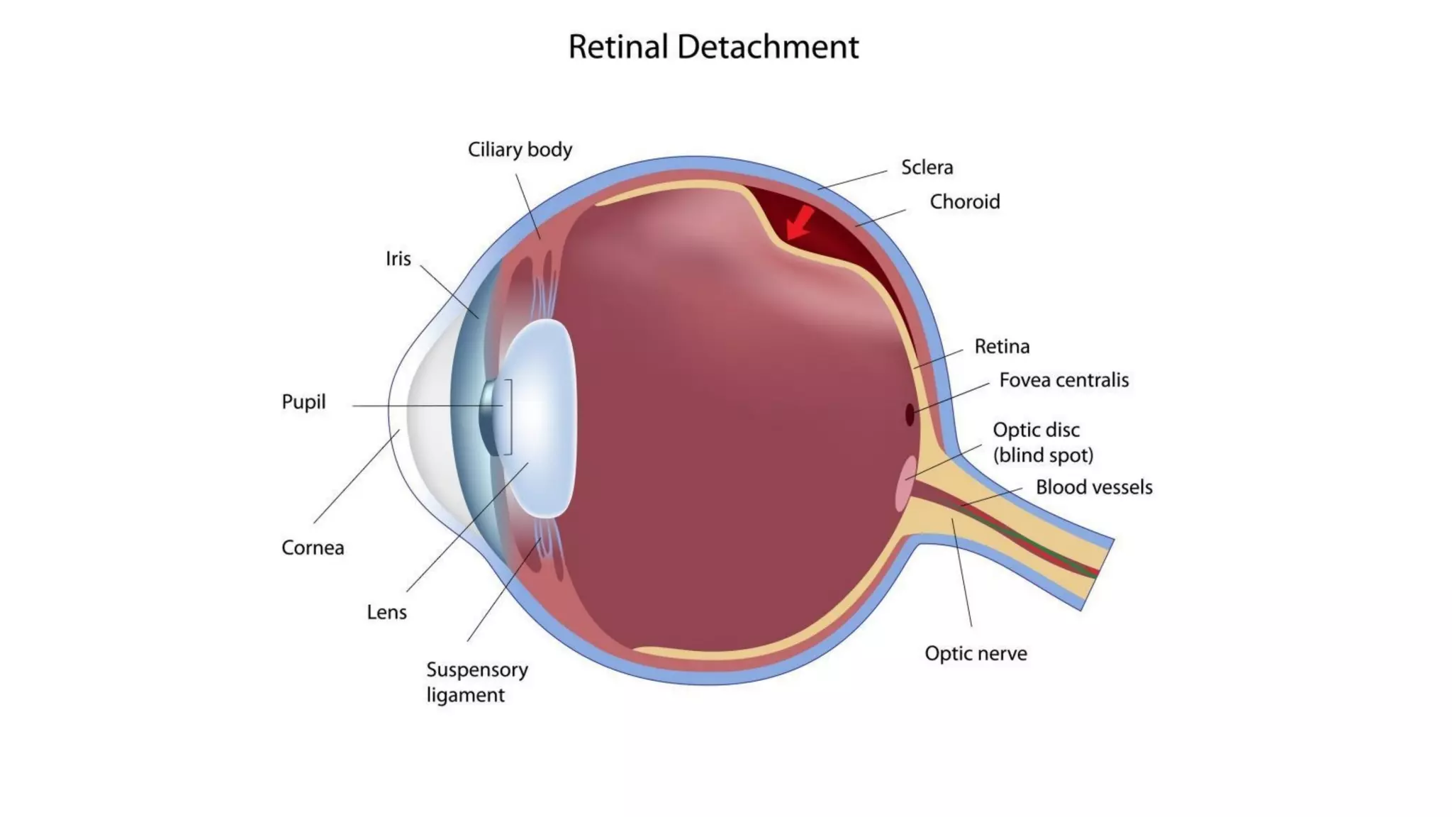
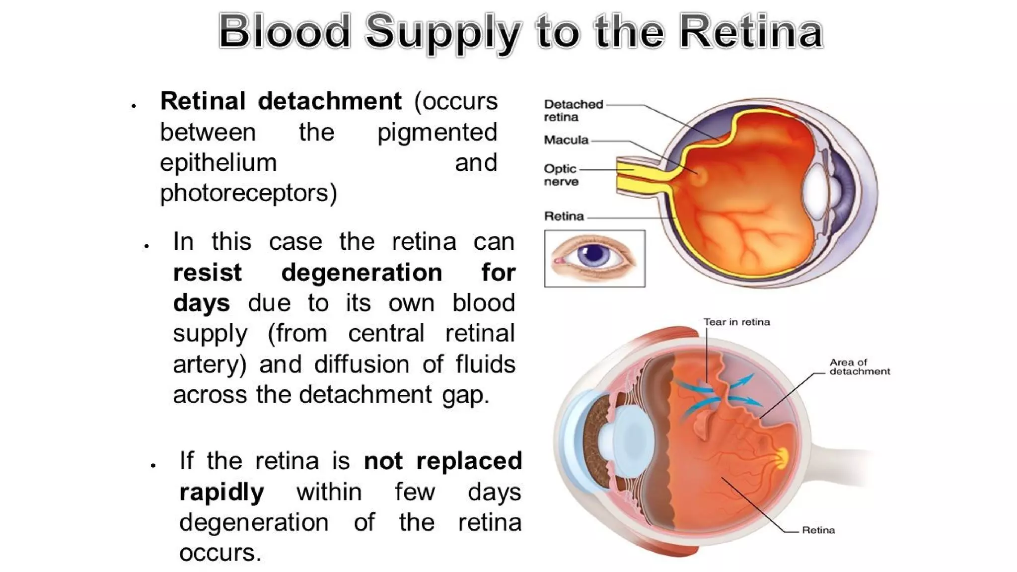
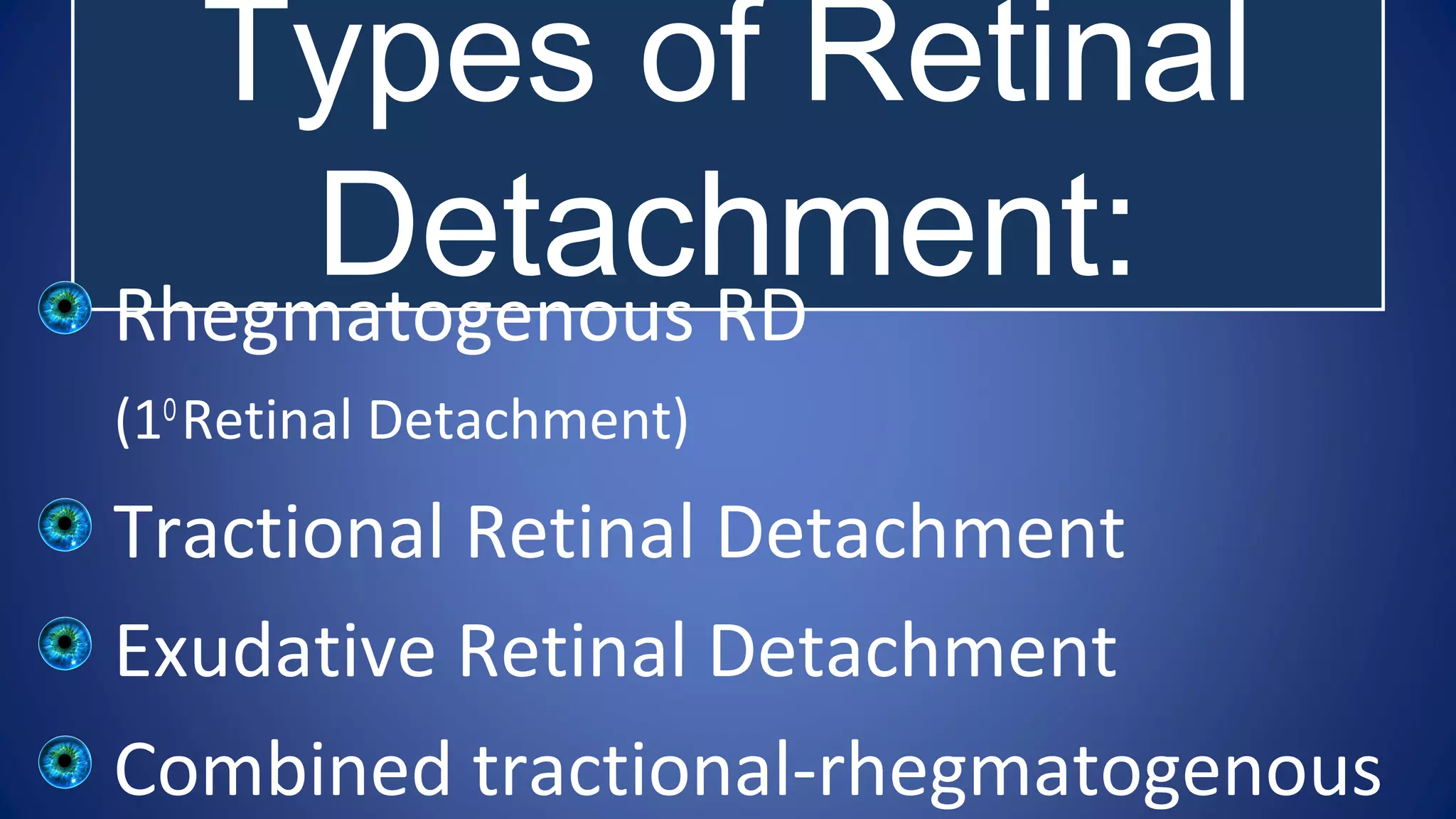
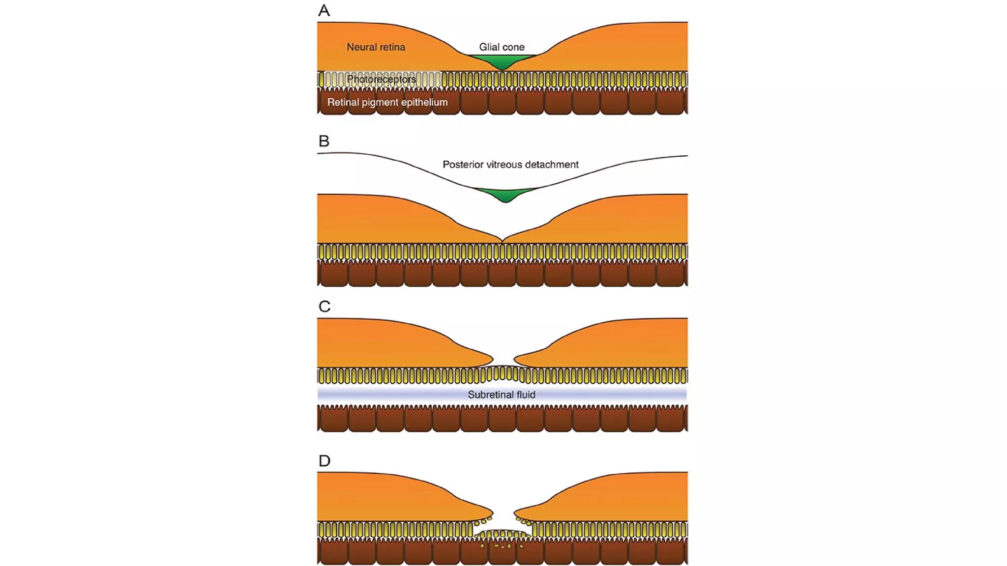
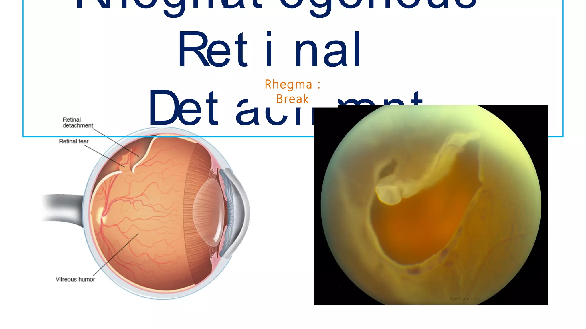
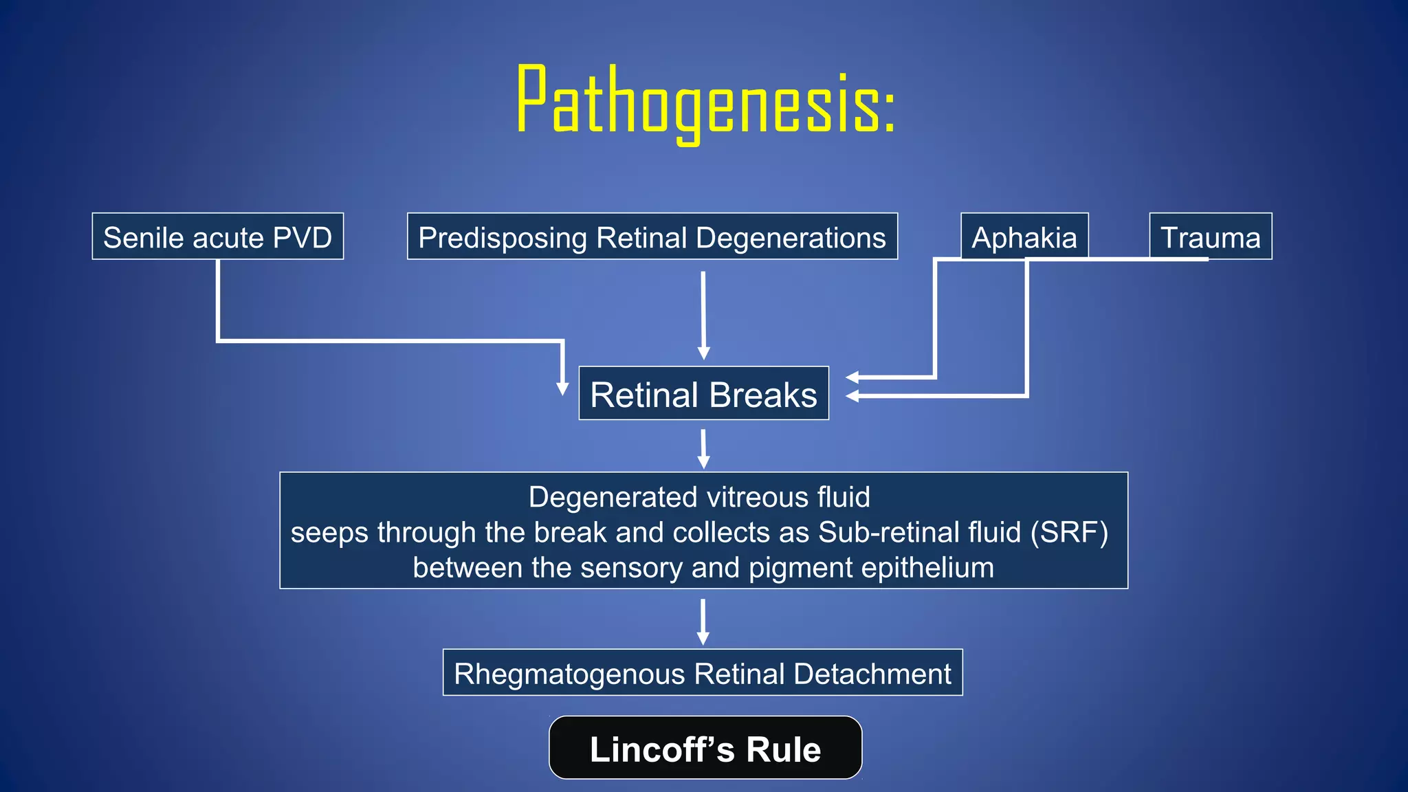
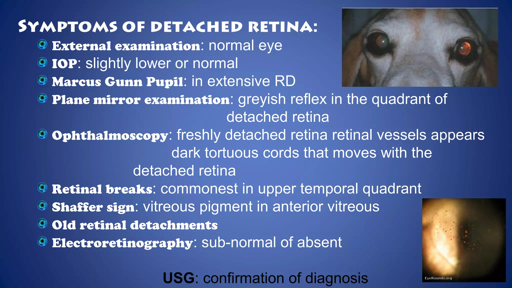
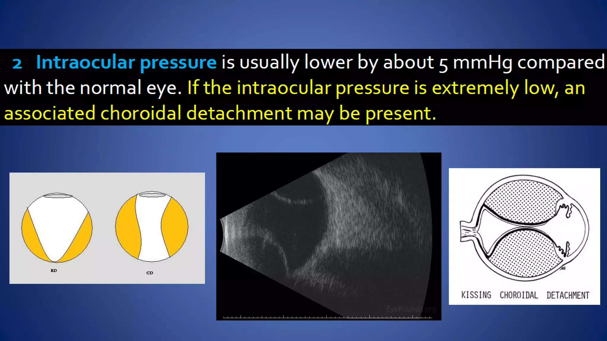
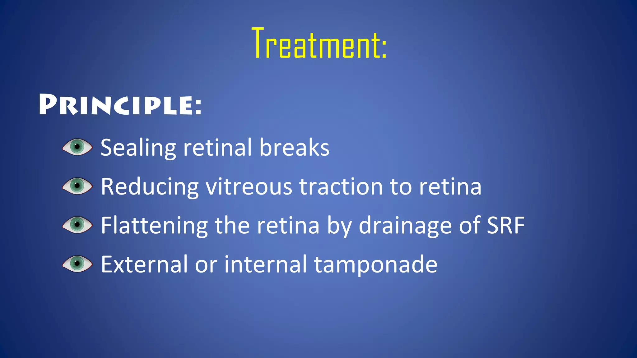
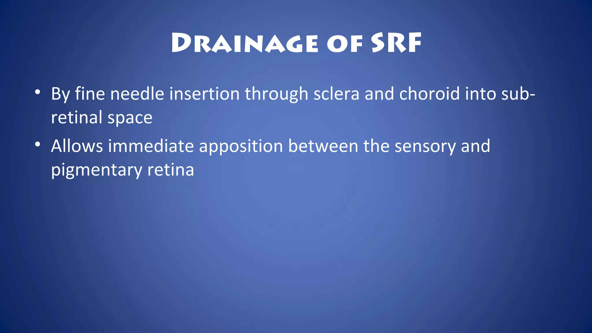
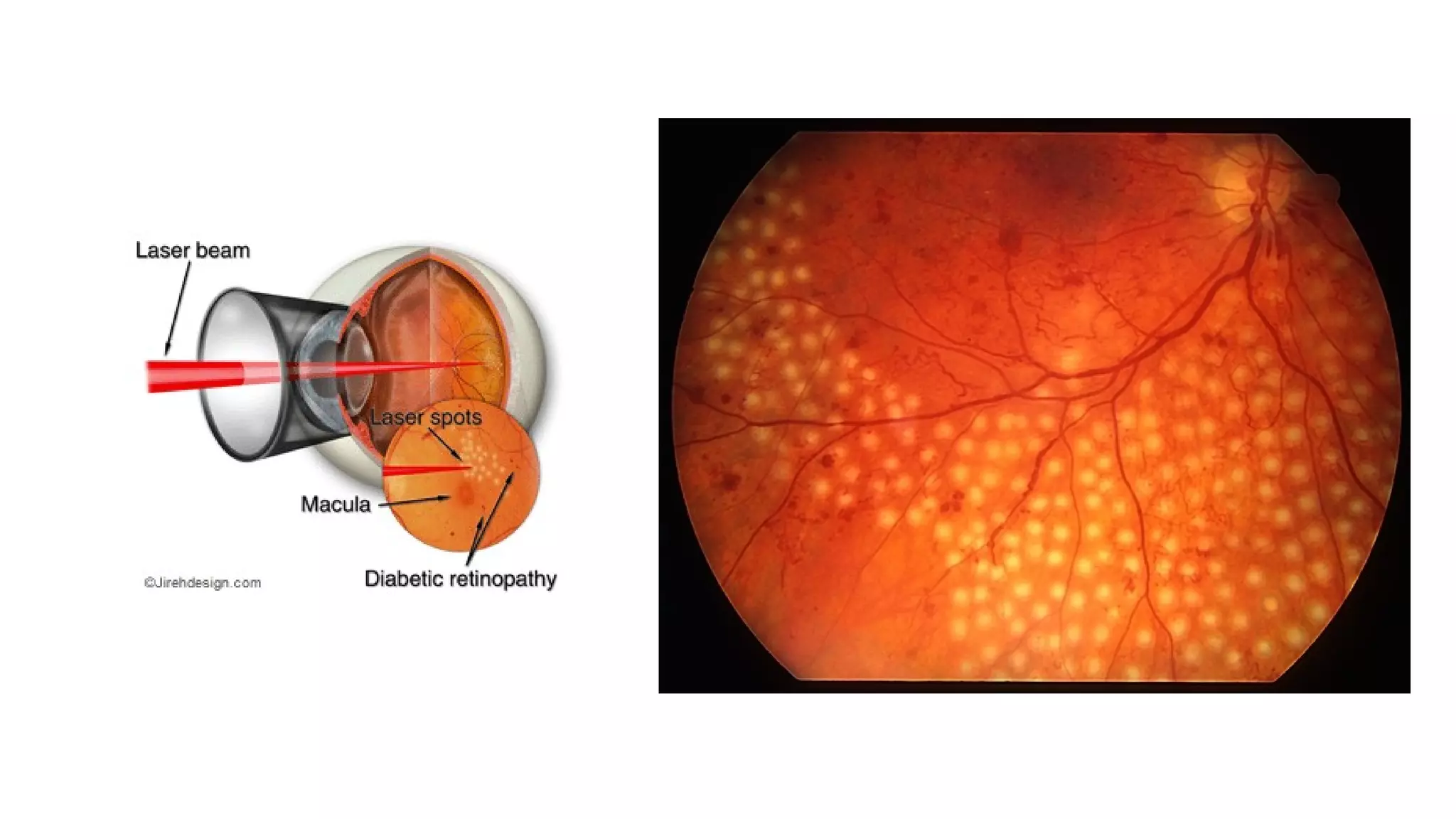
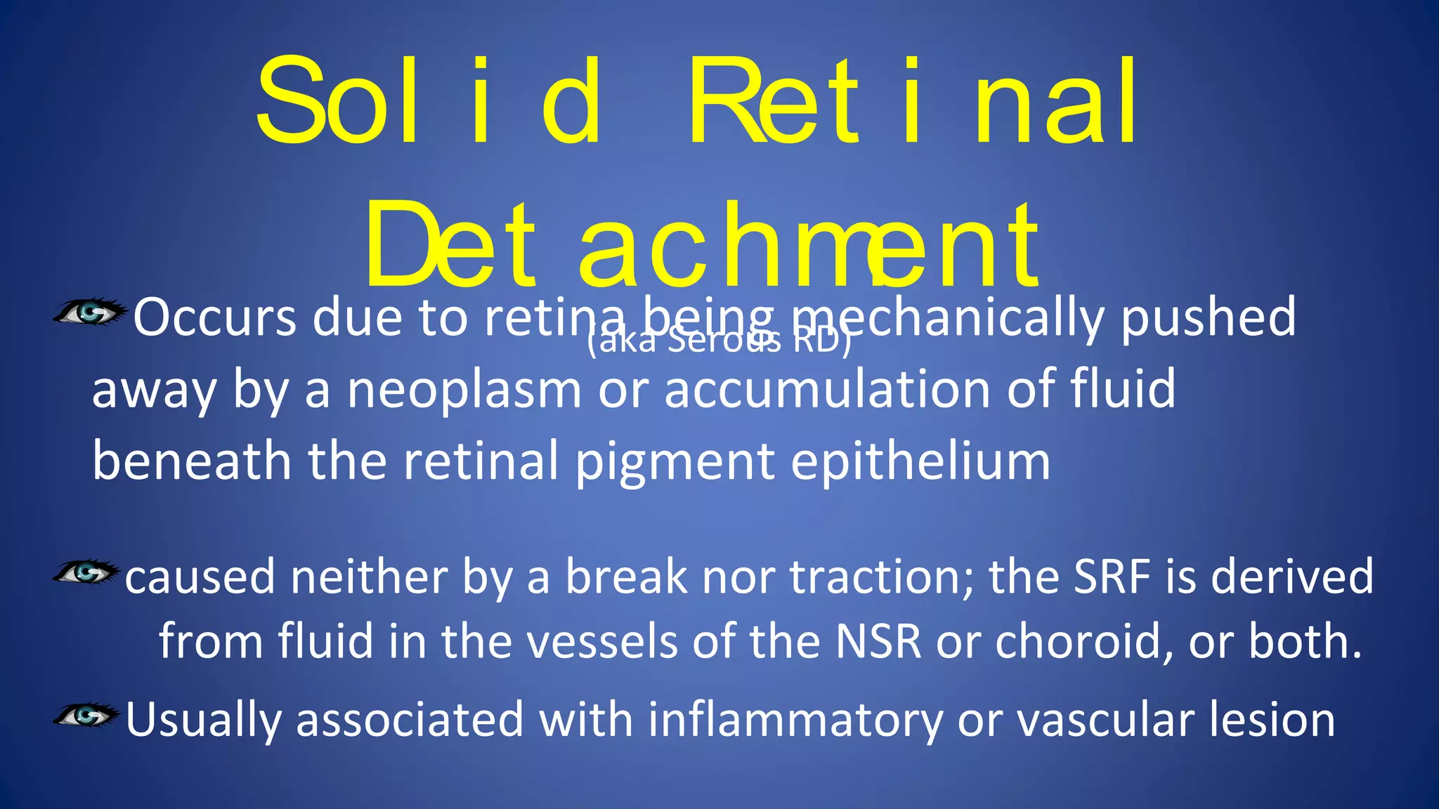
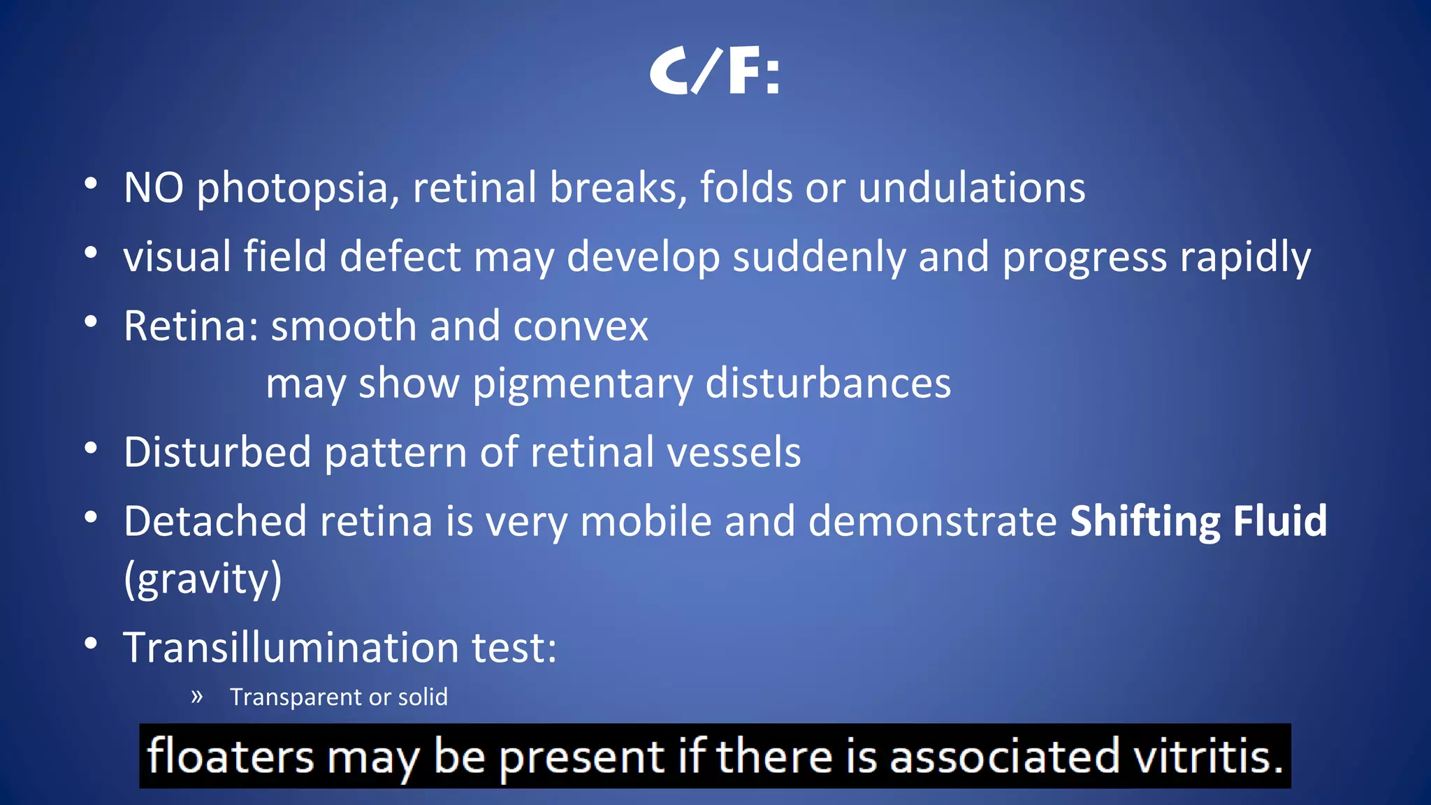
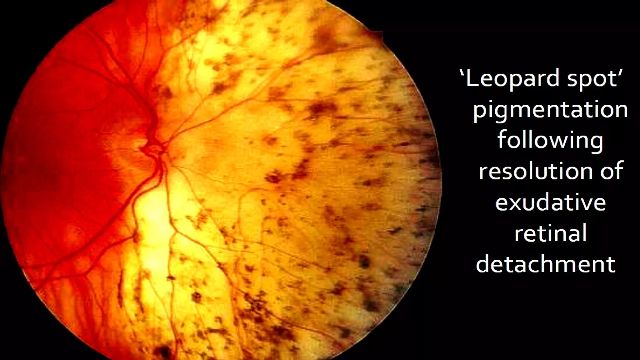
RETINAL
DETACHMENT
Niraj Phoju
RETINA
• light-sensitive layer of tissue
• sends visual messages through
the optic nerve
Retinal Detachment is the separation of the
neurosensory retina from the pigmented epithelium.
• Even normally potential space between the layers
• Thus, a misnomer------”Retinal Separation”
Types of Retinal
Detachment:Rhegmatogenous RD
(1O
Retinal Detachment)
Tractional Retinal Detachment
Exudative Retinal Detachment
Combined tractional-rhegmatogenous
Rhegmat ogenous
Ret i nal
Det achment
Rhegma :
Break
Ret i nal
Det achment
(Commonest variant)
occurs 20
to a full-thickness defect in the sensory retina,
which permits fluid derived from synchytic (liquefied)
vitreous to gain access to the subretinal space.
i.e. associated with retinal breaks (tear or hole) through
which sub-retinal fluid seeps into and separates the
sensory retina from the pigment epithelium
Etiology still not clear
Predisposing Factors:
Age: common in 40-60 yrs, however age is no bar
Sex: M>F
Myopia:
Aphakia and pseudophakia:
Retinal degenerations:
Trauma:
Senile posterior vitreous detachment:
Pathogenesis:
Senile acute PVD Predisposing Retinal Degenerations Aphakia
Retinal Breaks
Trauma
Degenerated vitreous fluid
seeps through the break and collects as Sub-retinal fluid (SRF)
between the sensory and pigment epithelium
Rhegmatogenous Retinal Detachment
Lincoff’s Rule
Prodromal Symptoms:
Floaters (d/t rapid vitreous degeneration)
Photopsia (d/t irritation of retina by vitreous movement)
Symptoms of detached retina:
Localised relative loss in visual field (black curtain)
Sudden appeareance of dark clouds in front of eye
Sudden painless loss of vision
(when detachment is large and central)
C/F:
Symptoms of detached retina:
External examination: normal eye
IOP: slightly lower or normal
Marcus Gunn Pupil: in extensive RD
Plane mirror examination: greyish reflex in the quadrant of
detached retina
Ophthalmoscopy: freshly detached retina retinal vessels appears
dark tortuous cords that moves with the
detached retina
Retinal breaks: commonest in upper temporal quadrant
Shaffer sign: vitreous pigment in anterior vitreous
Old retinal detachments
Electroretinography: sub-normal of absent
USG: confirmation of diagnosis
Proliferative vitreoretinopathy
Complicated cataract
Uveitis
Phithisis bulbi
Complications:
Principle:
Sealing retinal breaks
Reducing vitreous traction to retina
Flattening the retina by drainage of SRF
External or internal tamponade
Treatment:
Sealing retinal breaks
Detection
Accurate localization
Sealing (by aseptic chorioretinitis)
 Photocoagulation
 Cryocoagulation (prefered)
Drainage of SRF
• By fine needle insertion through sclera and choroid into sub-
retinal space
• Allows immediate apposition between the sensory and
pigmentary retina
Maintainence of
chorioretinal apposition
At least for couple of weeks
Many techniques:
 Scleral buckling (external tamponade)
 Pneumatic retinopexy
 Pars plana viterectomy
 Endolaser photocoagulation
 Internal tamponade
Pneumatic Retinopexy
Internal Tamponade
(Perfluorocarbon)
PPV
By preventing retinal break possibility
 Eg.: laser photocoagulation when lattice degenerations is diagnosed
By preventing high risk factors
 Eg.: myopia, aphakia…
Regular eye checkup
Prophylaxix:
Exudat i ve or Sol i d Ret i nal Det achment
Sol i d Ret i nal
Det achment
(aka Serous RD)Occurs due to retina being mechanically pushed
away by a neoplasm or accumulation of fluid
beneath the retinal pigment epithelium
caused neither by a break nor traction; the SRF is derived
from fluid in the vessels of the NSR or choroid, or both.
Usually associated with inflammatory or vascular lesion
Natural Response of the RPE
Etiology:
C/F:
• NO photopsia, retinal breaks, folds or undulations
• visual field defect may develop suddenly and progress rapidly
• Retina: smooth and convex
may show pigmentary disturbances
• Disturbed pattern of retinal vessels
• Detached retina is very mobile and demonstrate Shifting Fluid
(gravity)
• Transillumination test:
» Transparent or solid
Shifting Fluid:
Investigations:
• Complete ocular and systemic examination
• B-scan USG
• FFA:
• shows source of fluid
• CT/MRI
• Esp. useful in case of tumor
Treatment:
• Spontaneous regression after the absorption of the fluid
• Treatment of the cause:
• Enucleation
• Esp. if due to malignancy
Tr act i onal Ret i nal Det achment
• The NSR is pulled away from the RPE by contracting
vitreoretinal membranes in the absence of a retinal break.
• Here retina being mechanically pulled away from its bed by the
contraction of the fibrous tissue in the vitrous (vitreo-retinal
traction band)
Tr act i onal Ret i nal Det achment
Etiology:
• Post-traumatic (penetration trauma)
» Contraction of the scar tissue
• Proliferative diabetic retinopathy
• Post hemorrhagic retinitis proliferans
• ROP
• Retinal dysplasia
• Proliferative retinopathy of Eale’s disease
• Toxocariasis
C/F:
NO photopsia and floaters
Retinal breaks are usually absent
No shifting of fluid
Detached retina : Concave
Presence of vitreoretinal traction bands
Retinal mobility is severly reduced
Treatment:
Surgery
• Usually difficult
• PPV (to cut the traction bands)
• Then internal tamponade (long acting gas or silicon oil)
Usually bad prognosis
Thank You

Retinal Detachment | Ophthalmology

  • 1.
  • 2.
    RETINA • light-sensitive layerof tissue • sends visual messages through the optic nerve
  • 4.
    Retinal Detachment isthe separation of the neurosensory retina from the pigmented epithelium. • Even normally potential space between the layers • Thus, a misnomer------”Retinal Separation”
  • 8.
    Types of Retinal Detachment:RhegmatogenousRD (1O Retinal Detachment) Tractional Retinal Detachment Exudative Retinal Detachment Combined tractional-rhegmatogenous
  • 10.
    Rhegmat ogenous Ret inal Det achment Rhegma : Break
  • 11.
    Ret i nal Detachment (Commonest variant) occurs 20 to a full-thickness defect in the sensory retina, which permits fluid derived from synchytic (liquefied) vitreous to gain access to the subretinal space. i.e. associated with retinal breaks (tear or hole) through which sub-retinal fluid seeps into and separates the sensory retina from the pigment epithelium Etiology still not clear
  • 12.
    Predisposing Factors: Age: commonin 40-60 yrs, however age is no bar Sex: M>F Myopia: Aphakia and pseudophakia: Retinal degenerations: Trauma: Senile posterior vitreous detachment:
  • 13.
    Pathogenesis: Senile acute PVDPredisposing Retinal Degenerations Aphakia Retinal Breaks Trauma Degenerated vitreous fluid seeps through the break and collects as Sub-retinal fluid (SRF) between the sensory and pigment epithelium Rhegmatogenous Retinal Detachment Lincoff’s Rule
  • 14.
    Prodromal Symptoms: Floaters (d/trapid vitreous degeneration) Photopsia (d/t irritation of retina by vitreous movement) Symptoms of detached retina: Localised relative loss in visual field (black curtain) Sudden appeareance of dark clouds in front of eye Sudden painless loss of vision (when detachment is large and central) C/F:
  • 15.
    Symptoms of detachedretina: External examination: normal eye IOP: slightly lower or normal Marcus Gunn Pupil: in extensive RD Plane mirror examination: greyish reflex in the quadrant of detached retina Ophthalmoscopy: freshly detached retina retinal vessels appears dark tortuous cords that moves with the detached retina Retinal breaks: commonest in upper temporal quadrant Shaffer sign: vitreous pigment in anterior vitreous Old retinal detachments Electroretinography: sub-normal of absent USG: confirmation of diagnosis
  • 18.
  • 19.
    Principle: Sealing retinal breaks Reducingvitreous traction to retina Flattening the retina by drainage of SRF External or internal tamponade Treatment:
  • 20.
    Sealing retinal breaks Detection Accuratelocalization Sealing (by aseptic chorioretinitis)  Photocoagulation  Cryocoagulation (prefered)
  • 21.
    Drainage of SRF •By fine needle insertion through sclera and choroid into sub- retinal space • Allows immediate apposition between the sensory and pigmentary retina
  • 22.
    Maintainence of chorioretinal apposition Atleast for couple of weeks Many techniques:  Scleral buckling (external tamponade)  Pneumatic retinopexy  Pars plana viterectomy  Endolaser photocoagulation  Internal tamponade
  • 23.
  • 25.
  • 27.
    By preventing retinalbreak possibility  Eg.: laser photocoagulation when lattice degenerations is diagnosed By preventing high risk factors  Eg.: myopia, aphakia… Regular eye checkup Prophylaxix:
  • 28.
    Exudat i veor Sol i d Ret i nal Det achment
  • 29.
    Sol i dRet i nal Det achment (aka Serous RD)Occurs due to retina being mechanically pushed away by a neoplasm or accumulation of fluid beneath the retinal pigment epithelium caused neither by a break nor traction; the SRF is derived from fluid in the vessels of the NSR or choroid, or both. Usually associated with inflammatory or vascular lesion
  • 30.
  • 31.
  • 32.
    C/F: • NO photopsia,retinal breaks, folds or undulations • visual field defect may develop suddenly and progress rapidly • Retina: smooth and convex may show pigmentary disturbances • Disturbed pattern of retinal vessels • Detached retina is very mobile and demonstrate Shifting Fluid (gravity) • Transillumination test: » Transparent or solid
  • 33.
  • 34.
    Investigations: • Complete ocularand systemic examination • B-scan USG • FFA: • shows source of fluid • CT/MRI • Esp. useful in case of tumor
  • 35.
    Treatment: • Spontaneous regressionafter the absorption of the fluid • Treatment of the cause: • Enucleation • Esp. if due to malignancy
  • 37.
    Tr act ional Ret i nal Det achment
  • 38.
    • The NSRis pulled away from the RPE by contracting vitreoretinal membranes in the absence of a retinal break. • Here retina being mechanically pulled away from its bed by the contraction of the fibrous tissue in the vitrous (vitreo-retinal traction band) Tr act i onal Ret i nal Det achment
  • 39.
    Etiology: • Post-traumatic (penetrationtrauma) » Contraction of the scar tissue • Proliferative diabetic retinopathy • Post hemorrhagic retinitis proliferans • ROP • Retinal dysplasia • Proliferative retinopathy of Eale’s disease • Toxocariasis
  • 41.
    C/F: NO photopsia andfloaters Retinal breaks are usually absent No shifting of fluid Detached retina : Concave Presence of vitreoretinal traction bands Retinal mobility is severly reduced
  • 42.
    Treatment: Surgery • Usually difficult •PPV (to cut the traction bands) • Then internal tamponade (long acting gas or silicon oil) Usually bad prognosis
  • 43.

Editor's Notes

  • #12 The retinal breaks responsible for RD are caused by interplay between dynamic vitreoretinal traction and an underlying weakness in the peripheral retina referred to as predisposing degeneration.
  • #13 Retinal Degenerations: lattice degeneration snail tract degeneration