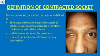
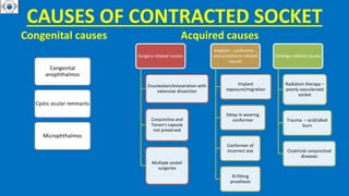
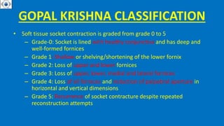
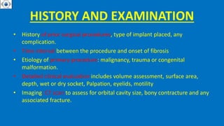
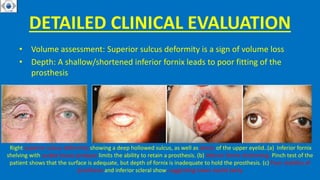
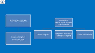
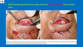
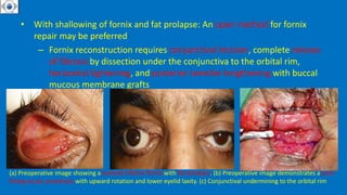
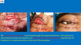
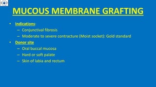
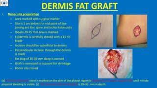
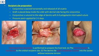
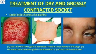
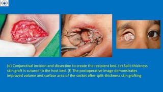
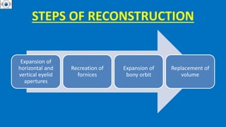
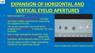
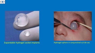
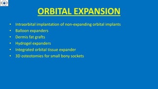
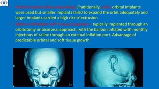
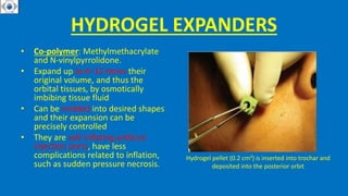
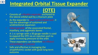
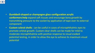
CONTRACTED SOCKET
DEFINITION OF IDEAL SOCKET
An Ideal Anophthalmic Socket
– A socket lined with healthy
conjunctival epithelium
– Centrally placed implant of adequate
volume
– Adequately deep inferior and
superior fornices to retain the
prosthesis
– Optimal implant-prosthesis coupling
to ensure motility of the artificial
eye
– Normal position and adequate tone
of eyelids
– Well-fitted prosthesis that looks
similar to the contralateral eye
DEFINITION OF CONTRACTED SOCKET
Contracted socket, or socket contracture, is defined
as
• Shrinkage and shortening of all or a part of
orbital tissues causing a decrease in depth of
the fornices and orbital volume
• Inability to retain an ocular prosthesis
• It can either be due to soft tissue or bony
contraction.
CAUSES OF CONTRACTED SOCKET
Congenital causes
Congenital
anophthalmos
Cystic ocular remnants
Microphthalmos
Acquired causes
Surgery-related causes
Enucleation/evisceration with
extensive dissection
Conjunctiva and
Tenon’s capsule
not preserved
Multiple socket
surgeries
Implant-, conformer-,
and prosthesis-related
causes
Implant
exposure/migration
Delay in wearing
conformer
Conformer of
incorrect size
Ill-fitting
prosthesis
Etiology-related causes
Radiation therapy –
poorly vascularized
socket
Trauma – acid/alkali
burn
Cicatricial conjunctival
diseases
IMPORTANCE OF PLACING IMPLANT
• Appropriately sized orbital implant help reducing the risk of post-
enucleation socket syndrome from volume loss after enucleation or
evisceration
– The average volume of the globe is 7.2 ml, and the combined volume
of the implant and prosthesis should equal the volume of the removed
globe
– The average volume of a prosthesis is 2.5 ml; thus the implant needs
to equal almost two thirds of the volume of the globe
– Increasing the volume of the prosthesis may result in mechanical
ectropion of the lower eyelid from the heavier weight
POST ENUCLEATION SOCKET
SYNDROME
• Deepening of the upper eyelid
sulcus (superior sulcus
deformity)
• Upper eyelid ptosis (with upper
eyelid dysfunction/
lagophthalmos)
• Lower eyelid laxity and
ectropion
GOPAL KRISHNA CLASSIFICATION
• Soft tissue socket contraction is graded from grade 0 to 5
– Grade-0: Socket is lined with healthy conjunctiva and has deep and
well-formed fornices
– Grade 1: Shallow or shelving/shortening of the lower fornix
– Grade 2: Loss of upper and lower fornices
– Grade 3: Loss of upper, lower, medial and lateral fornices
– Grade 4: Loss of all fornices and reduction of palpebral aperture in
horizontal and vertical dimensions
– Grade 5: Recurrence of socket contracture despite repeated
reconstruction attempts
OTHER CLASSIFICATION
• Mild: Only one fornix is involved (usually lower fornix) + shortening of
posterior lamellae of the eyelids
• Moderate: Both superior + inferior fornices are involved (decrease in area)
• Severe: All fornices are involved + phimosis of palpebral aperture (loss of
area + volume)
• Malignant: Severe loss of area, volume, with associated bony contracture
CLASSIFICATION OF SOCKET
CONTRACTURE WAS DESCRIBED BY
TAWFIK ET AL.
• Grade 1: Minimal or no actual contracture . Complains : Inability to retain
the prosthesis (horizontal eyelid laxity with subsequent prolapse of the
inferior fornix )
• Grade 2: Mild contracture of the inferior or superior fornix. Complain :
Rolling-in of the upper and lower eyelid margins
• Grade 3: Scarring is more advanced . Complaint : Impossible to wear the
prosthesis. Cicatrization involves the entire upper and lower fornices
• Grade 4: Severe phimosis of the palpebral fissure both vertically and
horizontally. H/o : Recurrent cases
HISTORY AND EXAMINATION
• History of prior surgical procedures, type of implant placed, any
complication.
• Time interval between the procedure and onset of fibrosis
• Etiology of primary procedure: malignancy, trauma or congenital
malformation.
• Detailed clinical evaluation includes volume assessment, surface area,
depth, wet or dry socket, Palpation, eyelids, motility
• Imaging: CT scan to assess for orbital cavity size, bony contracture and any
associated fracture.
DETAILED CLINICAL EVALUATION
• Volume assessment: Superior sulcus deformity is a sign of volume loss
• Depth: A shallow/shortened inferior fornix leads to poor fitting of the
prosthesis
Right superior sulcus deformity showing a deep hollowed sulcus, as well as ptosis of the upper eyelid..(a) Inferior fornix
shelving with socket tissue prolapse limits the ability to retain a prosthesis. (b) Inferior fornix shortening. Pinch test of the
patient shows that the surface is adequate, but depth of fornix is inadequate to hold the prosthesis. (c) Poor stability of
prosthesis and inferior scleral show, suggesting lower eyelid laxity
Surface area of the socket and depth of the fornices are noted .Assess the
socket lining
Fibrous bands and symblepharon in the socket.. Multiple granuloma formation in the socket may be secondary to poor
tissue closure technique, poor prosthesis fit with chronic surface irritation, or other inflammatory etiology…Large
exposure of a silicone implant
• Moisture: Dry or wet
• Palpation: Presence and position of the implant. An inferiorly displaced
implant can often obliterate the inferior fornix
• Eyelids: Excessive eyelid or canthal tendon laxity that should be addressed
• Motility: Extraocular movements and tone of the orbicularis muscle
• CT scan
– to assess for orbital cavity size (hypoplastic in congenital anophthalmos),
bony contracture, and associated orbital fractures contributing to a
sunken appearance (cases with previous trauma)
– to ascertain the presence, size, and position of an orbital implant
MANAGEMENT OF CONTRACTED
SOCKET
AIM :to create a healthy socket which is able to hold stable
ocular prosthesis along with reasonable symmetry of
palpebral apertures ,canthal angles and superior sulci
AQUIRED CONTRACTED SOCKET
MANAGEMENT
INADEQUATE SURFACE
FORNIX FORMATION SUTURES
LID LAXITY: TARSAL STRIP
PROCEDURE
MUCOUS MEMBRANE GRAFT
AMNIOTIC MEMBRANE
GRAFTING
SPLIT SKIN GRAFT
INADEQUATE VOLUME
Intraconal implant
Dermis fat graft
COMBINED-
INADEQUATE SURFACE
AND VOLUME
Dermis fat graft
Temporalis muscle flap
with split skin graft
Radial forearm flap
MILD CONTRACTED SOCKET:
– Inability to retain prosthesis
Cause: lower eyelid laxity with minimal shortening of fornix
With adequate fornix:
• Treatment options
– Transverse blepharotomy with marginal rotation of eyelid
– Horizontal eyelid shortening, if lid laxity coexist
• With shortening of fornix (usually inferior): Closed method fornix repair
done
• With shallowing of fornix and fat prolapse: An open method for fornix
repair may be preferred
– Fornix reconstruction requires conjunctival incision, complete release
of fibrosis by dissection under the conjunctiva to the orbital rim,
horizontal tightening, and posterior lamellar lengthening with buccal
mucous membrane grafts
(a) Preoperative image showing a shelved inferior fornix with fat prolapse. (b) Preoperative image demonstrates a poor
fitting ocular prosthesis with upward rotation and lower eyelid laxity. (c) Conjunctival undermining to the orbital rim
• .
(d)Fornix-deepening sutures are passed through the conjunctival fornix . The sutures are
externalized and tied over bolsters. (e) Lateral canthal tendon tightening is performed. (f)
Prosthesis is in optimal position at the end of the procedure
MUCOUS MEMBRANE GRAFTING
• Indications:
– Conjunctival fibrosis
– Moderate to severe contracture (Moist socket): Gold standard
• Donor site
– Oral buccal mucosa
– Hard or soft palate
– Skin of labia and rectum
• Preparation of donor site:
– Site is marked with a surgical pen
– Infiltrated with lidocaine plus adrenaline
– Stensen duct identified
– Full thickness graft is removed with a 15 Bard-Parker blade
– Ideal graft should be 25% larger than the defect
– Donor site closed
HARVESTING OF BUCCAL MUCOUS
MEMBRANE GRAFT
(a) Harvesting of buccal mucosal graft from inside the lower lip. (b) Submucosal tissue
is trimmed from the posterior aspect of the graft
• Preparation of Recipient site:
– Site is infiltrated with 2% lidocaine plus adrenaline
– Conjunctiva is incised horizontally below the tarsus
– Dissection of conjunctiva from underlying cicatrix and fibrotic tissue
– Subconjunctival dissection done until lower lid is freely mobile
– MMG is placed with mucosal side facing ocular surface
– Sutured to recipient fornix and conjunctiva with 6-0 polyglactin suture
– Fornix reformation sutures are passes and tied on the skin through
bolsters
• .
(c) Dissection is performed in the inferior fornix to release the scar tissue.(d) The
harvested mucous membrane graft is trimmed and sutured to the host bed with
polyglactin or chromic gut sutures (e) Postoperative photo demonstrates adequate
surface and improvement in fornix depth
DERMAL FAT GRAFTING
• Autologous implant
• It consist of the de-epithelialized dermis layer that is attached to and supplies
nutrients to the adjacent subcutaneous fat tissue
• Readily available, inexpensive and negligible allergy risk
• Indication
– Volume and surface loss in moderate to severe contracted socket
• Donor site
– Gluteal region (upper outer)
– Lower abdomen
– Inner thigh
DERMIS FAT GRAFT
• Donor site preparation
– Area marked with surgical marker
– Site is 5 cm below the mid point of line
joining ant iliac spine and ischial tuberosity
– Ideally 20-25 mm area is marked
– Epidermis is carefully shaved with a 15 no
blade
– Incision should be superficial to dermis
– Perpendicular incision through the dermis
is made
– Fat plug of 20-30 mm deep is excised
– Graft is oversized to account for shrinkage
– Donor site closed
(a) A 25 mm diameter circle is marked on the skin of the gluteal region(b) Epidermis is incised superficially until minute
pinpoint bleeding is visible. (c) The harvested dermis fat graft is 20–30 mm in depth.
• Recipient site preparation
– Conjunctiva is excised horizontally and released of all cicatrix
– Graft is placed deep inside the orbit with dermal side facing the conjunctiva
– Conjunctiva is sutured to the edge of dermis with 6-0 polygalactin interrupted suture
– Pressure patch applied for 2-3 days.
Subconjunctival dissection is performed to prepare the host bed. (e) The dermis fat graft is
transferred to the orbital recipient site. (f) The dermis fat graft completely sutured into the socket
without excessive tension
COMPLICATION OF DERMAL FAT
GRAFT
• Atrophy of fat graft
• Pressure necrosis of graft
• Donor site wound dehiscence
• Scar formation and deformity
GRADE V CONTRACTED SOCKET
• Difficult to manage: Recalcitrant/ Malignant socket
• Characterized by socket that has underwent multiple procedures
• Recommendations for management:
– Exenteration
– Customised osseo-integrated facial prosthesis
TREATMENT OF DRY AND GROSSLY
CONTRACTED SOCKET
• Socket Split-thickness skin grafting
(a) Split-thickness skin graft is harvested from the inner aspect of the thigh. (b)
Harvested split-thickness graft is demonstrated. (c) Grossly contracted socket
• .
(d) Conjunctival incision and dissection to create the recipient bed. (e) Split-thickness
skin graft is sutured to the host bed. (f) The postoperative image demonstrates
improved volume and surface area of the socket after split-thickness skin grafting
CONGENITAL CONTRACTED SOCKET
Congenital contracted socket
• In both Anophthalmos and Microphthalmos, there is
– Shortening of the vertical and horizontal dimensions of eyelids leading
to phimosis
– Shortened conjunctival fornices with deficiency of palpebral and
bulbar conjunctiva
– Hypoplasia of the bony orbit, and facial asymmetry due to reduction in
soft tissue volume
• Treatment should commence as early as possible as early intervention will
help stimulate the growth of the orbital bones and the periocular and
midfacial tissues
STEPS OF RECONSTRUCTION
Expansion of
horizontal and
vertical eyelid
apertures
Recreation of
fornices
Expansion of
bony orbit
Replacement of
volume
EXPANSION OF HORIZONTAL AND
VERTICAL EYELID APERTURES
• Gold standard of serial conformers
• Expanding hydrogel conformers can also
stimulate socket expansion for expanding
the palpebral fissures
• The socket expanders are small when dry,
ranging from 6 to 9 mm in diameter, for easy
insertion
• Due to high hydrophilic properties, they
expand to 11, 14, and 18 mm when fully
hydrated, which typically takes 2–4 weeks.
• Can be replaced with a larger expanding
conformer for additional results
Serial conformers used to expand socket
Expandable hydrogel socket implants Hydrogel sphere in conjunctival cul-de-sac
ORBITAL EXPANSION
• Intraorbital implantation of non-expanding orbital implants
• Balloon expanders
• Dermis fat grafts
• Hydrogel expanders
• Integrated orbital tissue expander
• 3D osteotomies for small bony sockets
• Orbital Implants (Non-expanding) :Traditionally, static orbital implants
were used but smaller implants failed to expand the orbit adequately and
larger implants carried a high risk of extrusion
• Balloon (Inflatable Soft Tissue) Expanders: typically implanted through an
orbitotomy or bicoronal approach, with the balloon inflated with monthly
injections of saline through an external inflation port. Advantage of
predictable orbital and soft tissue growth
HYDROGEL EXPANDERS
• Co-polymer: Methylmethacrylate
and N-vinylpyrrolidone.
• Expand up to 6–12 times their
original volume, and thus the
orbital tissues, by osmotically
imbibing tissue fluid
• Can be molded into desired shapes
and their expansion can be
precisely controlled
• They are self-inflating without
injection ports, have less
complications related to inflation,
such as sudden pressure necrosis.
Hydrogel pellet (0.2 cm3) is inserted into trochar and
deposited into the posterior orbit
Integrated Orbital Tissue Expander
(OTE)
• Consists of a flexible expander anchored to
the lateral orbital wall by a titanium plate
• As the expander is fixed to the orbital wall, it
has the advantage of sustained and
unidirectional expansion
• Induce growth of the associated frontal,
maxillary, and zygomatic bones
• A 1 cc syringe with a 30 gauge needle is used
to expand the OTE via an injection port to
apply increasing pressure to the orbit
• These expanders can therefore be inflated
and deflated, as needed
• Safe and effective in managing the
anophthalmic socket with good long-term
outcome
3D OSTEOTOMIES FOR SMALL BONY
SOCKETS
• C- and U-shaped osteotomies can be created to expand the roof, lateral
wall, and orbital floor
• The osteotomy segments are advanced forward and away from the center
of the orbital cavity
• The orbital floor and rim are placed at a higher level compared to the
normal side to provide better support to the prosthesis
• Bone grafts are used to fill the defect
THAKYOU 
REFERENCES:2019_BOOK_OCULOFACIALORBITALANDLACRIMAL
SURGERY
• Dumbbell-shaped or champagne glass configuration acrylic
conformers:help expand soft tissues and encourage bony growth by
transmitting pressure to the socket by application of tape over its external
component
• Custom scleral shells: can be useful in cases of microphthalmic eyes to
promote orbital growth. Custom clear shells can be made for mild to
moderate microphthalmia with positive response to visual evoked
potential testing, in order to allow the eye to achieve its maximum visual
potential

CONTRACTED_SOCKET presentation DHB .pptx

  • 1.
  • 2.
    DEFINITION OF IDEALSOCKET An Ideal Anophthalmic Socket – A socket lined with healthy conjunctival epithelium – Centrally placed implant of adequate volume – Adequately deep inferior and superior fornices to retain the prosthesis – Optimal implant-prosthesis coupling to ensure motility of the artificial eye – Normal position and adequate tone of eyelids – Well-fitted prosthesis that looks similar to the contralateral eye
  • 3.
    DEFINITION OF CONTRACTEDSOCKET Contracted socket, or socket contracture, is defined as • Shrinkage and shortening of all or a part of orbital tissues causing a decrease in depth of the fornices and orbital volume • Inability to retain an ocular prosthesis • It can either be due to soft tissue or bony contraction.
  • 4.
    CAUSES OF CONTRACTEDSOCKET Congenital causes Congenital anophthalmos Cystic ocular remnants Microphthalmos Acquired causes Surgery-related causes Enucleation/evisceration with extensive dissection Conjunctiva and Tenon’s capsule not preserved Multiple socket surgeries Implant-, conformer-, and prosthesis-related causes Implant exposure/migration Delay in wearing conformer Conformer of incorrect size Ill-fitting prosthesis Etiology-related causes Radiation therapy – poorly vascularized socket Trauma – acid/alkali burn Cicatricial conjunctival diseases
  • 5.
    IMPORTANCE OF PLACINGIMPLANT • Appropriately sized orbital implant help reducing the risk of post- enucleation socket syndrome from volume loss after enucleation or evisceration – The average volume of the globe is 7.2 ml, and the combined volume of the implant and prosthesis should equal the volume of the removed globe – The average volume of a prosthesis is 2.5 ml; thus the implant needs to equal almost two thirds of the volume of the globe – Increasing the volume of the prosthesis may result in mechanical ectropion of the lower eyelid from the heavier weight
  • 6.
    POST ENUCLEATION SOCKET SYNDROME •Deepening of the upper eyelid sulcus (superior sulcus deformity) • Upper eyelid ptosis (with upper eyelid dysfunction/ lagophthalmos) • Lower eyelid laxity and ectropion
  • 7.
    GOPAL KRISHNA CLASSIFICATION •Soft tissue socket contraction is graded from grade 0 to 5 – Grade-0: Socket is lined with healthy conjunctiva and has deep and well-formed fornices – Grade 1: Shallow or shelving/shortening of the lower fornix – Grade 2: Loss of upper and lower fornices – Grade 3: Loss of upper, lower, medial and lateral fornices – Grade 4: Loss of all fornices and reduction of palpebral aperture in horizontal and vertical dimensions – Grade 5: Recurrence of socket contracture despite repeated reconstruction attempts
  • 8.
    OTHER CLASSIFICATION • Mild:Only one fornix is involved (usually lower fornix) + shortening of posterior lamellae of the eyelids • Moderate: Both superior + inferior fornices are involved (decrease in area) • Severe: All fornices are involved + phimosis of palpebral aperture (loss of area + volume) • Malignant: Severe loss of area, volume, with associated bony contracture
  • 9.
    CLASSIFICATION OF SOCKET CONTRACTUREWAS DESCRIBED BY TAWFIK ET AL. • Grade 1: Minimal or no actual contracture . Complains : Inability to retain the prosthesis (horizontal eyelid laxity with subsequent prolapse of the inferior fornix ) • Grade 2: Mild contracture of the inferior or superior fornix. Complain : Rolling-in of the upper and lower eyelid margins • Grade 3: Scarring is more advanced . Complaint : Impossible to wear the prosthesis. Cicatrization involves the entire upper and lower fornices • Grade 4: Severe phimosis of the palpebral fissure both vertically and horizontally. H/o : Recurrent cases
  • 10.
    HISTORY AND EXAMINATION •History of prior surgical procedures, type of implant placed, any complication. • Time interval between the procedure and onset of fibrosis • Etiology of primary procedure: malignancy, trauma or congenital malformation. • Detailed clinical evaluation includes volume assessment, surface area, depth, wet or dry socket, Palpation, eyelids, motility • Imaging: CT scan to assess for orbital cavity size, bony contracture and any associated fracture.
  • 11.
    DETAILED CLINICAL EVALUATION •Volume assessment: Superior sulcus deformity is a sign of volume loss • Depth: A shallow/shortened inferior fornix leads to poor fitting of the prosthesis Right superior sulcus deformity showing a deep hollowed sulcus, as well as ptosis of the upper eyelid..(a) Inferior fornix shelving with socket tissue prolapse limits the ability to retain a prosthesis. (b) Inferior fornix shortening. Pinch test of the patient shows that the surface is adequate, but depth of fornix is inadequate to hold the prosthesis. (c) Poor stability of prosthesis and inferior scleral show, suggesting lower eyelid laxity
  • 12.
    Surface area ofthe socket and depth of the fornices are noted .Assess the socket lining Fibrous bands and symblepharon in the socket.. Multiple granuloma formation in the socket may be secondary to poor tissue closure technique, poor prosthesis fit with chronic surface irritation, or other inflammatory etiology…Large exposure of a silicone implant
  • 13.
    • Moisture: Dryor wet • Palpation: Presence and position of the implant. An inferiorly displaced implant can often obliterate the inferior fornix • Eyelids: Excessive eyelid or canthal tendon laxity that should be addressed • Motility: Extraocular movements and tone of the orbicularis muscle • CT scan – to assess for orbital cavity size (hypoplastic in congenital anophthalmos), bony contracture, and associated orbital fractures contributing to a sunken appearance (cases with previous trauma) – to ascertain the presence, size, and position of an orbital implant
  • 14.
    MANAGEMENT OF CONTRACTED SOCKET AIM:to create a healthy socket which is able to hold stable ocular prosthesis along with reasonable symmetry of palpebral apertures ,canthal angles and superior sulci
  • 15.
    AQUIRED CONTRACTED SOCKET MANAGEMENT INADEQUATESURFACE FORNIX FORMATION SUTURES LID LAXITY: TARSAL STRIP PROCEDURE MUCOUS MEMBRANE GRAFT AMNIOTIC MEMBRANE GRAFTING SPLIT SKIN GRAFT
  • 16.
    INADEQUATE VOLUME Intraconal implant Dermisfat graft COMBINED- INADEQUATE SURFACE AND VOLUME Dermis fat graft Temporalis muscle flap with split skin graft Radial forearm flap
  • 17.
    MILD CONTRACTED SOCKET: –Inability to retain prosthesis Cause: lower eyelid laxity with minimal shortening of fornix With adequate fornix: • Treatment options – Transverse blepharotomy with marginal rotation of eyelid – Horizontal eyelid shortening, if lid laxity coexist
  • 18.
    • With shorteningof fornix (usually inferior): Closed method fornix repair done
  • 19.
    • With shallowingof fornix and fat prolapse: An open method for fornix repair may be preferred – Fornix reconstruction requires conjunctival incision, complete release of fibrosis by dissection under the conjunctiva to the orbital rim, horizontal tightening, and posterior lamellar lengthening with buccal mucous membrane grafts (a) Preoperative image showing a shelved inferior fornix with fat prolapse. (b) Preoperative image demonstrates a poor fitting ocular prosthesis with upward rotation and lower eyelid laxity. (c) Conjunctival undermining to the orbital rim
  • 20.
    • . (d)Fornix-deepening suturesare passed through the conjunctival fornix . The sutures are externalized and tied over bolsters. (e) Lateral canthal tendon tightening is performed. (f) Prosthesis is in optimal position at the end of the procedure
  • 21.
    MUCOUS MEMBRANE GRAFTING •Indications: – Conjunctival fibrosis – Moderate to severe contracture (Moist socket): Gold standard • Donor site – Oral buccal mucosa – Hard or soft palate – Skin of labia and rectum
  • 22.
    • Preparation ofdonor site: – Site is marked with a surgical pen – Infiltrated with lidocaine plus adrenaline – Stensen duct identified – Full thickness graft is removed with a 15 Bard-Parker blade – Ideal graft should be 25% larger than the defect – Donor site closed
  • 23.
    HARVESTING OF BUCCALMUCOUS MEMBRANE GRAFT (a) Harvesting of buccal mucosal graft from inside the lower lip. (b) Submucosal tissue is trimmed from the posterior aspect of the graft
  • 24.
    • Preparation ofRecipient site: – Site is infiltrated with 2% lidocaine plus adrenaline – Conjunctiva is incised horizontally below the tarsus – Dissection of conjunctiva from underlying cicatrix and fibrotic tissue – Subconjunctival dissection done until lower lid is freely mobile – MMG is placed with mucosal side facing ocular surface – Sutured to recipient fornix and conjunctiva with 6-0 polyglactin suture – Fornix reformation sutures are passes and tied on the skin through bolsters
  • 25.
    • . (c) Dissectionis performed in the inferior fornix to release the scar tissue.(d) The harvested mucous membrane graft is trimmed and sutured to the host bed with polyglactin or chromic gut sutures (e) Postoperative photo demonstrates adequate surface and improvement in fornix depth
  • 26.
    DERMAL FAT GRAFTING •Autologous implant • It consist of the de-epithelialized dermis layer that is attached to and supplies nutrients to the adjacent subcutaneous fat tissue • Readily available, inexpensive and negligible allergy risk • Indication – Volume and surface loss in moderate to severe contracted socket • Donor site – Gluteal region (upper outer) – Lower abdomen – Inner thigh
  • 27.
    DERMIS FAT GRAFT •Donor site preparation – Area marked with surgical marker – Site is 5 cm below the mid point of line joining ant iliac spine and ischial tuberosity – Ideally 20-25 mm area is marked – Epidermis is carefully shaved with a 15 no blade – Incision should be superficial to dermis – Perpendicular incision through the dermis is made – Fat plug of 20-30 mm deep is excised – Graft is oversized to account for shrinkage – Donor site closed (a) A 25 mm diameter circle is marked on the skin of the gluteal region(b) Epidermis is incised superficially until minute pinpoint bleeding is visible. (c) The harvested dermis fat graft is 20–30 mm in depth.
  • 28.
    • Recipient sitepreparation – Conjunctiva is excised horizontally and released of all cicatrix – Graft is placed deep inside the orbit with dermal side facing the conjunctiva – Conjunctiva is sutured to the edge of dermis with 6-0 polygalactin interrupted suture – Pressure patch applied for 2-3 days. Subconjunctival dissection is performed to prepare the host bed. (e) The dermis fat graft is transferred to the orbital recipient site. (f) The dermis fat graft completely sutured into the socket without excessive tension
  • 29.
    COMPLICATION OF DERMALFAT GRAFT • Atrophy of fat graft • Pressure necrosis of graft • Donor site wound dehiscence • Scar formation and deformity
  • 30.
    GRADE V CONTRACTEDSOCKET • Difficult to manage: Recalcitrant/ Malignant socket • Characterized by socket that has underwent multiple procedures • Recommendations for management: – Exenteration – Customised osseo-integrated facial prosthesis
  • 32.
    TREATMENT OF DRYAND GROSSLY CONTRACTED SOCKET • Socket Split-thickness skin grafting (a) Split-thickness skin graft is harvested from the inner aspect of the thigh. (b) Harvested split-thickness graft is demonstrated. (c) Grossly contracted socket
  • 33.
    • . (d) Conjunctivalincision and dissection to create the recipient bed. (e) Split-thickness skin graft is sutured to the host bed. (f) The postoperative image demonstrates improved volume and surface area of the socket after split-thickness skin grafting
  • 34.
  • 35.
    Congenital contracted socket •In both Anophthalmos and Microphthalmos, there is – Shortening of the vertical and horizontal dimensions of eyelids leading to phimosis – Shortened conjunctival fornices with deficiency of palpebral and bulbar conjunctiva – Hypoplasia of the bony orbit, and facial asymmetry due to reduction in soft tissue volume • Treatment should commence as early as possible as early intervention will help stimulate the growth of the orbital bones and the periocular and midfacial tissues
  • 36.
    STEPS OF RECONSTRUCTION Expansionof horizontal and vertical eyelid apertures Recreation of fornices Expansion of bony orbit Replacement of volume
  • 37.
    EXPANSION OF HORIZONTALAND VERTICAL EYELID APERTURES • Gold standard of serial conformers • Expanding hydrogel conformers can also stimulate socket expansion for expanding the palpebral fissures • The socket expanders are small when dry, ranging from 6 to 9 mm in diameter, for easy insertion • Due to high hydrophilic properties, they expand to 11, 14, and 18 mm when fully hydrated, which typically takes 2–4 weeks. • Can be replaced with a larger expanding conformer for additional results Serial conformers used to expand socket
  • 38.
    Expandable hydrogel socketimplants Hydrogel sphere in conjunctival cul-de-sac
  • 39.
    ORBITAL EXPANSION • Intraorbitalimplantation of non-expanding orbital implants • Balloon expanders • Dermis fat grafts • Hydrogel expanders • Integrated orbital tissue expander • 3D osteotomies for small bony sockets
  • 40.
    • Orbital Implants(Non-expanding) :Traditionally, static orbital implants were used but smaller implants failed to expand the orbit adequately and larger implants carried a high risk of extrusion • Balloon (Inflatable Soft Tissue) Expanders: typically implanted through an orbitotomy or bicoronal approach, with the balloon inflated with monthly injections of saline through an external inflation port. Advantage of predictable orbital and soft tissue growth
  • 41.
    HYDROGEL EXPANDERS • Co-polymer:Methylmethacrylate and N-vinylpyrrolidone. • Expand up to 6–12 times their original volume, and thus the orbital tissues, by osmotically imbibing tissue fluid • Can be molded into desired shapes and their expansion can be precisely controlled • They are self-inflating without injection ports, have less complications related to inflation, such as sudden pressure necrosis. Hydrogel pellet (0.2 cm3) is inserted into trochar and deposited into the posterior orbit
  • 42.
    Integrated Orbital TissueExpander (OTE) • Consists of a flexible expander anchored to the lateral orbital wall by a titanium plate • As the expander is fixed to the orbital wall, it has the advantage of sustained and unidirectional expansion • Induce growth of the associated frontal, maxillary, and zygomatic bones • A 1 cc syringe with a 30 gauge needle is used to expand the OTE via an injection port to apply increasing pressure to the orbit • These expanders can therefore be inflated and deflated, as needed • Safe and effective in managing the anophthalmic socket with good long-term outcome
  • 43.
    3D OSTEOTOMIES FORSMALL BONY SOCKETS • C- and U-shaped osteotomies can be created to expand the roof, lateral wall, and orbital floor • The osteotomy segments are advanced forward and away from the center of the orbital cavity • The orbital floor and rim are placed at a higher level compared to the normal side to provide better support to the prosthesis • Bone grafts are used to fill the defect
  • 45.
  • 46.
    • Dumbbell-shaped orchampagne glass configuration acrylic conformers:help expand soft tissues and encourage bony growth by transmitting pressure to the socket by application of tape over its external component • Custom scleral shells: can be useful in cases of microphthalmic eyes to promote orbital growth. Custom clear shells can be made for mild to moderate microphthalmia with positive response to visual evoked potential testing, in order to allow the eye to achieve its maximum visual potential

Editor's Notes

  • #3 Acquired socket contracture results from shrinkage and shortening of part or all of the orbital tissues in the anophthalmic orbit, resulting in conjunctival fornices that are inadequate to allow retention of a prosthesis
  • #6 Placing a small implant of less than 20  mm will result in more noticeable post enucleation socket syndrome
  • #36 Consider both soft tissue and bony hypoplasia. BLEPHAROPTOSIS
  • #41 The ideal expander diameter is 22 mm, and the target inflation period ranges from 20 to 36 weeks.
  • #43 Complications include difficulty in locating the injection port, displacement of the titanium plate, tissue necrosis around the plate, and spontaneous deflation of the expander
  • #48 . If maximal expansion has been achieved with serial conformers and above options, then surgical lengthening of the fornices and posterior lamella may be required with mucous membrane grafting.