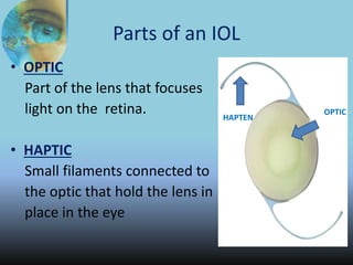
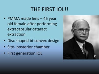
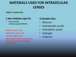
RECENT ADVANCES IN IOLS
Dr. Laltanpuia Chhangte
GMC ,HALDWANI
What is intraocular lens?
• An artificial lens implanted in the eyes
• Replaces the crystalline lens
• To correct the optical power of the eyes
following:-
Lens being
clouded by
cataract
Refractive surgery
Primary vs secondary implantation
• Primary implantation – use of IOLs during
surgery for cataract
• Secondary implantation – implantation of IOL
to correct aphakia in a previosly operated eye
Parts of an IOL
• OPTIC
Part of the lens that focuses
light on the retina.
• HAPTIC
Small filaments connected to
the optic that hold the lens in
place in the eye
HAPTEN
OPTIC
HISTORY OF IOL
• K
THE FIRST IOL!!
• PMMA made lens – 45 year
old female after performing
extracapsular cataract
extraction
• Disc shaped bi-convex design
• Site- posterior chamber
• First generation IOL
EVOLUTION OF IOLs
1. First generation IOLs
• Ridley lenses
• Disadvantages – posterior dislocation
poor surgical technique
2. Second generation IOLs
• Rigid and semi-rigid anterior chamber IOLs
• Advantages – reduce posterior dislocation
• Disadvantage – corneal decompensation
UGH syndrome
3. Third generation IOLs
• Iris supported lens
• Advantages- less corneal decompensation
• Disadvantages – iris chaffing
pupillary distortion
c/c inflammation
cystoid macular edema
4. Fourth generation IOLs
• Modern anterior chamber lens
• Flexible loops and multiple point fixation
• Advantages – more stable, better design, less
complications
• Disadvantages – anterior chamber is not the
physiological site for IOL
5. Fifth generation IOLs
• PMMA lenses
• Foldable and small incision lenses
MATERIALS USED FOR INTRAOCULAR
LENSES
Optic materials
1.Non-foldable-rigid IOL
• Poly-methyl
methacrylate(PMMA)
Water content <1%
Refractive index 1.49
Usually single piece
May be penetrated by aqueous
humor known as ‘glistenings’
(very rare)
2.Flexible IOLs
• Silicones
• Hydrophobic acrylic
• Hydrophilic acrylic
• Hydrogel
• Collamer
Features of IOL
INTRAOCULAR LENS DESIGN
• Single piece / monobloc
• Plate haptic / open loop haptics
• Angulated / planar haptics
• Edge design
• Optic design
Different types of haptic angulation
relative to the plane of optic:-
For posterior chamber lens:-
• 100 anterior angulation to keep the optic
part away from the pupil.
For anterior chamber lens:-
• Posteriorly angulated lens to vault the
intraocular lens away from the pupil
ANGULATED HAPTICS ALLOW FOR ADEQUATE
PUPILLARY CLEARANCE AND ADHESION TO THE
POSTERIOR CAPSULE
………
Square, truncated optic edge
Suitable position for implanting IOL in
eye
• Best
placed in
posterior
chamber
in the
capsular
bag.
ADVANTAGES OF IN-THE-BAG
PLACEMENT
• Proper anatomical site
• Symmetrical loop placement
• Intraoperative stretching or tearing of zonules is avoided
• Minimal magnification (<2%); (20-30% aphakic glasses, 7-12% aphakic
contact lens, ACIOL 2-5% )
• Low incidence of lens decentration and dislocation
• Maximal distance from the posterior iris pigment epithelium, iris root, and
ciliary processes
• Reduced posterior capsular opacification
POSITIONING OF IOL
1. Posterior chamber
implantation
• Ciliary sulcus fixation
• In the bag fixation
• Scleral fixation
Eg:- modified C loop type
IOL
In-the-bag fixation
OTHER POSITIONS:
• Posterior capsule tear
• zonular dialysis
Ciliary sulcus fixation
Scleral IOL
2. Anterior chamber
implantation
• Angle supported IOLs
• Iris fixated
e.g Kelman multiflex type
IOL
3. Iris- fixated lens
• Fixed on the iris with
claws,loops or sutures
• Eg- Singh and Worst’s
iris claw lens
BASED ON OPTICAL CORRECTION
1. Mono-focal intraocular lens
• Lens with uni-focal power
• This common IOL type has been used for
several decades .
• Mono-focals are set to provide best corrected
vision at near, intermediate or far distances.
• Most people who choose mono-focals have
their IOLs set for distance vision and use
reading glasses for near activities.
• Some who choose mono-focals decide to have
the IOL for one eye set for distance vision, and
the other set for near vision, a strategy called
"monovision."
……..
• The brain adapts and synthesizes the information from
both eyes to provide vision at intermediate distances.
People who regularly use computers, PDAs or other digital
devices may find this especially useful.
• Individuals considering monovision may be able to try this
technique with contact lenses first to see how well they can
adapt to monovision.
• Those who require crisp, detailed vision may decide
monovision is not for them. People with appropriate vision
prescriptions may find that monovision allows them see
well at most distances with little or no need for eyeglasses.
2.Multifocal IOLs
• Single IOL with two or
more focal points
• Refractive
• Diffractive
• Combination of both
• Simultaneous vision
lenses
• Pseudo-accommodation
Multifocal IOLs
• Multifocal iols have the advantage of providing
patients with functional vision at near ,far and at
intermediate distances.
• Patients can have a traditional monofocal iol with a
refractive target of emmetropia,mild myopia,or
monovision(1 eye distance,1eye near)
• The Array (AMO),Santa ana CA multifocal silicone
PCIOL was the first multifocal IOL to be granted fda
approval in US.
• Three mutifocal lenses are currently FDA approved
for use after cataract extraction:
• ReZoom lens(AMO),The Acrysof ResTOR(alcon) and
Technis(AMO) multifocal iols
ReZoom lens
• Flexible,3-piece , acrylic, distance dominant
zonal refractive IOL
• Insertion through 2.8 mm CCI
• 5 expanded refractive zones within the 6 mm
optic & produces 2.80 D of near power
• Pupil must be 2 mm to reach the near effect
Multiple focal points of a refractive
multifocal IOL
DIFFRACTIVE MULTIFOCAL IOLs
• Anterior aspheric surface : basic refractive
power
• Multiple grooves on posterior surface :
diffractive power
• 41% of light : distance
• 41% : near vision
• Pupil independent
Multiple focal points of a diffractive
multifocal IOL
Acrysof ReSTOR lens(alcon lab)
• FDA approved in 2005
• 1/3 piece ,acrylic ,aspheric ,foldable ,diffractive lens that
simultaneously focuses light on both near and distance
targets
• Central 3.6mm difrractive zone is mainly for near and outside
that is for distant vision
• Loss of 6%-19% light which affects contrast sensitivity esp in
patients with smaller pupils
• ReSTOR IOL is manufactured with +4.00 D+3.00D add at IOL
plane.
• Patients who received +4.00D expressed dissatisfaction with
the near focal point being too close
Tecnis Multifocal IOLs (AMO)
• FDA approved in 2009
• Foldable,hydrophobic,diffractive aspheric
acrylic lens
• The diffractive steps are on the posterior
aspect on the lens and extend fully to the
periphery.
• Diffractive Power +4.0 diopters of near
addition (+3.0 Diopters at spectacle plane)
Patient selection
• Relatively easy going people who place a high value on
reducing dependence on glasses or contact lenses
• Good potential vision
• <1.00D preexisting corneal astigmatism
• Demanding patients strictly avoided
Special considerations:
• Counselling (most important)
• Accurate Biometry
• IOL master strongly recommended
Surgical technique
• Same as in standard SICS with a foldable acrylic
IOL
• Round, centered CCC completely overlapping the
lens optic
• Removal of all viscoelastic from behind the lens
• Optimal refractive effects depend upon good
centration
• Discouraged in case of inadequate centration or if
posterior capsule is not intact
Side effects and complications
• Capsular opacification
• Glare,halos,and ghosting than a monofocal
IOL or an accommodating IOL
• Decreased contrast sensitivity
• Intermediate vision may be weaker with
multifocal IOLs than an accommodating one
• Requires Visual-Cortical Neuro-adaptation
Preferances
ASPHERIC IOLs
• Human eye : Aspheric Optics
• Cornea : Positive spherical aberration
• Young crystalline lens : Negative spherical aberration
• Ageing crystalline lens : Increased positive spherical
aberration
• spherical aberration ~ fourth power of the pupil diameter
Anterior prolate surface
• Tecnis, Advanced Medical Optics (AMO)
Posterior prolate surface
• Acrysof IQ, Alcon Laboratories
Both Anterior and Posterior prolate surfaces
• Akreos AO, SofPort AO and L161 AO, Bausch &
Lomb
ASPHERIC IOLs
Advantages Disadvantages
ACCOMMODATIVE IOLs
• Monofocal IOL
• To restore a significant amount of true accommodation
to patients with surgically induced pseudophakia.
• During ciliary muscle contraction,forward displacement
of the IOL led to an increase in the IOLs effective power
and thus an improvement in near vision.
• 1 mm of anterior movement of lens = 1.80 D of
accommodation
• Mimicking the eye's natural ability to focus
Crytalens ( Baush & Lomb):modified silicone, plate
haptic(hinged),biconvex optic which potentially allow
anterior movement of the lens during accommodation.
Accomodative IOLs
• Another theory is ciliary muscle
contraction causes a steepening of
anterior optic surface allowing for
better near vision.
• Only FDA approved IOL for
correction of presbyopia
• Although the exact cause of movement is unclear,it appears to be
the a combination of posterior chamber pressure on the back
surface of iol and Cilliary Body pressure on the iol haptics that
vaults the optic forward.
• As the ciliary body contracts,increased pressure is transmitted via
vitreous to polyamide haptics & the compression betwwen the
haptics causes the optic to bow forward
• Accomodative arching of the lens enhances depth of focus
Hydrophilic Acrylic,single optic
• BioComFold type 43E (Morcher GmbH)
• 1CU (HumanOptics AG)
• Tetraflex (Lenstec Inc.)
Development is also currently underway for dual optics IOLs
eg,Sychrony IOL(visiogen,Irvine,CA) and deformable IOLs(smart IOL)
Synchrony Dual-Optic IOL (Visiogen)
• One piece Silicon foldable IOL
• Two optics
• high plus anterior
• posterior minus lens
• connected by spring like haptics.
• Zonular tension is released –
compression of optic-spring haptic
releases anterior optic forward.
TORIC IOLs
• 15-29% of patients undergoing cataract surgery
have substantial corneal astigmatism >=1.50 D
• Either we go for arcuate keratotomy or limbal
relaxing procedures or alternatively a toric IOL
can incorporate the astigmatic correction into
the spherical IOL power.
INSTRUMENTATION
• STAAR toric IOL
• First FDA approved (in 1998) toric IOL
• Single piece,plate haptic,foldable silicone IOL
designed to be placed in capsular bag using an
injector through a 3mm incision.once in the eye
,it must be oriented with its long axis precisely in
the steep meridian.
• 6mm biconvex optic with a spherocyndrical
anterior and spherical posterior surface.
• Spherical power in the range of =9.5 to =28.5 D
with a choice of cylindrical powers of 2.00 and
3.50D which correct 1.50 and 2.25 D of
astigmatism respectively
………
• AcrySof IQ Toric IOL (Alcon Labs, USA)
• Recently approved by FDA
• 6mm biconvex acrlic optic,available in the
range of +6 to +30 D & 3 astigmatism powers
:+1.50 D,+2.25D and +3.00 D which correct
+1.03,+1.55&+2.06 D respectively
• Posterior surface has added cylindrical power
and axis markings on the lens optic
Patient selection
• CANDIDATES > or = 1.0D i.e mild to moderate
regular corneal astigmatism
• Patients with astigmatism in amounts exceeding
the upper correction limits of these lenses may
require additional measures to obtain full
correction
• Limitations of toric iols must be explained before
hand
• Patients should be infrmed as the lens may
rotate within capsular bag
• Inappropriate for the Patient who may require
silicone oil for RD repair in future
Planning and surgical technique
• Amount and axis of astigmatism should be measured
accurately with a keratometer or if possible coneal
topography
• AcrySof Toric IOL Calculator generates correct power and
model lens as well as orientation of the lens alignment
markers
• compensates for surgically induced astigmatism
……..
• Vertical& horizontal meridians should be marked on the
cornea with the pt in an upright position.
• reference marks at the 3- and 9-o’clock
• After the IOL is injected into the capsular bag viscoelastic
material behind the IOL is aspirated and the IOL is rotated
into the position on the steep meridian
• It may roate when balanced saline solution is injected to
reform the AC
• Final alignment
Complications
• Possibility of IOL rotation as full correction is not
achieved unless the IOL is properly aligned in the axis
of astigmatism
According to STAAR,a 10 degree off axis rotation reduces
the correction by approx. 1/3rd
• 20degree by 2/3rd
• And an off axis correction of more than 30 dregree can
actually increase the cylindrical refractive error.
• When a plate haptic toric IOL is used,surgeon should
take care when performing Nd:Yag capsulotomy .if the
capsulotomy is too large a plate haptic may prolapse
posteriorly
Typically a misaligned IOL is recognized within days of
surgery,it should be repositioned before permanent
fibrosis occurs within capsular bag.
ROLLABLE IOLs
• Ultrathin ~100 µ
• Hydrophilic material
• Front surface curved
• Back surface: series of steps with concentric rings
• Open up gradually
• Implanted by phakonit technique
Eg:
• Acrismart
• Thin Optx ultrachoice
• Slimflex lens
ANIRIDIA IOLS
• Cover the defects of aniridia or partial iris loss
as in trauma
• Has a black diaphragm over the optic
Phakic IOLS
PHAKIC IOLS
• Implantation of IOL without removing natural
crystalline lens.
• ADVANTAGE: Preserves natural accommodation
• Mostly used in Myopic eyes: -5 to -20 DS
• Also used in Hyperopic eyes
• Concern in Hyperopes:
• More chances of endothelial damage
• Increased risk of angle closure glaucoma
• Life-long regular follow up required.
Types
• Posterior Chamber
• Iris fixated
• Angle fixated
Patient selection
• Patients who are near or beyond the FDA approved
limits for laser vision correction
• Patient with extreme corneal curvatures(flatter than 34
D in myopics corrections and steeper than 50 D in
hyperopic corrections after laser refractive surgeries)
CONTRAINDICATIONS:
Patients with compromised corneal
endothelium,iritis,rubeosis iridis,cataract & glaucoma
• Informed consent must be taken
Ancillary tests :specular microscopy,corneal pachymetry
and AC depth
SURGICAL TECHNIQUE
• TA with an intracameral supplement is appropriate and
PIOL is inserted through a small incision
• A perpipheral iridotomy is recommended for all
currently FDA approved PIOLs reduce the risk of
puppilary block and angle closure.
• Preoperative iridotomy is preferable when small
incision implant surgery is performed bcoz surgical
iridotomy or iridectomy is technically difficult to
perform through beveled clear corneal incision
• Viscoelastic material should be meticulously removed
at the conclusion of surgery to prevent post op IOP
spike.
Posterior chamber phakic lens
• Implantable collamer lens (ICL) (VISIAN;
STAAR)
• Sticklens
Correct IOL length is selected by using the white
to white measurement between 3 and 9 o clock
positions.
Implantable Collamer Lens (ICL)
• Pre-crystalline lens made of silicone or
collamer
• Co-polymer of HEMA(99%)+Porcine
collagen(1%)
Overall size- 11-13 mm
Optical zone - 4.5-5.5 mm
• Optic of the PCPIOL is vaulted both to
avoid contact with the crystalline lens ans
to allow aqueous to flow over the
crystalline lens
• Currently approved for myopes with -
3.00D TO -20.00D in adults 21-45 age
group with a minimum AC dephth=3 mm
Avoid touching the central aprt of crystalline
lens
COMPLICATIONS:
• Endothelial cell damage
• Inflammation
• Pigment dispersal
• Elevated IOP
• Cataract
IRIS FIXATED PHAKIC IOL
• VERISYSE/ARTISAN (AMO/OPTECH):
• Myopia -5.00 to -20.00 D in patients 21
yrs or older ,min. AC depth 3.2 mm
• Made of PMMA,convexo-
concave,Length = 8.5 mm,Optic size =
5-6 mm
• Claw Haptics fixed to iris by a
encavation process
• Generally inserted through a superior
limbal incision with side ports are
made at 10 & 2 o clock positions.
• Specialised encavation needle is
introduced from one of the side ports
and a knucle of iris is brought up into
the claw haptic
ADVANTAGES OVER ICL:
• Customized smaller size possible
• Easier examination from end-to-end
COMPLICATIONS-
• Early post op AC inflammation
• Glaucoma
• Iris atrophy on fixation sites
• Implant dislocation
• De-centration
• Endothelial cell loss
ANGLE FIXATED PHAKIC IOL
• TWO TYPES-
-4 point fixation
• Baikoffs modification of Kelman type haptic
design
-3 point fixation
• Vivarte(IOLtech)
• Can be inserted through a temporal clear
corneal incision or a scleral pocket
• No ACPIOL is currently FDA approved
Complications
Endothelial cell loss
Irregular pupil
Iris depigmentation
Post-op inflammation
Halos and glare
Surgical induced astigmatism
PIGGYBACK IOLs
• An intraocular lens that “piggybacks” onto an
existing intraocular lens or two IOLs are
implanted simultaneously.
• First IOL is placed in the capsular bag.
• The second (piggyback) IOL is placed in the
bag or sulcus.
• Easier to place 2nd IOL than to explant IOL &
replace it
• Lesser risk
• More predictable
• Can change power with time-by adding IOL or
explanting an IOL
• Better image quality
• Increased depth of focus
COMPLICATIONS
• Interlenticular opacification
• (Interpseudophakos Elshnig’s pearls)
(RED ROCK SYNDROME)
• Unpredictable final IOL position
THE FUTURE IOLs
LIGHT ADJUSTABLE IOL
TELESCOPIC IOLs
SMART IOLs
LIGHT ADJUSTABLE IOLS
Calhoun Vision
• 3 piece Silicone optic PCIOL with two C-PMMA haptics
• Silicone matrix has been embedded with silicone subunits called
macromers.
• Photosensitive to the near-ultraviolet wavelength of energy in a
specific pattern,when irradiated with UV light through slit lamp
delivery system ,the macromere polymerise and are depleted.
• Monomers from the non irradiated part of IOL optic are in higher
centre bcoz of induced osmotic gradient move to the centre and
cause the IOL to swell up and increases the power of IOL
• myopic adjustment : periphery of the lens
• Final irradiation step locks in is done & IOL optic is diffusely
irradiated within 1-2 weeks post op once the desired power has
been acheived
• Nomograms have been developed that can reportedly correct over
a 5.00D range of myopia,hyperopia,astigmatism.
• Sunglasses for about three weeks
BLUE LIGHT FILTERING IOLs
• Lenses are yellow colored to screen out UV
Light and blue spectrum
• Reduced risk of age related macular
degeneration
PHOTOCHROMATIC IOLs
• UV absorbing photo chromes
IMPLANTABLE MINIATURE
TELESCOPE
• Miniature implantable Galilean
telescope
• Implanted in posterior chamber
• Contains number of micro lenses
in the optic which gives a
magnification of about 3 at the
central visual field
• Used to treat macular
degeneration
TELESCOPIC IOLs
• New generation miniature
telescopes which uses 2 mirrors
instead of lens to provide
magnification with minimum
loss of peripheral vision
BIOMETRY
THEOREITICAL FORMULAE
• Binkhorst formula
• Colenbrander’s formula
• Gull strand’s formula
• Hoffer’s formula
EMPERICAL FORMULAE
• Modified Sanders-Retzlaff-Kraff formula
• SRK II
• SRK III
• SRK T
• Haigis formula
Modified SRK( Sanders-Retzlaff-Kraff
formula)
• E=A - 2.5L - 0.9K
• Parameters used in the formula are estimated
by
• A-scan ultrasonographic sonometry and
keratometry
• Instrument - biometer
Based on the statistical correlation between
calculated and observed refractive error after
ocular implantation.
E=A - 2.5L - 0.9K
• Power ( I )required to produce Required post
operative refraction ( R ) ,
I = E – cr R
• cr – empirical constant
cr = 1 if E < 14
cr = 1.25 if E > 14
Smart lens(medennium,irwine,CA)
• Thermoplastic acrylic gel that can be customized into any
size,shape,or power specified by the physician would
allow the patients to retain accommodation.
• The hydrophobic acrylic material is chemically bonded to
wax,which melts inside the eye at body temperature &
allows the predetermined shape,powe of the material to
emerge.
• Theoretically,compression of this pliable lens by the
capsular bag would allow adjustment of its effective
power in a manner similar to the way the crystalline lens
adjusts.
• Other examples of deformable IOLs that are still in
preliminary stage of development
are:Flexoptic(AMO),Fluidvision IOL(Power vision,Belmont
,CA),NuLens(NuLens,Israel)
RECENT ADVANCES IN INTRAOCULAR LENS

RECENT ADVANCES IN INTRAOCULAR LENS

  • 1.
    RECENT ADVANCES INIOLS Dr. Laltanpuia Chhangte GMC ,HALDWANI
  • 2.
    What is intraocularlens? • An artificial lens implanted in the eyes • Replaces the crystalline lens • To correct the optical power of the eyes following:- Lens being clouded by cataract Refractive surgery
  • 3.
    Primary vs secondaryimplantation • Primary implantation – use of IOLs during surgery for cataract • Secondary implantation – implantation of IOL to correct aphakia in a previosly operated eye
  • 4.
    Parts of anIOL • OPTIC Part of the lens that focuses light on the retina. • HAPTIC Small filaments connected to the optic that hold the lens in place in the eye HAPTEN OPTIC
  • 5.
  • 6.
    THE FIRST IOL!! •PMMA made lens – 45 year old female after performing extracapsular cataract extraction • Disc shaped bi-convex design • Site- posterior chamber • First generation IOL
  • 7.
    EVOLUTION OF IOLs 1.First generation IOLs • Ridley lenses • Disadvantages – posterior dislocation poor surgical technique 2. Second generation IOLs • Rigid and semi-rigid anterior chamber IOLs • Advantages – reduce posterior dislocation • Disadvantage – corneal decompensation UGH syndrome
  • 8.
    3. Third generationIOLs • Iris supported lens • Advantages- less corneal decompensation • Disadvantages – iris chaffing pupillary distortion c/c inflammation cystoid macular edema
  • 9.
    4. Fourth generationIOLs • Modern anterior chamber lens • Flexible loops and multiple point fixation • Advantages – more stable, better design, less complications • Disadvantages – anterior chamber is not the physiological site for IOL
  • 10.
    5. Fifth generationIOLs • PMMA lenses • Foldable and small incision lenses
  • 11.
    MATERIALS USED FORINTRAOCULAR LENSES Optic materials 1.Non-foldable-rigid IOL • Poly-methyl methacrylate(PMMA) Water content <1% Refractive index 1.49 Usually single piece May be penetrated by aqueous humor known as ‘glistenings’ (very rare) 2.Flexible IOLs • Silicones • Hydrophobic acrylic • Hydrophilic acrylic • Hydrogel • Collamer
  • 13.
  • 14.
    INTRAOCULAR LENS DESIGN •Single piece / monobloc • Plate haptic / open loop haptics • Angulated / planar haptics • Edge design • Optic design
  • 15.
    Different types ofhaptic angulation relative to the plane of optic:- For posterior chamber lens:- • 100 anterior angulation to keep the optic part away from the pupil. For anterior chamber lens:- • Posteriorly angulated lens to vault the intraocular lens away from the pupil
  • 16.
    ANGULATED HAPTICS ALLOWFOR ADEQUATE PUPILLARY CLEARANCE AND ADHESION TO THE POSTERIOR CAPSULE
  • 17.
  • 19.
  • 21.
    Suitable position forimplanting IOL in eye • Best placed in posterior chamber in the capsular bag.
  • 22.
    ADVANTAGES OF IN-THE-BAG PLACEMENT •Proper anatomical site • Symmetrical loop placement • Intraoperative stretching or tearing of zonules is avoided • Minimal magnification (<2%); (20-30% aphakic glasses, 7-12% aphakic contact lens, ACIOL 2-5% ) • Low incidence of lens decentration and dislocation • Maximal distance from the posterior iris pigment epithelium, iris root, and ciliary processes • Reduced posterior capsular opacification
  • 23.
    POSITIONING OF IOL 1.Posterior chamber implantation • Ciliary sulcus fixation • In the bag fixation • Scleral fixation Eg:- modified C loop type IOL
  • 24.
  • 25.
    OTHER POSITIONS: • Posteriorcapsule tear • zonular dialysis
  • 26.
  • 27.
  • 28.
    2. Anterior chamber implantation •Angle supported IOLs • Iris fixated e.g Kelman multiflex type IOL
  • 29.
    3. Iris- fixatedlens • Fixed on the iris with claws,loops or sutures • Eg- Singh and Worst’s iris claw lens
  • 30.
    BASED ON OPTICALCORRECTION 1. Mono-focal intraocular lens • Lens with uni-focal power • This common IOL type has been used for several decades . • Mono-focals are set to provide best corrected vision at near, intermediate or far distances. • Most people who choose mono-focals have their IOLs set for distance vision and use reading glasses for near activities. • Some who choose mono-focals decide to have the IOL for one eye set for distance vision, and the other set for near vision, a strategy called "monovision."
  • 31.
    …….. • The brainadapts and synthesizes the information from both eyes to provide vision at intermediate distances. People who regularly use computers, PDAs or other digital devices may find this especially useful. • Individuals considering monovision may be able to try this technique with contact lenses first to see how well they can adapt to monovision. • Those who require crisp, detailed vision may decide monovision is not for them. People with appropriate vision prescriptions may find that monovision allows them see well at most distances with little or no need for eyeglasses.
  • 32.
    2.Multifocal IOLs • SingleIOL with two or more focal points • Refractive • Diffractive • Combination of both • Simultaneous vision lenses • Pseudo-accommodation
  • 33.
    Multifocal IOLs • Multifocaliols have the advantage of providing patients with functional vision at near ,far and at intermediate distances. • Patients can have a traditional monofocal iol with a refractive target of emmetropia,mild myopia,or monovision(1 eye distance,1eye near) • The Array (AMO),Santa ana CA multifocal silicone PCIOL was the first multifocal IOL to be granted fda approval in US. • Three mutifocal lenses are currently FDA approved for use after cataract extraction: • ReZoom lens(AMO),The Acrysof ResTOR(alcon) and Technis(AMO) multifocal iols
  • 34.
    ReZoom lens • Flexible,3-piece, acrylic, distance dominant zonal refractive IOL • Insertion through 2.8 mm CCI • 5 expanded refractive zones within the 6 mm optic & produces 2.80 D of near power • Pupil must be 2 mm to reach the near effect
  • 36.
    Multiple focal pointsof a refractive multifocal IOL
  • 37.
    DIFFRACTIVE MULTIFOCAL IOLs •Anterior aspheric surface : basic refractive power • Multiple grooves on posterior surface : diffractive power • 41% of light : distance • 41% : near vision • Pupil independent
  • 38.
    Multiple focal pointsof a diffractive multifocal IOL
  • 40.
    Acrysof ReSTOR lens(alconlab) • FDA approved in 2005 • 1/3 piece ,acrylic ,aspheric ,foldable ,diffractive lens that simultaneously focuses light on both near and distance targets • Central 3.6mm difrractive zone is mainly for near and outside that is for distant vision • Loss of 6%-19% light which affects contrast sensitivity esp in patients with smaller pupils • ReSTOR IOL is manufactured with +4.00 D+3.00D add at IOL plane. • Patients who received +4.00D expressed dissatisfaction with the near focal point being too close
  • 41.
    Tecnis Multifocal IOLs(AMO) • FDA approved in 2009 • Foldable,hydrophobic,diffractive aspheric acrylic lens • The diffractive steps are on the posterior aspect on the lens and extend fully to the periphery. • Diffractive Power +4.0 diopters of near addition (+3.0 Diopters at spectacle plane)
  • 44.
    Patient selection • Relativelyeasy going people who place a high value on reducing dependence on glasses or contact lenses • Good potential vision • <1.00D preexisting corneal astigmatism • Demanding patients strictly avoided Special considerations: • Counselling (most important) • Accurate Biometry • IOL master strongly recommended
  • 45.
    Surgical technique • Sameas in standard SICS with a foldable acrylic IOL • Round, centered CCC completely overlapping the lens optic • Removal of all viscoelastic from behind the lens • Optimal refractive effects depend upon good centration • Discouraged in case of inadequate centration or if posterior capsule is not intact
  • 46.
    Side effects andcomplications • Capsular opacification • Glare,halos,and ghosting than a monofocal IOL or an accommodating IOL • Decreased contrast sensitivity • Intermediate vision may be weaker with multifocal IOLs than an accommodating one • Requires Visual-Cortical Neuro-adaptation
  • 47.
  • 48.
    ASPHERIC IOLs • Humaneye : Aspheric Optics • Cornea : Positive spherical aberration • Young crystalline lens : Negative spherical aberration • Ageing crystalline lens : Increased positive spherical aberration • spherical aberration ~ fourth power of the pupil diameter
  • 53.
    Anterior prolate surface •Tecnis, Advanced Medical Optics (AMO) Posterior prolate surface • Acrysof IQ, Alcon Laboratories Both Anterior and Posterior prolate surfaces • Akreos AO, SofPort AO and L161 AO, Bausch & Lomb
  • 54.
  • 55.
    ACCOMMODATIVE IOLs • MonofocalIOL • To restore a significant amount of true accommodation to patients with surgically induced pseudophakia. • During ciliary muscle contraction,forward displacement of the IOL led to an increase in the IOLs effective power and thus an improvement in near vision. • 1 mm of anterior movement of lens = 1.80 D of accommodation • Mimicking the eye's natural ability to focus Crytalens ( Baush & Lomb):modified silicone, plate haptic(hinged),biconvex optic which potentially allow anterior movement of the lens during accommodation.
  • 56.
    Accomodative IOLs • Anothertheory is ciliary muscle contraction causes a steepening of anterior optic surface allowing for better near vision. • Only FDA approved IOL for correction of presbyopia
  • 57.
    • Although theexact cause of movement is unclear,it appears to be the a combination of posterior chamber pressure on the back surface of iol and Cilliary Body pressure on the iol haptics that vaults the optic forward. • As the ciliary body contracts,increased pressure is transmitted via vitreous to polyamide haptics & the compression betwwen the haptics causes the optic to bow forward • Accomodative arching of the lens enhances depth of focus Hydrophilic Acrylic,single optic • BioComFold type 43E (Morcher GmbH) • 1CU (HumanOptics AG) • Tetraflex (Lenstec Inc.) Development is also currently underway for dual optics IOLs eg,Sychrony IOL(visiogen,Irvine,CA) and deformable IOLs(smart IOL)
  • 59.
    Synchrony Dual-Optic IOL(Visiogen) • One piece Silicon foldable IOL • Two optics • high plus anterior • posterior minus lens • connected by spring like haptics. • Zonular tension is released – compression of optic-spring haptic releases anterior optic forward.
  • 60.
    TORIC IOLs • 15-29%of patients undergoing cataract surgery have substantial corneal astigmatism >=1.50 D • Either we go for arcuate keratotomy or limbal relaxing procedures or alternatively a toric IOL can incorporate the astigmatic correction into the spherical IOL power.
  • 61.
    INSTRUMENTATION • STAAR toricIOL • First FDA approved (in 1998) toric IOL • Single piece,plate haptic,foldable silicone IOL designed to be placed in capsular bag using an injector through a 3mm incision.once in the eye ,it must be oriented with its long axis precisely in the steep meridian. • 6mm biconvex optic with a spherocyndrical anterior and spherical posterior surface. • Spherical power in the range of =9.5 to =28.5 D with a choice of cylindrical powers of 2.00 and 3.50D which correct 1.50 and 2.25 D of astigmatism respectively
  • 62.
    ……… • AcrySof IQToric IOL (Alcon Labs, USA) • Recently approved by FDA • 6mm biconvex acrlic optic,available in the range of +6 to +30 D & 3 astigmatism powers :+1.50 D,+2.25D and +3.00 D which correct +1.03,+1.55&+2.06 D respectively • Posterior surface has added cylindrical power and axis markings on the lens optic
  • 63.
    Patient selection • CANDIDATES> or = 1.0D i.e mild to moderate regular corneal astigmatism • Patients with astigmatism in amounts exceeding the upper correction limits of these lenses may require additional measures to obtain full correction • Limitations of toric iols must be explained before hand • Patients should be infrmed as the lens may rotate within capsular bag • Inappropriate for the Patient who may require silicone oil for RD repair in future
  • 64.
    Planning and surgicaltechnique • Amount and axis of astigmatism should be measured accurately with a keratometer or if possible coneal topography • AcrySof Toric IOL Calculator generates correct power and model lens as well as orientation of the lens alignment markers • compensates for surgically induced astigmatism
  • 65.
    …….. • Vertical& horizontalmeridians should be marked on the cornea with the pt in an upright position. • reference marks at the 3- and 9-o’clock • After the IOL is injected into the capsular bag viscoelastic material behind the IOL is aspirated and the IOL is rotated into the position on the steep meridian • It may roate when balanced saline solution is injected to reform the AC • Final alignment
  • 67.
    Complications • Possibility ofIOL rotation as full correction is not achieved unless the IOL is properly aligned in the axis of astigmatism According to STAAR,a 10 degree off axis rotation reduces the correction by approx. 1/3rd • 20degree by 2/3rd • And an off axis correction of more than 30 dregree can actually increase the cylindrical refractive error. • When a plate haptic toric IOL is used,surgeon should take care when performing Nd:Yag capsulotomy .if the capsulotomy is too large a plate haptic may prolapse posteriorly Typically a misaligned IOL is recognized within days of surgery,it should be repositioned before permanent fibrosis occurs within capsular bag.
  • 68.
    ROLLABLE IOLs • Ultrathin~100 µ • Hydrophilic material • Front surface curved • Back surface: series of steps with concentric rings • Open up gradually • Implanted by phakonit technique Eg: • Acrismart • Thin Optx ultrachoice • Slimflex lens
  • 70.
    ANIRIDIA IOLS • Coverthe defects of aniridia or partial iris loss as in trauma • Has a black diaphragm over the optic
  • 71.
  • 72.
    PHAKIC IOLS • Implantationof IOL without removing natural crystalline lens. • ADVANTAGE: Preserves natural accommodation • Mostly used in Myopic eyes: -5 to -20 DS • Also used in Hyperopic eyes • Concern in Hyperopes: • More chances of endothelial damage • Increased risk of angle closure glaucoma • Life-long regular follow up required.
  • 73.
    Types • Posterior Chamber •Iris fixated • Angle fixated
  • 74.
    Patient selection • Patientswho are near or beyond the FDA approved limits for laser vision correction • Patient with extreme corneal curvatures(flatter than 34 D in myopics corrections and steeper than 50 D in hyperopic corrections after laser refractive surgeries) CONTRAINDICATIONS: Patients with compromised corneal endothelium,iritis,rubeosis iridis,cataract & glaucoma • Informed consent must be taken Ancillary tests :specular microscopy,corneal pachymetry and AC depth
  • 75.
    SURGICAL TECHNIQUE • TAwith an intracameral supplement is appropriate and PIOL is inserted through a small incision • A perpipheral iridotomy is recommended for all currently FDA approved PIOLs reduce the risk of puppilary block and angle closure. • Preoperative iridotomy is preferable when small incision implant surgery is performed bcoz surgical iridotomy or iridectomy is technically difficult to perform through beveled clear corneal incision • Viscoelastic material should be meticulously removed at the conclusion of surgery to prevent post op IOP spike.
  • 76.
    Posterior chamber phakiclens • Implantable collamer lens (ICL) (VISIAN; STAAR) • Sticklens Correct IOL length is selected by using the white to white measurement between 3 and 9 o clock positions.
  • 77.
    Implantable Collamer Lens(ICL) • Pre-crystalline lens made of silicone or collamer • Co-polymer of HEMA(99%)+Porcine collagen(1%) Overall size- 11-13 mm Optical zone - 4.5-5.5 mm • Optic of the PCPIOL is vaulted both to avoid contact with the crystalline lens ans to allow aqueous to flow over the crystalline lens • Currently approved for myopes with - 3.00D TO -20.00D in adults 21-45 age group with a minimum AC dephth=3 mm Avoid touching the central aprt of crystalline lens
  • 78.
    COMPLICATIONS: • Endothelial celldamage • Inflammation • Pigment dispersal • Elevated IOP • Cataract
  • 79.
    IRIS FIXATED PHAKICIOL • VERISYSE/ARTISAN (AMO/OPTECH): • Myopia -5.00 to -20.00 D in patients 21 yrs or older ,min. AC depth 3.2 mm • Made of PMMA,convexo- concave,Length = 8.5 mm,Optic size = 5-6 mm • Claw Haptics fixed to iris by a encavation process • Generally inserted through a superior limbal incision with side ports are made at 10 & 2 o clock positions. • Specialised encavation needle is introduced from one of the side ports and a knucle of iris is brought up into the claw haptic
  • 80.
    ADVANTAGES OVER ICL: •Customized smaller size possible • Easier examination from end-to-end COMPLICATIONS- • Early post op AC inflammation • Glaucoma • Iris atrophy on fixation sites • Implant dislocation • De-centration • Endothelial cell loss
  • 81.
    ANGLE FIXATED PHAKICIOL • TWO TYPES- -4 point fixation • Baikoffs modification of Kelman type haptic design -3 point fixation • Vivarte(IOLtech) • Can be inserted through a temporal clear corneal incision or a scleral pocket • No ACPIOL is currently FDA approved
  • 82.
    Complications Endothelial cell loss Irregularpupil Iris depigmentation Post-op inflammation Halos and glare Surgical induced astigmatism
  • 83.
    PIGGYBACK IOLs • Anintraocular lens that “piggybacks” onto an existing intraocular lens or two IOLs are implanted simultaneously. • First IOL is placed in the capsular bag. • The second (piggyback) IOL is placed in the bag or sulcus.
  • 84.
    • Easier toplace 2nd IOL than to explant IOL & replace it • Lesser risk • More predictable • Can change power with time-by adding IOL or explanting an IOL • Better image quality • Increased depth of focus
  • 85.
    COMPLICATIONS • Interlenticular opacification •(Interpseudophakos Elshnig’s pearls) (RED ROCK SYNDROME) • Unpredictable final IOL position
  • 86.
    THE FUTURE IOLs LIGHTADJUSTABLE IOL TELESCOPIC IOLs SMART IOLs
  • 87.
    LIGHT ADJUSTABLE IOLS CalhounVision • 3 piece Silicone optic PCIOL with two C-PMMA haptics • Silicone matrix has been embedded with silicone subunits called macromers. • Photosensitive to the near-ultraviolet wavelength of energy in a specific pattern,when irradiated with UV light through slit lamp delivery system ,the macromere polymerise and are depleted. • Monomers from the non irradiated part of IOL optic are in higher centre bcoz of induced osmotic gradient move to the centre and cause the IOL to swell up and increases the power of IOL • myopic adjustment : periphery of the lens • Final irradiation step locks in is done & IOL optic is diffusely irradiated within 1-2 weeks post op once the desired power has been acheived • Nomograms have been developed that can reportedly correct over a 5.00D range of myopia,hyperopia,astigmatism.
  • 88.
    • Sunglasses forabout three weeks
  • 89.
    BLUE LIGHT FILTERINGIOLs • Lenses are yellow colored to screen out UV Light and blue spectrum • Reduced risk of age related macular degeneration PHOTOCHROMATIC IOLs • UV absorbing photo chromes
  • 90.
    IMPLANTABLE MINIATURE TELESCOPE • Miniatureimplantable Galilean telescope • Implanted in posterior chamber • Contains number of micro lenses in the optic which gives a magnification of about 3 at the central visual field • Used to treat macular degeneration
  • 91.
    TELESCOPIC IOLs • Newgeneration miniature telescopes which uses 2 mirrors instead of lens to provide magnification with minimum loss of peripheral vision
  • 92.
    BIOMETRY THEOREITICAL FORMULAE • Binkhorstformula • Colenbrander’s formula • Gull strand’s formula • Hoffer’s formula EMPERICAL FORMULAE • Modified Sanders-Retzlaff-Kraff formula • SRK II • SRK III • SRK T • Haigis formula
  • 93.
    Modified SRK( Sanders-Retzlaff-Kraff formula) •E=A - 2.5L - 0.9K • Parameters used in the formula are estimated by • A-scan ultrasonographic sonometry and keratometry • Instrument - biometer Based on the statistical correlation between calculated and observed refractive error after ocular implantation.
  • 94.
    E=A - 2.5L- 0.9K
  • 95.
    • Power (I )required to produce Required post operative refraction ( R ) , I = E – cr R • cr – empirical constant cr = 1 if E < 14 cr = 1.25 if E > 14
  • 96.
    Smart lens(medennium,irwine,CA) • Thermoplasticacrylic gel that can be customized into any size,shape,or power specified by the physician would allow the patients to retain accommodation. • The hydrophobic acrylic material is chemically bonded to wax,which melts inside the eye at body temperature & allows the predetermined shape,powe of the material to emerge. • Theoretically,compression of this pliable lens by the capsular bag would allow adjustment of its effective power in a manner similar to the way the crystalline lens adjusts. • Other examples of deformable IOLs that are still in preliminary stage of development are:Flexoptic(AMO),Fluidvision IOL(Power vision,Belmont ,CA),NuLens(NuLens,Israel)

Editor's Notes

  • #3 In conditions
  • #33  BASED ON EITHER REFRACTIVE OR DIFFRACTIVE OPTICS
  • #36 Zone 1,3 and 5 : distance 60% distance, 40% near and intermediate Zone 2 and 4 : near
  • #41 Depending upon pupil size,
  • #44 Apodization literally means "removing the foot“ To remove or smooth a discontinuity at the edges
  • #45 Better to avoid demanding patients
  • #46 Use is discouraged..and a monofocal lens should be placed instead
  • #47 Cap opacification is of greater concern as minimal changes in the capsule may lead to early deterioration of vision Complaints of halos tend to subside over months due to patients neural adaptation.
  • #54 Lengh is more at poles than equatorial axis ..improves image quality and visual performance
  • #56 Its an attempt to restore That’s y ACIOLS have lower A constant than PCIOL. However few studies have questioned the amplitude of true accommodation being d sole reason as ,,pupil zsize,with the rule astigmatism & mild myopid may also contribute to near vision
  • #57 Plate hinged haptics
  • #61 Either v go for arcuate keratotomy or limbal relaxing procedures during or after cat surgery.ie lasik,
  • #62 Iols are available with spherical power in the range
  • #64 Like implantation of toric iol will not eliminate the need for reading glasses And may require anither surgery to reposition it As staar is a silicone iol..while acrysof would still b an option
  • #66 As any misalignment resulting from the torsional globe rotation that sometimes may occur wih movement in supine position
  • #77 Alternative methods beng high f USG/anterior segment OCT ,slit beam or laser inferometry
  • #78 Vaulting can be confir biomicroscopymed at the slit lamp as well as with USG..
  • #88 In Mypoia instead of periphery ,central part is irradiated bczo of reduction in central thickness of iol
  • #92 No loss of peripheral vision