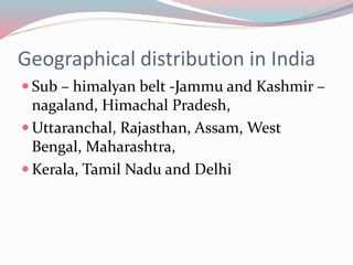
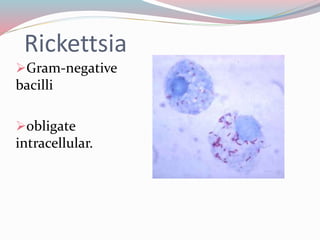
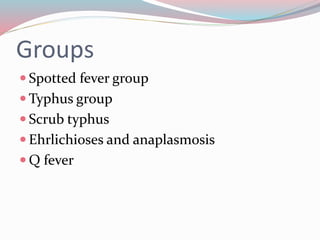
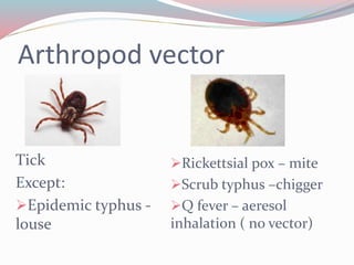
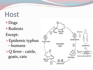
Geographical distribution in India 
 Sub – himalyan belt -Jammu and Kashmir – 
nagaland, Himachal Pradesh, 
Uttaranchal, Rajasthan, Assam, West 
Bengal, Maharashtra, 
Kerala, Tamil Nadu and Delhi
Rickettsia 
Gram-negative 
bacilli 
obligate 
intracellular.
Groups 
 Spotted fever group 
Typhus group 
 Scrub typhus 
 Ehrlichioses and anaplasmosis 
 Q fever
Arthropod vector 
Tick 
Except: 
Epidemic typhus - 
louse 
Rickettsial pox – mite 
Scrub typhus –chigger 
Q fever – aeresol 
inhalation ( no vector)
Host 
 Dogs 
Rodents 
Except: 
Epidemic typhus 
- humans 
Q fever - cattle, 
goats, cats
Pathogenesis 
Bite 
Blood stream 
Endothelium/vascular smooth muscles 
Target cells 
 Host defenses – T cell, TNF α, γ interferon
History 
Removal of an attached tick ( 60%) 
 Endemic area/ wooded area 
 Season – July to October 
 Similar illness in close contacts 
Contact with dog
Clinical features 
Incubation period – 2 – 14 day 
Common symptoms: 
Clinical triad 
 Fever 
 Rash - centripetal 
Headache
Rash 
Early stage: 
Discrete, pale, rose red, 
blanching maculopapular 
on extremities 
Late stage: 
Petechial /haemorragic / 
purpuric/Ecchymosies/ 
necrotic/ulcer
Eschar 
red induration 
Enlarge 
Vesiculates 
Ruptures 
Eschar 
3 – 4 wks 
Resolves
Others symptoms: 
 Lympadenopathy 
 Hepatospleenomegaly 
 Edema 
 Conjunctival congestion 
 Gastrointestinal symptoms – emesis, pain 
 CNS involvement 
 Pulmonary infiltrates – Q fever 
 Infectious mononucleosis like syndrome - ehrelichia
Complication 
 CNS - Cerebral edema, Meningoencephalitis 
Neurologic sequelae, 
 RS -Noncardiogenic pulmonary edema, 
consolidation 
CVS - vascular collapse, Myocarditis 
RENAL - acute renal failure 
 LIVER - Hepatic dysfunction 
Multiorgan damage
Fulminant RMSF 
Death< 5 days 
G6PD deficiency individuals 
DIC 
Liver/ renal/ respiratory failure
Common lab abnormalities 
 Increased AST and ALT 
 Decreased platelets 
 Decreased Na (+/-) 
 Decreased WBC (+/-), left shift 
 Interstital infiltrate ( Qfever) 
 Atypical lymphocytosis (sennetsu 
ehirlichiosis)
Diagnostic tests 
Early 
Immunohistochemistry of skin biopsy 
 Direct fluorescent antibody test 
PCR 
After 1 wk 
 Indirect fluorescent antibody test – Gold 
standard 
4 fold increase/ titre of >64
Other investigations 
Immunoperoxidase assay 
 Lattex agglutination test 
 Indirect haemagglutination test 
 ELISA 
 Dot – blot immunoassay 
Weil – Felix – high specific/low sensitive
Treatment 
Doxycycline (2.2 mg/kg/dose bid PO or IV, 
maximum 200 mg/day) - DOC 
Tetracycline (25–50 mg/kg/day divided 
every 6 hr PO, maximum 2 g/day) 
 Chloramphenicol (50–100 mg/kg/day 
divided every 6 hr IV, maximum 3 g/day)
 Azithromycin – doxycycline resistant 
 Clarithromycin 
 Fluroquinolones 
Trimethoprim – sulfamethoxazole 
 Rifampicin – scrub typhus

Rickettsial infections

  • 2.
    Geographical distribution inIndia  Sub – himalyan belt -Jammu and Kashmir – nagaland, Himachal Pradesh, Uttaranchal, Rajasthan, Assam, West Bengal, Maharashtra, Kerala, Tamil Nadu and Delhi
  • 3.
    Rickettsia Gram-negative bacilli obligate intracellular.
  • 4.
    Groups  Spottedfever group Typhus group  Scrub typhus  Ehrlichioses and anaplasmosis  Q fever
  • 5.
    Arthropod vector Tick Except: Epidemic typhus - louse Rickettsial pox – mite Scrub typhus –chigger Q fever – aeresol inhalation ( no vector)
  • 6.
    Host  Dogs Rodents Except: Epidemic typhus - humans Q fever - cattle, goats, cats
  • 7.
    Pathogenesis Bite Bloodstream Endothelium/vascular smooth muscles Target cells  Host defenses – T cell, TNF α, γ interferon
  • 8.
    History Removal ofan attached tick ( 60%)  Endemic area/ wooded area  Season – July to October  Similar illness in close contacts Contact with dog
  • 9.
    Clinical features Incubationperiod – 2 – 14 day Common symptoms: Clinical triad  Fever  Rash - centripetal Headache
  • 10.
    Rash Early stage: Discrete, pale, rose red, blanching maculopapular on extremities Late stage: Petechial /haemorragic / purpuric/Ecchymosies/ necrotic/ulcer
  • 11.
    Eschar red induration Enlarge Vesiculates Ruptures Eschar 3 – 4 wks Resolves
  • 12.
    Others symptoms: Lympadenopathy  Hepatospleenomegaly  Edema  Conjunctival congestion  Gastrointestinal symptoms – emesis, pain  CNS involvement  Pulmonary infiltrates – Q fever  Infectious mononucleosis like syndrome - ehrelichia
  • 13.
    Complication  CNS- Cerebral edema, Meningoencephalitis Neurologic sequelae,  RS -Noncardiogenic pulmonary edema, consolidation CVS - vascular collapse, Myocarditis RENAL - acute renal failure  LIVER - Hepatic dysfunction Multiorgan damage
  • 14.
    Fulminant RMSF Death<5 days G6PD deficiency individuals DIC Liver/ renal/ respiratory failure
  • 15.
    Common lab abnormalities  Increased AST and ALT  Decreased platelets  Decreased Na (+/-)  Decreased WBC (+/-), left shift  Interstital infiltrate ( Qfever)  Atypical lymphocytosis (sennetsu ehirlichiosis)
  • 16.
    Diagnostic tests Early Immunohistochemistry of skin biopsy  Direct fluorescent antibody test PCR After 1 wk  Indirect fluorescent antibody test – Gold standard 4 fold increase/ titre of >64
  • 17.
    Other investigations Immunoperoxidaseassay  Lattex agglutination test  Indirect haemagglutination test  ELISA  Dot – blot immunoassay Weil – Felix – high specific/low sensitive
  • 18.
    Treatment Doxycycline (2.2mg/kg/dose bid PO or IV, maximum 200 mg/day) - DOC Tetracycline (25–50 mg/kg/day divided every 6 hr PO, maximum 2 g/day)  Chloramphenicol (50–100 mg/kg/day divided every 6 hr IV, maximum 3 g/day)
  • 19.
     Azithromycin –doxycycline resistant  Clarithromycin  Fluroquinolones Trimethoprim – sulfamethoxazole  Rifampicin – scrub typhus