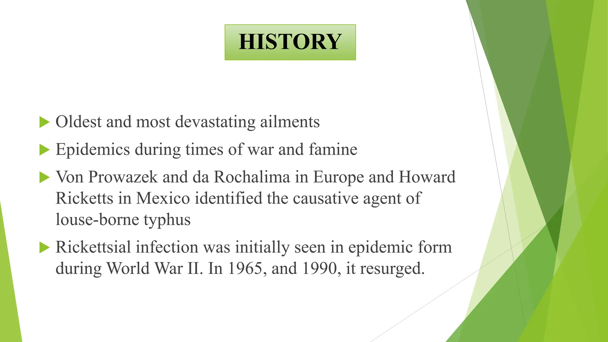
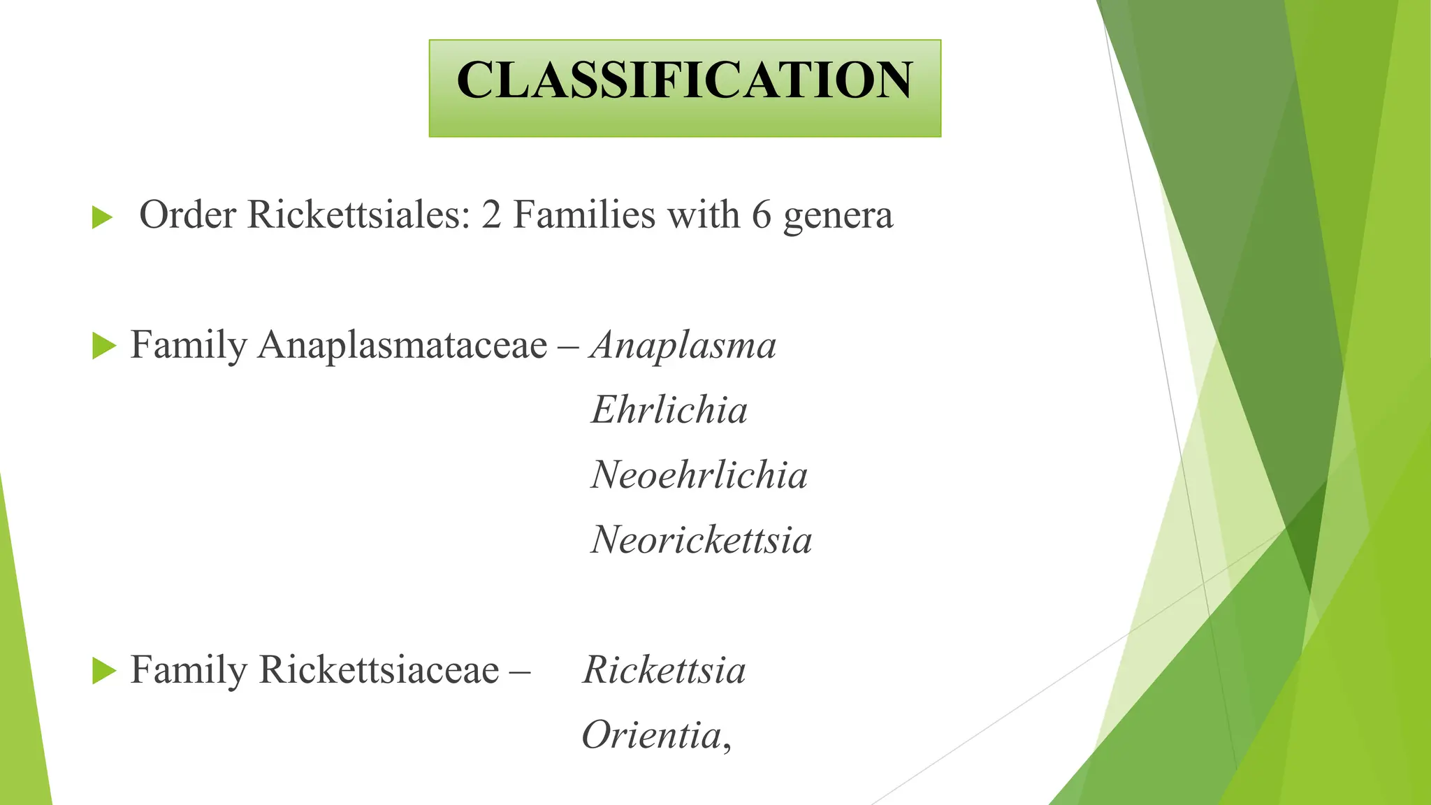
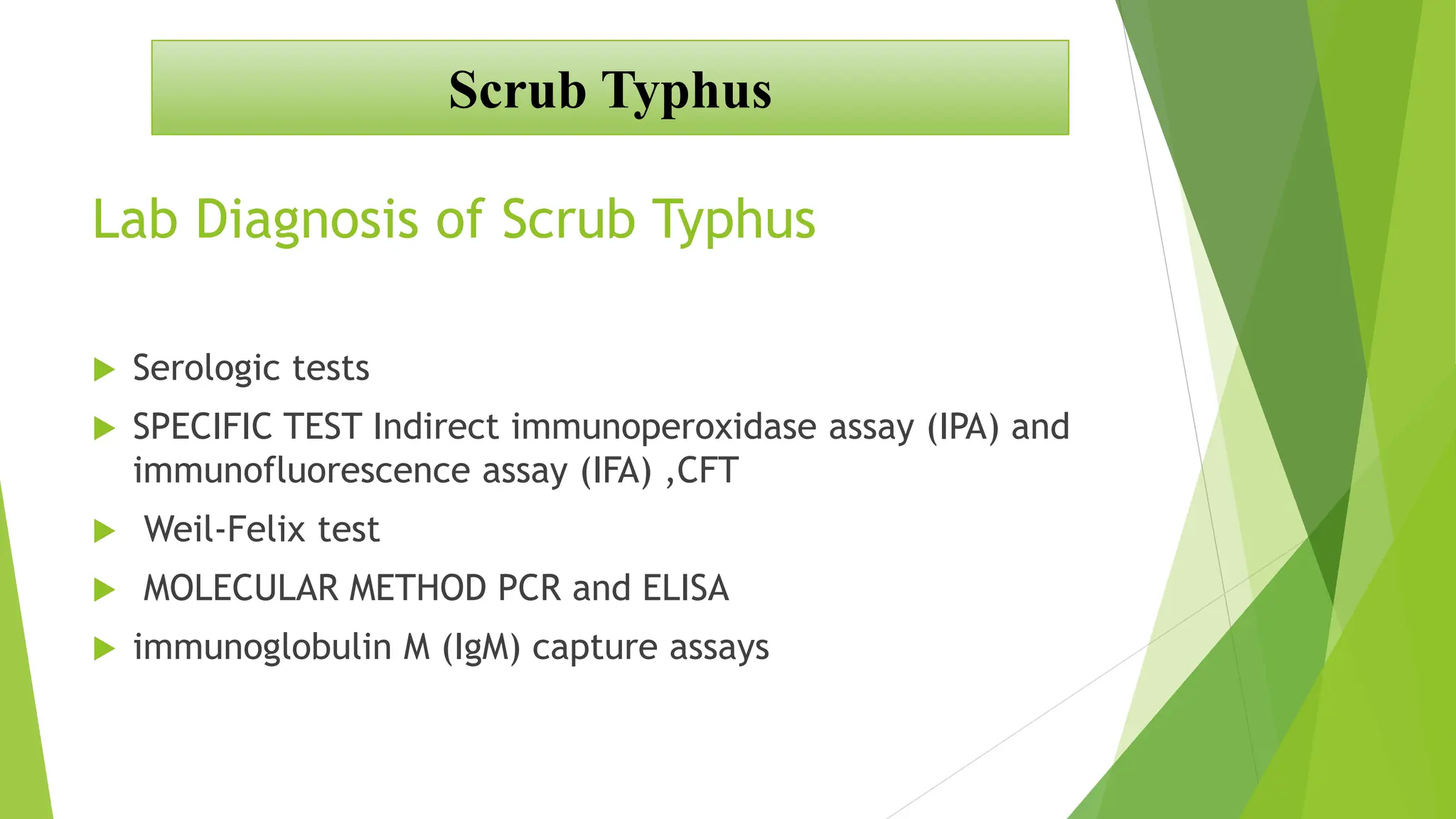
RICKETTSIAL DISEASE IN
INDIA
DR PREETI ( PRESENTER)
DR ARTI (COLLABORATOR)
DR JUTANG (MODERATER)
OUTLINES
 INTRODUCTION
 HISTORY
 CLASSIFICATION
 EPIDEMIOLOGICAL DETERMINANT
 GLOBAL BURDEN
 STATE STATUS
 OUTBREAKS IN INDIA
 CLINICAL PRESENTATION
 SCRUB TYPHUS VS NON SCRUB
 LAB DIAGNOSIS
 TREATMENT
INTRODUCTION
 Gram-negative cocco bacilli
 Obligate Intracellular
 Non cultivable
 Life cycle circulating between mammalian
hosts and hematophagous arthropod vectors
 Human accidental host
 Contain both DNA and RNA
 Cell Wall contains - Outer Membrane +
Phoshoglyceride + Lipopolysaccharide
 Antigen Surface cell Ag
OMP A and B
LPS Ag
HISTORY
 Hippocrates (460 BC)
 Typhus‘confused state of the intellect’associated with fevers.
– As ‘febrile, exanthematic illness, associated with nervous system’
described in L’epidemion.
 Napoleone’s Grande Armee (in 1812)
– Reduced over 42 fold (from 422000 men to 10000men) during
invasion of Russia
– Majority dying from typhus rather than combat.
HISTORY
 The Second World War (1942‐1945)
– Scrub typhus: 18000 cases and 639 deaths
– Murine typyphus: 787 cases and 15 deaths
During outbreaks in armies, ships and prison,
– Burning of clothes
– Changing of bedding
– Crude quarantine measures
HISTORY
 Charles Nicole (in 1909)
– Demonstrated Pediculus corporis (body louse) was
the vector of epidemic typhus
In 19th Century, ill‐defined entity of ‘typhus’was dissected into triad
of
– Typhus
– Typhoid
– Relapsing fevers
HISTORY
 Oldest and most devastating ailments
 Epidemics during times of war and famine
 Von Prowazek and da Rochalima in Europe and Howard
Ricketts in Mexico identified the causative agent of
louse-borne typhus
 Rickettsial infection was initially seen in epidemic form
during World War II. In 1965, and 1990, it resurged.
The famous Rickettsiologists
Stanislaus Von Prowazek Howard Taylor Ricketts Charles Nicolle with collaborators
Helen Sparrow and Rudolph Weig
 Order Rickettsiales: 2 Families with 6 genera
 Family Anaplasmataceae – Anaplasma
Ehrlichia
Neoehrlichia
Neorickettsia
 Family Rickettsiaceae – Rickettsia
Orientia,
CLASSIFICATION
CLASSIFICATION
CLASSIFICATION
PATHOGENESIS
VECTOR
EPIDEMIOLOGICAL DETERMINANTS
history of animal contact (dog/cattle) or insect bite.
Rural area
Age
EPIDEMIOLOGICAL DETERMINANTS
Case definition
Definition of suspected/clinical case:
• AUFI of 5 days or more with or without eschar should
be suspected as a case of rickettsial infection.
• Other presenting features may be headache
• Rash ( more often seen in fair persons),
• lymphadenopathy, multi-organ involvement and
acute respiratory distress.
EPIDEMIOLOGICAL DETERMINANTS
Definition of probable case:
A suspected clinical case showing titres of 1:80 or above
in OX2, OX19 and OXK antigens by weil-Felix test and an
optical density(Od) > 0.5 for IgM by ElISA is considered
positive for members of typhus and spotted fever groups
of Rickettsiae
EPIDEMIOLOGICAL DETERMINANTS
Definition of confirmed case:
A confirmed case is the one in which
(a) Rickettsial dNA is detected in eschar
samples or whole blood by PCR, or
(b) Rising antibody titres on acute and
convalescent serum samples detected by
indirect immune fluorescecnce assay (IFA).
EPIDEMIOLOGICAL DETERMINANTS
 Scrub typhus is the commonest occurring rickettsial
infection in India.
 The infection is transmitted through the larval mites or
‘chiggers’ belonging to the family Trombiculidae.
 Only the larval stages take blood meal.
 Small rodents particularly wild rats of subgenus Rattus
are natural hosts .
 The field rodent and vector mites act as reservoir and
between the two the infection perpetuates in nature.
Scrub Typhus
Risk Factors
 Outdoor activities
 Travelers agricultural workers, forestry personnel
 Increased human-animal interactions
 sandy beaches, scrub vegetation, mountains, and equatorial rain
forests.
Scrub Typhus
Scrub Typhus
Scrub Typhus
Clinical Presentation
 CLASSICAL TRIAD Rash , Eschar and lymphadenopathy
 Incubation period can be from 6 to 20 days (Average 10 days)
 Papule followed by an eschar at the site of chigger feeding (only
in 50%)
Scrub Typhus
Epidemiology Globally
 They are endemic across extensive part of Asia, South Asia, Australia and
the Pacific.
 It is found in India, Pakistan, China, Thailand, Malaysia, Taiwan, Tibet,
Japan, Russia, South Korea and Nepal.
 These regions in which scrub typhus is endemic is referred as the
tsutsugamushi triangle.
 They were clinically described in the Far East more than 1500 years ago.
 Over 1 Billion people are living in endemic areas and as many as 1 million
are infected annually.
Scrub Typhus
 Equatorial rain forests, semi deserts and Alpine subarctic terrains
in the Himalayan regions
 Specific habitats such as abandoned plantations, gardens or rice
fields, overgrown forest clearings, shrubby fringes of fields and
forests, river banks and grassy fields. These ecological patches
which attract the natural host of mite vectors are called ‘mite
islands
Scrub Typhus
Epidemiology Globally – Mite Islands
 Scrub typhus is the commonest worldwide, causing an estimated
1,000,000 cases per year.
 Reported from several States in India including Jammu and
Kashmir, Himachal Pradesh, Uttaranchal (now Uttrakhand),
Bihar, West Bengal, Meghalaya, Rajasthan, Maharashtra,
Karnataka, Tamil Nadu and Kerala.
 In some regions scrub typhus accounts for upto 50 per cent of
undifferentiated fever presenting to hospital.
Scrub Typhus
Epidemiology India
 In this review, the epidemiology of scrub typhus within the
country and the regional distribution were heterogeneous with the
maximum proportion of cases being reported from South India
(55.5%) followed by North India (31.5%)
Scrub Typhus
Epidemiology India
 Eleven outbreaks have been reported from 2000 to 2011, with >900
cases and 42 deaths (case-fatality ratio 5%–17%) in Himachal
Pradesh, Manipur, and one each from Jammu to Kashmir, Tamil
Nadu, Pondicherry, West Bengal, and Meghalaya. Scrub typhus
caused all the outbreaks (exception Kangra: epidemic typhus).
 Cases have also been reported from Rajasthan, Uttaranchal, Assam,
Maharashtra, Kerala, and Karnataka.[8]
Scrub Typhus
Epidemiology India - Outbreaks
Devasagayam E, Dayanand
D, Kundu D, Kamath MS,
Kirubakaran R, Varghese GM
(2021) The burden of scrub
typhus in India: A
systematic review. PLoS
Negl Trop Dis 15(7):
e0009619.
https://doi.org/10.1371/jo
urnal.pntd.0009619
Complication
1. Most common: hepatitis (40.5% of cases of scrub typhus)
thrombocytopenia (28.4%),
2. acute respiratory distress syndrome or ARDS (20.5%),
3. acute kidney injury (19.2%),
4. meningitis (16.4%), shock (16.2%), and
5. myocarditis (15.5%).
Scrub Typhus
Devasagayam E, Dayanand D, Kundu D, Kamath MS, Kirubakaran R, Varghese GM (2021) The burden of scrub typhus in India: A systematic
review. PLoS Negl Trop Dis 15(7): e0009619. https://doi.org/10.1371/journal.pntd.0009619
Lab Diagnosis of Scrub Typhus
 Serologic tests
 SPECIFIC TEST Indirect immunoperoxidase assay (IPA) and
immunofluorescence assay (IFA) ,CFT
 Weil-Felix test
 MOLECULAR METHOD PCR and ELISA
 immunoglobulin M (IgM) capture assays
Scrub Typhus
Weil Felix Test
 Slide/Tube Agglutination test
 Heterophile Antigen
 This test should be carried out only after 5-7 days of
onset of fever. Titre of 1:80 considered possible infection.
Scrub Typhus
Scrub Typhus
Immunochromography Test
Scrub Typhus
 Rapid diagnostic test
 Detect IG M
Enzyme-linked immunosorbent assay
(ELISA)
 IgM and IgG ELISA:immunoglobulin M (IgM) capture assay
 Presence of IgM antibodies, indicates recent infection with
Rickettsia.
 IgM antibody titre is observed at the end of 1st week, whereas
IgG antibodies appear at the end of 2nd week. The cut-off value is
optical density of 0.5. Baseline titres need to be established
keeping in view the regional variations.
Scrub Typhus
 Polymerase chain reaction (PCR):
 Rapid and specific test. It detect rickettsial DNA in whole blood,
buffy coat fraction or tissue specimen.
 RTPCR is used to target the gene encoding the major 56 kDa
and/or 47 kDa surface antigens.
 Sensitive in first week for blood samples (because of presence of
rickettsemia (O. tsutsugamushi, R. rickettsii, R. typhi and R.
prowazekii) in first 7-10 days.)
Scrub Typhus
 Immunufluoroscence assay (IFA): ‘Gold standard’
 Recommended only for research and in areas where
seroprevalence of rickettsial diseases has been established
and a reference facility is already available
Scrub Typhus
 Indirect immunoperoxidase assay (IPA): It gives comparable result
as IFA but requires special instrument and experienced personnel
for interpretation of the test.
 Modification of IFA
Scrub Typhus
Supportive laboratory Investigations
 Leucocytosis is seen, i.e. WBC count > 11,000/μl.
 Thrombocytopenia (i.e. < 1,00,000/μl)
 Biochemistry: Raised transaminase levels
 Imaging: Chest X-ray shows infilterates, mostly
bilateral
Scrub Typhus
 Spotted fever group (SFG) rickettsioses
 Typhus group (TG)
NON Scrub Typhus Rickettsial Diseases
Epidemic Typhus
Louseborne typhus, caused by R. prowazekii
Endemic Typhus
Fleaborne typhus, caused by R.typhi
Rocky Mountain spotted Fever
Tickborne SF, caused by R. rickettsia
NON Scrub Typhus Rickettsial Diseases
Epidemic Typhus
Louseborne typhus, caused by R. prowazekii
Endemic Typhus
Fleaborne typhus, caused by R.typhi
Rocky Mountain spotted Fever
Tickborne SF, caused by R. rickettsia
NON Scrub Typhus Rickettsial Diseases
Rocky Mountain spotted Fever
Tickborne SF, caused by R. rickettsia
fever, rash, headache, malaise, myalgia, nausea
history of tick exposure
NON Scrub Typhus Rickettsial Diseases
Epidemic Typhus
Louseborne typhus, caused by R. prowazekii
Starts with fever and chills.
• A characteristic rash appears
– on the fourth or fifth day,
– Starting on the trunk and spreading over the limbs but
sparing the face, palms and soles
NON Scrub Typhus Rickettsial Diseases
Epidemic Typhus
In some who recover from the disease, the
rickettsiae may remain latent in the lymphoid tissues
or organs for years
Such latent infection may at times be reactivated
leading to recrudescent typhus or Brill Zinsser disease
NON Scrub Typhus Rickettsial Diseases
Rickettsial disease (RDs) other than scrub
typhus (ST) in INDIA
 Documented before the centenary by Megaw (1917) in a
febrile case of a European male bitten by a tick on his
travels from Almora to Lucknow
 An outbreak of 12 cases recorded by Major E.S. Phipson
(1922) in Shimla investigated and documented using the
Weil–Felix reaction with the Bacillus proteus X19 strain
 Study from Kashmir in 1951 documented that up to 91% of
RDs were due to murine typhus
NON Scrub Typhus Rickettsial Diseases
PREVALENCE
Risk Factor
 Crowded Condition
 Refugees housed in camps, incarcerated populations)
 rarely reported among tourists
NON Scrub Typhus Rickettsial Diseases
Giemsa stain of Rickettsia rickettsia
LAB DIADNOSIS
 Serology traditionally the mainstay of diagnosis, although
this has been limited by cross-reactions among closely
related members and diminished sensitivity/utility in the
acute phase of illness
 Nucleic acid amplification tests using blood specimens or
tissue swabs/biopsy specimens, sequencing, and
 Mass spectrometry, have emerged in recent years for both
pathogen and vector identification
TREATMENT
TREATMENT
RIKETTSIAL DISEASE IN INDIA [Autosaved].pptx

RIKETTSIAL DISEASE IN INDIA [Autosaved].pptx

  • 1.
    RICKETTSIAL DISEASE IN INDIA DRPREETI ( PRESENTER) DR ARTI (COLLABORATOR) DR JUTANG (MODERATER)
  • 2.
    OUTLINES  INTRODUCTION  HISTORY CLASSIFICATION  EPIDEMIOLOGICAL DETERMINANT  GLOBAL BURDEN  STATE STATUS  OUTBREAKS IN INDIA  CLINICAL PRESENTATION  SCRUB TYPHUS VS NON SCRUB  LAB DIAGNOSIS  TREATMENT
  • 3.
    INTRODUCTION  Gram-negative coccobacilli  Obligate Intracellular  Non cultivable  Life cycle circulating between mammalian hosts and hematophagous arthropod vectors  Human accidental host  Contain both DNA and RNA  Cell Wall contains - Outer Membrane + Phoshoglyceride + Lipopolysaccharide  Antigen Surface cell Ag OMP A and B LPS Ag
  • 4.
    HISTORY  Hippocrates (460BC)  Typhus‘confused state of the intellect’associated with fevers. – As ‘febrile, exanthematic illness, associated with nervous system’ described in L’epidemion.  Napoleone’s Grande Armee (in 1812) – Reduced over 42 fold (from 422000 men to 10000men) during invasion of Russia – Majority dying from typhus rather than combat.
  • 5.
    HISTORY  The SecondWorld War (1942‐1945) – Scrub typhus: 18000 cases and 639 deaths – Murine typyphus: 787 cases and 15 deaths During outbreaks in armies, ships and prison, – Burning of clothes – Changing of bedding – Crude quarantine measures
  • 6.
    HISTORY  Charles Nicole(in 1909) – Demonstrated Pediculus corporis (body louse) was the vector of epidemic typhus In 19th Century, ill‐defined entity of ‘typhus’was dissected into triad of – Typhus – Typhoid – Relapsing fevers
  • 7.
    HISTORY  Oldest andmost devastating ailments  Epidemics during times of war and famine  Von Prowazek and da Rochalima in Europe and Howard Ricketts in Mexico identified the causative agent of louse-borne typhus  Rickettsial infection was initially seen in epidemic form during World War II. In 1965, and 1990, it resurged.
  • 8.
    The famous Rickettsiologists StanislausVon Prowazek Howard Taylor Ricketts Charles Nicolle with collaborators Helen Sparrow and Rudolph Weig
  • 9.
     Order Rickettsiales:2 Families with 6 genera  Family Anaplasmataceae – Anaplasma Ehrlichia Neoehrlichia Neorickettsia  Family Rickettsiaceae – Rickettsia Orientia, CLASSIFICATION
  • 10.
  • 11.
  • 12.
  • 13.
  • 14.
    history of animalcontact (dog/cattle) or insect bite. Rural area Age EPIDEMIOLOGICAL DETERMINANTS
  • 15.
    Case definition Definition ofsuspected/clinical case: • AUFI of 5 days or more with or without eschar should be suspected as a case of rickettsial infection. • Other presenting features may be headache • Rash ( more often seen in fair persons), • lymphadenopathy, multi-organ involvement and acute respiratory distress. EPIDEMIOLOGICAL DETERMINANTS
  • 16.
    Definition of probablecase: A suspected clinical case showing titres of 1:80 or above in OX2, OX19 and OXK antigens by weil-Felix test and an optical density(Od) > 0.5 for IgM by ElISA is considered positive for members of typhus and spotted fever groups of Rickettsiae EPIDEMIOLOGICAL DETERMINANTS
  • 17.
    Definition of confirmedcase: A confirmed case is the one in which (a) Rickettsial dNA is detected in eschar samples or whole blood by PCR, or (b) Rising antibody titres on acute and convalescent serum samples detected by indirect immune fluorescecnce assay (IFA). EPIDEMIOLOGICAL DETERMINANTS
  • 19.
  • 20.
     Scrub typhusis the commonest occurring rickettsial infection in India.  The infection is transmitted through the larval mites or ‘chiggers’ belonging to the family Trombiculidae.  Only the larval stages take blood meal.  Small rodents particularly wild rats of subgenus Rattus are natural hosts .  The field rodent and vector mites act as reservoir and between the two the infection perpetuates in nature. Scrub Typhus
  • 21.
    Risk Factors  Outdooractivities  Travelers agricultural workers, forestry personnel  Increased human-animal interactions  sandy beaches, scrub vegetation, mountains, and equatorial rain forests. Scrub Typhus
  • 22.
  • 23.
  • 24.
    Clinical Presentation  CLASSICALTRIAD Rash , Eschar and lymphadenopathy  Incubation period can be from 6 to 20 days (Average 10 days)  Papule followed by an eschar at the site of chigger feeding (only in 50%) Scrub Typhus
  • 25.
    Epidemiology Globally  Theyare endemic across extensive part of Asia, South Asia, Australia and the Pacific.  It is found in India, Pakistan, China, Thailand, Malaysia, Taiwan, Tibet, Japan, Russia, South Korea and Nepal.  These regions in which scrub typhus is endemic is referred as the tsutsugamushi triangle.  They were clinically described in the Far East more than 1500 years ago.  Over 1 Billion people are living in endemic areas and as many as 1 million are infected annually. Scrub Typhus
  • 26.
     Equatorial rainforests, semi deserts and Alpine subarctic terrains in the Himalayan regions  Specific habitats such as abandoned plantations, gardens or rice fields, overgrown forest clearings, shrubby fringes of fields and forests, river banks and grassy fields. These ecological patches which attract the natural host of mite vectors are called ‘mite islands Scrub Typhus Epidemiology Globally – Mite Islands
  • 27.
     Scrub typhusis the commonest worldwide, causing an estimated 1,000,000 cases per year.  Reported from several States in India including Jammu and Kashmir, Himachal Pradesh, Uttaranchal (now Uttrakhand), Bihar, West Bengal, Meghalaya, Rajasthan, Maharashtra, Karnataka, Tamil Nadu and Kerala.  In some regions scrub typhus accounts for upto 50 per cent of undifferentiated fever presenting to hospital. Scrub Typhus Epidemiology India
  • 28.
     In thisreview, the epidemiology of scrub typhus within the country and the regional distribution were heterogeneous with the maximum proportion of cases being reported from South India (55.5%) followed by North India (31.5%) Scrub Typhus Epidemiology India
  • 29.
     Eleven outbreakshave been reported from 2000 to 2011, with >900 cases and 42 deaths (case-fatality ratio 5%–17%) in Himachal Pradesh, Manipur, and one each from Jammu to Kashmir, Tamil Nadu, Pondicherry, West Bengal, and Meghalaya. Scrub typhus caused all the outbreaks (exception Kangra: epidemic typhus).  Cases have also been reported from Rajasthan, Uttaranchal, Assam, Maharashtra, Kerala, and Karnataka.[8] Scrub Typhus Epidemiology India - Outbreaks
  • 30.
    Devasagayam E, Dayanand D,Kundu D, Kamath MS, Kirubakaran R, Varghese GM (2021) The burden of scrub typhus in India: A systematic review. PLoS Negl Trop Dis 15(7): e0009619. https://doi.org/10.1371/jo urnal.pntd.0009619
  • 33.
    Complication 1. Most common:hepatitis (40.5% of cases of scrub typhus) thrombocytopenia (28.4%), 2. acute respiratory distress syndrome or ARDS (20.5%), 3. acute kidney injury (19.2%), 4. meningitis (16.4%), shock (16.2%), and 5. myocarditis (15.5%). Scrub Typhus
  • 34.
    Devasagayam E, DayanandD, Kundu D, Kamath MS, Kirubakaran R, Varghese GM (2021) The burden of scrub typhus in India: A systematic review. PLoS Negl Trop Dis 15(7): e0009619. https://doi.org/10.1371/journal.pntd.0009619
  • 35.
    Lab Diagnosis ofScrub Typhus  Serologic tests  SPECIFIC TEST Indirect immunoperoxidase assay (IPA) and immunofluorescence assay (IFA) ,CFT  Weil-Felix test  MOLECULAR METHOD PCR and ELISA  immunoglobulin M (IgM) capture assays Scrub Typhus
  • 36.
    Weil Felix Test Slide/Tube Agglutination test  Heterophile Antigen  This test should be carried out only after 5-7 days of onset of fever. Titre of 1:80 considered possible infection. Scrub Typhus
  • 37.
  • 38.
    Immunochromography Test Scrub Typhus Rapid diagnostic test  Detect IG M
  • 39.
    Enzyme-linked immunosorbent assay (ELISA) IgM and IgG ELISA:immunoglobulin M (IgM) capture assay  Presence of IgM antibodies, indicates recent infection with Rickettsia.  IgM antibody titre is observed at the end of 1st week, whereas IgG antibodies appear at the end of 2nd week. The cut-off value is optical density of 0.5. Baseline titres need to be established keeping in view the regional variations. Scrub Typhus
  • 40.
     Polymerase chainreaction (PCR):  Rapid and specific test. It detect rickettsial DNA in whole blood, buffy coat fraction or tissue specimen.  RTPCR is used to target the gene encoding the major 56 kDa and/or 47 kDa surface antigens.  Sensitive in first week for blood samples (because of presence of rickettsemia (O. tsutsugamushi, R. rickettsii, R. typhi and R. prowazekii) in first 7-10 days.) Scrub Typhus
  • 41.
     Immunufluoroscence assay(IFA): ‘Gold standard’  Recommended only for research and in areas where seroprevalence of rickettsial diseases has been established and a reference facility is already available Scrub Typhus
  • 42.
     Indirect immunoperoxidaseassay (IPA): It gives comparable result as IFA but requires special instrument and experienced personnel for interpretation of the test.  Modification of IFA Scrub Typhus
  • 43.
    Supportive laboratory Investigations Leucocytosis is seen, i.e. WBC count > 11,000/μl.  Thrombocytopenia (i.e. < 1,00,000/μl)  Biochemistry: Raised transaminase levels  Imaging: Chest X-ray shows infilterates, mostly bilateral Scrub Typhus
  • 44.
     Spotted fevergroup (SFG) rickettsioses  Typhus group (TG) NON Scrub Typhus Rickettsial Diseases
  • 45.
    NON Scrub TyphusRickettsial Diseases
  • 46.
    Epidemic Typhus Louseborne typhus,caused by R. prowazekii Endemic Typhus Fleaborne typhus, caused by R.typhi Rocky Mountain spotted Fever Tickborne SF, caused by R. rickettsia NON Scrub Typhus Rickettsial Diseases
  • 47.
    Epidemic Typhus Louseborne typhus,caused by R. prowazekii Endemic Typhus Fleaborne typhus, caused by R.typhi Rocky Mountain spotted Fever Tickborne SF, caused by R. rickettsia NON Scrub Typhus Rickettsial Diseases
  • 48.
    Rocky Mountain spottedFever Tickborne SF, caused by R. rickettsia fever, rash, headache, malaise, myalgia, nausea history of tick exposure NON Scrub Typhus Rickettsial Diseases
  • 49.
    Epidemic Typhus Louseborne typhus,caused by R. prowazekii Starts with fever and chills. • A characteristic rash appears – on the fourth or fifth day, – Starting on the trunk and spreading over the limbs but sparing the face, palms and soles NON Scrub Typhus Rickettsial Diseases
  • 50.
    Epidemic Typhus In somewho recover from the disease, the rickettsiae may remain latent in the lymphoid tissues or organs for years Such latent infection may at times be reactivated leading to recrudescent typhus or Brill Zinsser disease NON Scrub Typhus Rickettsial Diseases
  • 51.
    Rickettsial disease (RDs)other than scrub typhus (ST) in INDIA  Documented before the centenary by Megaw (1917) in a febrile case of a European male bitten by a tick on his travels from Almora to Lucknow  An outbreak of 12 cases recorded by Major E.S. Phipson (1922) in Shimla investigated and documented using the Weil–Felix reaction with the Bacillus proteus X19 strain  Study from Kashmir in 1951 documented that up to 91% of RDs were due to murine typhus NON Scrub Typhus Rickettsial Diseases
  • 52.
    NON Scrub TyphusRickettsial Diseases
  • 53.
    NON Scrub TyphusRickettsial Diseases
  • 54.
  • 55.
    Risk Factor  CrowdedCondition  Refugees housed in camps, incarcerated populations)  rarely reported among tourists NON Scrub Typhus Rickettsial Diseases
  • 61.
    Giemsa stain ofRickettsia rickettsia
  • 62.
    LAB DIADNOSIS  Serologytraditionally the mainstay of diagnosis, although this has been limited by cross-reactions among closely related members and diminished sensitivity/utility in the acute phase of illness  Nucleic acid amplification tests using blood specimens or tissue swabs/biopsy specimens, sequencing, and  Mass spectrometry, have emerged in recent years for both pathogen and vector identification
  • 63.
  • 64.