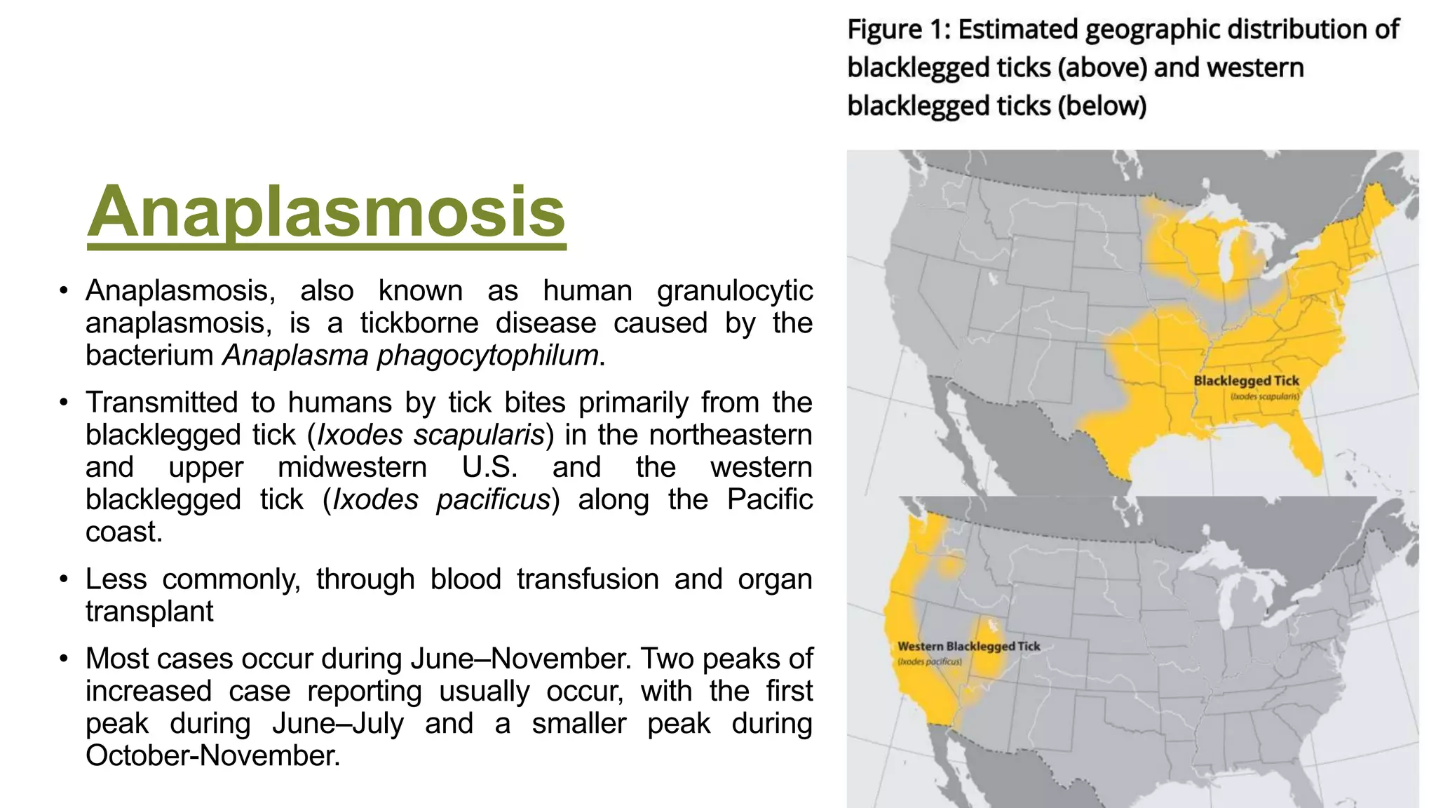
Tick borne
diseases
Anaplasmosis
• Anaplasmosis, also known as human granulocytic
anaplasmosis, is a tickborne disease caused by the
bacterium Anaplasma phagocytophilum.
• Transmitted to humans by tick bites primarily from the
blacklegged tick (Ixodes scapularis) in the northeastern
and upper midwestern U.S. and the western
blacklegged tick (Ixodes pacificus) along the Pacific
coast.
• Less commonly, through blood transfusion and organ
transplant
• Most cases occur during June–November. Two peaks of
increased case reporting usually occur, with the first
peak during June–July and a smaller peak during
October-November.
Presentation
• Anaplasmosis typically causes an acute febrile illness.
• Signs and symptoms vary greatly from person to person.
• Most people will have mild or moderate illness, though severe illness and death are
possible.
• Signs and symptoms typically begin within 5–14 days after the bite of an infected tick.
However, because tick bites are not painful, many people will not remember a tick bite.
• Healthcare providers should consider anaplasmosis or other tickborne infections in
the differential diagnosis for patients with a non-specific febrile illness of unknown
origin, particularly during spring and summer months when ticks are most active.
• Ehrlichiosis and anaplasmosis should be suspected in febrile patients with leukopenia with
an exposure history. The clinical signs are similar to those of the rickettsioses.
Early Illness
• Fever, chills, rigors
• Severe headache
• Malaise
• Myalgia
• Gastrointestinal symptoms (nausea, vomiting, diarrhea, anorexia) in about 20% of
cases
• Rash (<10% of cases) in patients with anaplasmosis.
• The presence of a rash might indicate a coinfection with Lyme disease, or another tickborne
disease.
• Nervous system involvement (meningoencephalitis, focal paralysis, etc.) rare
Late Illness
• Renal or respiratory failure
• Peripheral neuropathies
• Disseminated intravascular
coagulation (DIC)-like
coagulopathies
• Rhabdomyolysis
• Hemorrhage
Risk factors for severe disease:
• Delayed treatment
• Older age
• Immune compromising conditions,
e.g., advanced HIV, persons
receiving chemotherapy, or other
immune suppressing medications
General labs
• General laboratory findings can include mild anemia, thrombocytopenia,
leukopenia (characterized by relative and absolute lymphopenia and a left
shift) and mild to moderate elevations in hepatic transaminases.
• Abnormal laboratory findings can appear in the first week of illness; however, normal
laboratory findings do not rule out possible infection.
Clinical Diagnosis
• Early recognition and treatment is key.
• Maintain a high level of clinical suspicion for anaplasmosis or other tickborne diseases in
cases of non-specific febrile illness of unknown origin, particularly during spring and summer
months when ticks are most active. Early recognition and presumptive treatment is
important to prevent severe illness.
• Because anaplasmosis can be difficult to diagnose, particularly in the early stages of illness,
patients should treated presumptively based on clinical suspicion.
• Always take a thorough patient history, including:
• Recent tick bite. NOTE: Tick bites are often painless. Many people do not remember being bitten. Do
not rule out a tickborne infection if your patient does not remember a tick bite.
• Exposure to areas where ticks are commonly found, including wooded areas or brushy areas with high
grasses and leaf litter.
• Travel history (domestic and international) to areas where anaplasmosis is endemic.
• Laboratory confirmation is helpful for disease surveillance and understanding burden of
anaplasmosis infection in the United States.
Clinical Diagnosis
• The diagnosis of anaplasmosis often is made based on clinical signs
and symptoms, and can later be confirmed using specialized
confirmatory laboratory tests. Treatment should not be delayed
pending the receipt of laboratory test results, or withheld on the basis
of an initial negative laboratory result.
Laboratory Diagnosis
• PCR is performed on DNA extracted from whole blood specimens.
• Most sensitive in the first week of illness and decreases in sensitivity following the
administration of appropriate antibiotics (within 24–48 hours).
• Although a positive PCR result is helpful, a negative result does not rule out the
diagnosis, and treatment should not be withheld due to a negative result.
• PCR might also be used to amplify DNA in solid tissue and bone marrow specimens.
Laboratory Diagnosis
• The standard serologic test for diagnosis of anaplasmosis is the indirect
immunofluorescence antibody (IFA) assay for IgG using A.
phagocytophilum antigen. (high sensitivity and specificity)
• Paired acute and convalescent serum samples collected 2–4 weeks apart to
demonstrate evidence of a fourfold seroconversion.
• Antibody titers are frequently negative in the first week of illness. Anaplasmosis cannot
be confirmed using single acute antibody results.
• IgM IFA assays may also be offered by reference laboratories, however are not
necessarily indicators of acute infection and might be less specific than IgG antibodies.
Might remain elevated in patients for whom no other supportive evidence of a recent
rickettsiosis infection exists. For these reasons, IgM antibody titers alone should not be
used for laboratory diagnosis.
Laboratory Diagnosis
• Antibodies to A. phagocytophilum might remain elevated for many months after the
disease has resolved.
• In certain people, high titers of antibodies against A. phagocytophilum have been observed
up to four years after the acute illness.
• Between 5–10% of healthy people in some areas might have elevated antibody
titers due to past exposure to A. phagocytophilum or similar organisms.
• Comparison of paired, and appropriately timed, serologic assays provides the best
evidence of recent infection.
• Single or inappropriately timed serologic tests, in relation to clinical illness, can lead to
misinterpretation of results.
• In general, serology could be false positive due to cross reactivity with Ehrlichia
chaffeensis
Laboratory Diagnosis
• Blood microscopy:
• During the first week of illness a microscopic examination of a peripheral blood smear
might reveal morulae (microcolonies of anaplasmae) in the cytoplasm of granulocytes
and is highly suggestive of a diagnosis.
• However, blood smear examination is relatively insensitive and should not be relied
upon solely to diagnose anaplasmosis.
• The observance of morulae in a particular cell type cannot conclusively differentiate
between Anaplasma and Ehrlichia species.
• Giemsa or wright stains of peripheral blood or buffy coat is positive in 60%
Laboratory Diagnosis
• Culture:
• Blood is inoculated onto HL-60 or other myelocytic cell lines
• Positive between 3-30 days
• If a bone marrow biopsy is performed as part of the investigation of cytopenias,
immunostaining of the bone marrow biopsy specimen can diagnose A.
phagocytophilum infection.
Co-Infection
• Borrelia burgdorferi (the causative agent of Lyme disease),
• Babesia microti (the primary cause of human babesiosis),
• Borrelia mayonii (a newly discovered cause of borreliosis),
• Borrelia miyamotoi (a cause of tickborne relapsing fever)
• Powassan virus (cause of Powassan virus disease), and
• Ehrlichia muris eauclairensis
Treatment
• Doxycycline is the treatment of choice for anaplasmosis, and all other tickborne
rickettsial diseases.
• Doxycycline is most effective at preventing severe complications from developing if it is started early in
the course of disease.
• When treated with doxycycline, fever generally subsides within 24–48 hours.
• Patients with suspected anaplasmosis should be treated with doxycycline for 10–14 days to provide
appropriate length of therapy for possible concurrent Lyme disease infection.
• Lack of a clinical response to doxycycline suggests that the patient’s condition might not be
due to anaplasmosis or might be caused by other infections not responsive to doxycycline
(see coinfections).
• Resistance to doxycycline or relapses in symptoms after the completion of the
recommended course have not been documented.
• Rifampin is Alternative
Prophylaxis
• Post-tick bite antibiotic prophylaxis is not recommended to prevent
anaplasmosis.
• People who have been bitten by a tick should watch for signs and
symptoms. They should see their healthcare provider if fever, rash, or other
symptoms develop within two weeks of tick bite.
• Asymptomatic treatment for tick bites is not currently recommended.
Lyme Disease
• A systemic, tick-borne disease with protean manifestations, including
dermatologic, rheumatologic, neurologic, and cardiac abnormalities. The
most common clinical marker for the disease is erythema migrans (EM), the
initial skin lesion that occurs in 60%-80% of patients.
• Caused by spirochete Borrelia burgdorferi, B. mayonii
• Lyme disease is most frequently reported from the Upper Midwestern and
northeastern United States. Some cases are also reported in northern
California, Oregon, and Washington.
• Incubation Period is 3-30 days
Presentation (Localized Stage)
• Erythema migrans (EM)—red ring-like or homogenous expanding
rash; classic rash not present in all cases. See examples on following
pages.
• Flu-like symptoms—malaise, headache, fever, myalgia, arthralgia
• Lymphadenopathy
• During the localized (early) stage of illness, Lyme disease may be
diagnosed clinically in patients who present with an EM rash.
• Serologic tests may be insensitive at this stage. During disseminated disease,
however, serologic tests should be positive.
Presentation
Disseminated Stage
• Multiple secondary
annular rashes
• Flu-like symptoms
• Lymphadenopathy
Rheumatologic
Manifestations
• Transient, migratory
arthritis and effusion in
one or multiple joints
• Migratory pain in tendons,
bursae, muscle, and
bones
• Baker’s cyst
Cardiac Manifestations
• Conduction abnormalities,
e.g. atrioventricular node
block
• Myocarditis, pericarditis
Presentation
Neurologic Manifestations
• Bell’s palsy or other cranial
neuropathy
• Meningitis
• Motor and sensory
radiculoneuropathy,
mononeuritis multiplex
• Subtle cognitive difficulties
• Encephalitis,
encephalomyelitis, subtle
encephalopathy,
pseudotumor cerebri (all
rare)
Additional Manifestations
• Conjunctivitis, keratitis,
uveitis
• Mild hepatitis
• Splenomegaly
General Laboratory Findings
• Elevated erythrocyte sedimentation rate
• Mildly elevated hepatic transaminases
• Microscopic hematuria or proteinuria
• In Lyme meningitis, CSF typically shows lymphocytic pleocytosis, slightly
elevated protein, and normal glucose.
Diagnosis
• Observation of an EM rash with a history of recent travel to an endemic area (with or without
history of tick bite) is sufficient.
• For patients with evidence of disseminated infection (musculoskeletal, neurologic, or cardiac
manifestations),
• 2-tiered serologic testing, consisting of an ELISA/ IFA and confirmatory WB, is recommended.
• Two-tier test is defined as a positive or equivocal enzyme immunoassay (EIA) or immunofluorescent
assay (IFA) followed by a positive Immunoglobulin M (IgM) or Immunoglobulin G (IgG) western
immunoblot (WB) for Lyme disease
• If the first step is negative, no further testing is recommended.
• If the first step is positive or indeterminate (sometimes called “equivocal”), the second step should be performed.
• The overall result is positive only when the first step is positive (or equivocal) and the second step is positive (or
for some tests, equivocal).
• A positive single-tier IgG WB test for Lyme disease is also confirmatory for diagnosis
Notes on Serologic Tests for Lyme
Disease
• Serologic tests are insensitive during the first few weeks of infection. During this stage,
patients with an EM rash may be diagnosed clinically.
• In persons with illness duration of more than 1 month, only IgG or combined IgG/IgM testing
should be performed (not IgM alone). A positive IgM test alone in a patient with illness
duration of more than 1 month is not reliable for diagnosing current disease.
• Due to antibody persistence, single positive serologic test results cannot distinguish
between active and past infection.
• Serologic tests cannot be used to measure treatment response.
• Prompt use of Abx decrease abs response
• Infection with other diseases, including some tickborne diseases, or some viral, bacterial,
syphilis or autoimmune diseases, can result in false positive test results.
Diagnosis
• Isolation of organism from a clinical specimen is a confirmatory test:
• EM biopsy in liquid BSK, modified Kelly pettenkofer media for 2-6 weeks incubation
• For neuroborrelosis
• PCR of CSF (10-30% sensitivity)
• Serology is almost always positive
Early-stage skin manifestations,
arthritis, or Bell’s palsy
• Doxycycline or amoxicillin PO for 14–21 days.
• If arthritis persists, repeat the course, or consider IV ceftriaxone for 14–21 days.
Azithromycin is a third-line agent and associated with treatment failures.
late or neurological disease
• Acrodermatitis chronica atrophicans, 21–28 days of doxycycline;
• Isolated meningitis, 14–21 days of doxycycline;
• Encephalitis/myelitis, 14 days of IV ceftriaxone;
• Third-degree heart block, 14–21 days of IV ceftriaxone;
• Late neuroborreliosis, 14–28 days of IV ceftriaxone.
Prevention
• Avoid tick habitats, use insect repellent on exposed skin and clothing, and
carefully check every day for attached ticks. Minimize areas of exposed skin
by wearing long-sleeved shirts, long pants, and closed shoes; tucking shirts
in and tucking pants into socks can help reduce risk
Rocky Mountain
spotted fever
• Rocky Mountain spotted fever (RMSF) is a
tickborne disease caused by the intracellular
bacterium Rickettsia rickettsii.
• Primarily transmitted, through tick bites.
• Blood transfusion Transmission of R. rickettsii
is extremely rare.
• Organ transplant acquired RMSF has not been
documented, but is physiologically possible.
• Although cases are reported in every month of
the year, most cases occur during May–August
Signs and Symptoms
• RMSF is a rapidly progressive disease and without early
administration of doxycycline can be fatal within days.
• Signs and symptoms of RMSF begin 3-12 days after the bite of an infected
tick. However, because tick bites are not painful, many people do not
remember being bitten.
• Illness generally begins with sudden onset of fever and headache and most
people visit a healthcare provider during the first few days of symptoms.
Early illness (days 1-4)
• Fever, Myalgia
• Headache, Edema around the eyes and on the back of hands
• Gastrointestinal symptoms (nausea, vomiting, anorexia)
• Abdominal pain (may mimic appendicitis or acute abdominal pain)
• Rash (typically occurs 2-4 days after the onset of fever)
• A classic case of early RMSF involves a rash that appears 2-4 days after the onset of fever
as small, flat, pink, macules on the wrists, forearms, and ankles and spreads to include the
trunk and sometimes the palms of hands and soles of feet.
• Rash can be highly variable and people who fail to develop a rash, or develop an atypical
rash, are at increased risk of being misdiagnosed.
• While most (90%) people with
RMSF have some type of rash
during the course of illness, less
than 50% of patients have a rash
during the first 3 days of illness,
when most people first seek
medical care. For this reason,
clinicians should consider RMSF
if other signs and symptoms
support a diagnosis, even if a
rash is not present.
Late illness (day 5 or later)
• Altered mental status, coma, cerebral edema
• Respiratory compromise (pulmonary edema, acute respiratory distress
syndrome)
• Necrosis, often requiring amputation
• Multiorgan system damage (CNS, renal failure)
• Rash:
• The petechial rash of RMSF does not typically appear until day 5-6 of illness.
• NOTE: Petechiae are a sign of severe disease. Every attempt should be made to treat
before petechiae develop.
Risk factors for severe illness
• Delayed treatment
• Children < 10 years
• Persons with glucose-6-phosphate dehydrogenase (G6PD) deficiency
Infection in Children
• Children represent less than 6% of spotted fever cases, but 22% of spotted fever
fatalities.
• Children with RMSF frequently report experiencing nausea, vomiting, loss of
appetite, and rash, but are less likely to report a headache than adults.
• Other frequently observed signs and symptoms in children with RMSF include
abdominal pain, altered mental status, and edema involving the dorsum of the
hands or around the eyes.
• Children < 10 years are five times more likely than adults to die from RMSF.
• Early treatment with doxycycline saves lives.
• Doxycycline is the treatment of choice for adults and children of all ages.
General lab
• General laboratory findings can include thrombocytopenia, hyponatremia, or
elevated levels of hepatic transaminases although these tests may not
appear abnormal in early stages of illness.
Serology
• The standard serologic test for diagnosis of RMSF is the indirect immunofluorescence antibody (IFA) assay
for immunoglobulin G (IgG) using R. rickettsii antigen.
• IgG IFA assays should be performed on paired serum samples collected 2–4 weeks apart to demonstrate
evidence of a fourfold seroconversion.
• Antibody titers are frequently negative in the first week of illness. RMSF cannot be confirmed using single acute antibody
results.
• Immunoglobulin M (IgM) IFA assays are available through some reference laboratories, however results
might be less specific than IgG IFA assays for diagnosing a recent infection.
• R. rickettsii is closely related to other pathogenic spotted fever group Rickettsia (SFGR) species,
including R. akari, R. parkeri, and Rickettsia 364D. Closely related species of SFGR share similar antigens
such that antibodies directed to one of these antigens can cross-react with other heterologous spotted fever
group antigens.
• Most commercial labs are unable to differentiate one spotted fever infection from another using these serologic methods.
Points in serology
• Abs to R. rickettsii might remain elevated for months after the disease has resolved.
• In certain people, high titers of antibodies against R. rickettsii have been observed up
to four years after the acute illness.
• Ten percent or more of healthy people in some areas might have elevated antibody
titers due to past exposure to R. rickettsii or other SFGR.
• Comparison of paired, and appropriately timed, serologic assays provides the best
evidence of recent infection.
• Single or inappropriately timed serologic tests, in relation to clinical illness, can lead to
misinterpretation of results.
PCR
• It is performed on DNA extracted from whole blood.
• R. rickettsii infect the endothelial cells that line blood vessels and may not
circulate in large numbers in the blood until the disease has progressed to a
severe phase of infection.
• Although a positive PCR result is helpful,
• Negative result does not R/O Dx, and TTT should not be stopped.
• PCR might also be used to amplify DNA from a skin biopsy of a rash lesion,
or in post-mortem tissue specimens.
Culture and immunohistochemistry
(IHC) assays
• Can be performed on skin biopsies of a rash lesion, or post-mortem tissue
specimens.
• Culture isolation and IHC assays of R. rickettsii are only available at specialized
laboratories; routine hospital blood cultures cannot detect the organism.
Treatment
• Order diagnostic tests for patients with illness clinically compatible with
RMSF. However, do not delay treatment while waiting for diagnostic test
results.
• Doxycycline is the treatment of choice for RMSF, and all other
tickborne rickettsial diseases. Use of antibiotics other than doxycycline is
associated with a higher risk of fatal outcome from RMSF.
• Patients with suspected RMSF should be treated with doxycycline for at least 3 days
after the fever subsides and there is evidence of clinical improvement. Minimum course
of treatment is 5-7 days.
• Chloramphenicol is the only alternative drug used to treat RMSF;
Post-tick bite antibiotic prophylaxis
• not recommended to prevent RMSF.
• People who were bitten by a tick should be advised to watch for signs and
symptoms and see their healthcare provider if fever, rash, or other
symptoms develop within two weeks of tick bite.
• Treatment for asymptomatic individuals is not currently recommended.
Diagnosis of Ehrlichiosis (serology)
• The reference standard serologic test for diagnosis of ehrlichiosis is the indirect
immunofluorescence antibody (IFA) assay for immunoglobulin G (IgG).
• IgG IFA assays should be performed on paired acute and convalescent serum samples
collected 2–4 weeks apart to demonstrate evidence of a fourfold seroconversion.
• Antibody titers are frequently negative in the first week of illness. Ehrlichiosis cannot be
confirmed using single acute antibody results.
• Immunoglobulin M (IgM) IFA assays may also be offered by reference laboratories, however,
are not necessarily indicators of acute infection and might be less specific than IgG
antibodies.
• Antibodies, particularly IgM antibodies, might remain elevated in patients for whom no other
supportive evidence of a recent rickettsiosis infection exists. For these reasons, IgM
antibody titers alone should not be used for laboratory diagnosis.
Persistent Antibodies
• Antibodies against Ehrlichia species might remain elevated for many months
after disease has resolved.
• Comparison of paired, and appropriately timed, serologic assays provides
the best evidence of recent infection.
• Single or inappropriately timed serologic tests, in relation to clinical illness,
can lead to misinterpretation of results.
Cross Reactivity
• Closely related organisms, including those in
the Ehrlichia and Anaplasma genera, share similar antigens such that
antibodies directed to one of these antigens can cross-react.
• Most commercial labs are unable to differentiate between Ehrlichia species.
• In areas endemic for Ehrlichiosis and Anaplasmosis, IFA using antigen from
both Ehrlichia and Anaplasma species should be run side-by-side.

Tick borne diseases

  • 1.
  • 2.
    Anaplasmosis • Anaplasmosis, alsoknown as human granulocytic anaplasmosis, is a tickborne disease caused by the bacterium Anaplasma phagocytophilum. • Transmitted to humans by tick bites primarily from the blacklegged tick (Ixodes scapularis) in the northeastern and upper midwestern U.S. and the western blacklegged tick (Ixodes pacificus) along the Pacific coast. • Less commonly, through blood transfusion and organ transplant • Most cases occur during June–November. Two peaks of increased case reporting usually occur, with the first peak during June–July and a smaller peak during October-November.
  • 3.
    Presentation • Anaplasmosis typicallycauses an acute febrile illness. • Signs and symptoms vary greatly from person to person. • Most people will have mild or moderate illness, though severe illness and death are possible. • Signs and symptoms typically begin within 5–14 days after the bite of an infected tick. However, because tick bites are not painful, many people will not remember a tick bite. • Healthcare providers should consider anaplasmosis or other tickborne infections in the differential diagnosis for patients with a non-specific febrile illness of unknown origin, particularly during spring and summer months when ticks are most active. • Ehrlichiosis and anaplasmosis should be suspected in febrile patients with leukopenia with an exposure history. The clinical signs are similar to those of the rickettsioses.
  • 4.
    Early Illness • Fever,chills, rigors • Severe headache • Malaise • Myalgia • Gastrointestinal symptoms (nausea, vomiting, diarrhea, anorexia) in about 20% of cases • Rash (<10% of cases) in patients with anaplasmosis. • The presence of a rash might indicate a coinfection with Lyme disease, or another tickborne disease. • Nervous system involvement (meningoencephalitis, focal paralysis, etc.) rare
  • 5.
    Late Illness • Renalor respiratory failure • Peripheral neuropathies • Disseminated intravascular coagulation (DIC)-like coagulopathies • Rhabdomyolysis • Hemorrhage Risk factors for severe disease: • Delayed treatment • Older age • Immune compromising conditions, e.g., advanced HIV, persons receiving chemotherapy, or other immune suppressing medications
  • 6.
    General labs • Generallaboratory findings can include mild anemia, thrombocytopenia, leukopenia (characterized by relative and absolute lymphopenia and a left shift) and mild to moderate elevations in hepatic transaminases. • Abnormal laboratory findings can appear in the first week of illness; however, normal laboratory findings do not rule out possible infection.
  • 7.
    Clinical Diagnosis • Earlyrecognition and treatment is key. • Maintain a high level of clinical suspicion for anaplasmosis or other tickborne diseases in cases of non-specific febrile illness of unknown origin, particularly during spring and summer months when ticks are most active. Early recognition and presumptive treatment is important to prevent severe illness. • Because anaplasmosis can be difficult to diagnose, particularly in the early stages of illness, patients should treated presumptively based on clinical suspicion. • Always take a thorough patient history, including: • Recent tick bite. NOTE: Tick bites are often painless. Many people do not remember being bitten. Do not rule out a tickborne infection if your patient does not remember a tick bite. • Exposure to areas where ticks are commonly found, including wooded areas or brushy areas with high grasses and leaf litter. • Travel history (domestic and international) to areas where anaplasmosis is endemic. • Laboratory confirmation is helpful for disease surveillance and understanding burden of anaplasmosis infection in the United States.
  • 8.
    Clinical Diagnosis • Thediagnosis of anaplasmosis often is made based on clinical signs and symptoms, and can later be confirmed using specialized confirmatory laboratory tests. Treatment should not be delayed pending the receipt of laboratory test results, or withheld on the basis of an initial negative laboratory result.
  • 9.
    Laboratory Diagnosis • PCRis performed on DNA extracted from whole blood specimens. • Most sensitive in the first week of illness and decreases in sensitivity following the administration of appropriate antibiotics (within 24–48 hours). • Although a positive PCR result is helpful, a negative result does not rule out the diagnosis, and treatment should not be withheld due to a negative result. • PCR might also be used to amplify DNA in solid tissue and bone marrow specimens.
  • 10.
    Laboratory Diagnosis • Thestandard serologic test for diagnosis of anaplasmosis is the indirect immunofluorescence antibody (IFA) assay for IgG using A. phagocytophilum antigen. (high sensitivity and specificity) • Paired acute and convalescent serum samples collected 2–4 weeks apart to demonstrate evidence of a fourfold seroconversion. • Antibody titers are frequently negative in the first week of illness. Anaplasmosis cannot be confirmed using single acute antibody results. • IgM IFA assays may also be offered by reference laboratories, however are not necessarily indicators of acute infection and might be less specific than IgG antibodies. Might remain elevated in patients for whom no other supportive evidence of a recent rickettsiosis infection exists. For these reasons, IgM antibody titers alone should not be used for laboratory diagnosis.
  • 11.
    Laboratory Diagnosis • Antibodiesto A. phagocytophilum might remain elevated for many months after the disease has resolved. • In certain people, high titers of antibodies against A. phagocytophilum have been observed up to four years after the acute illness. • Between 5–10% of healthy people in some areas might have elevated antibody titers due to past exposure to A. phagocytophilum or similar organisms. • Comparison of paired, and appropriately timed, serologic assays provides the best evidence of recent infection. • Single or inappropriately timed serologic tests, in relation to clinical illness, can lead to misinterpretation of results. • In general, serology could be false positive due to cross reactivity with Ehrlichia chaffeensis
  • 12.
    Laboratory Diagnosis • Bloodmicroscopy: • During the first week of illness a microscopic examination of a peripheral blood smear might reveal morulae (microcolonies of anaplasmae) in the cytoplasm of granulocytes and is highly suggestive of a diagnosis. • However, blood smear examination is relatively insensitive and should not be relied upon solely to diagnose anaplasmosis. • The observance of morulae in a particular cell type cannot conclusively differentiate between Anaplasma and Ehrlichia species. • Giemsa or wright stains of peripheral blood or buffy coat is positive in 60%
  • 13.
    Laboratory Diagnosis • Culture: •Blood is inoculated onto HL-60 or other myelocytic cell lines • Positive between 3-30 days • If a bone marrow biopsy is performed as part of the investigation of cytopenias, immunostaining of the bone marrow biopsy specimen can diagnose A. phagocytophilum infection.
  • 14.
    Co-Infection • Borrelia burgdorferi(the causative agent of Lyme disease), • Babesia microti (the primary cause of human babesiosis), • Borrelia mayonii (a newly discovered cause of borreliosis), • Borrelia miyamotoi (a cause of tickborne relapsing fever) • Powassan virus (cause of Powassan virus disease), and • Ehrlichia muris eauclairensis
  • 15.
    Treatment • Doxycycline isthe treatment of choice for anaplasmosis, and all other tickborne rickettsial diseases. • Doxycycline is most effective at preventing severe complications from developing if it is started early in the course of disease. • When treated with doxycycline, fever generally subsides within 24–48 hours. • Patients with suspected anaplasmosis should be treated with doxycycline for 10–14 days to provide appropriate length of therapy for possible concurrent Lyme disease infection. • Lack of a clinical response to doxycycline suggests that the patient’s condition might not be due to anaplasmosis or might be caused by other infections not responsive to doxycycline (see coinfections). • Resistance to doxycycline or relapses in symptoms after the completion of the recommended course have not been documented. • Rifampin is Alternative
  • 16.
    Prophylaxis • Post-tick biteantibiotic prophylaxis is not recommended to prevent anaplasmosis. • People who have been bitten by a tick should watch for signs and symptoms. They should see their healthcare provider if fever, rash, or other symptoms develop within two weeks of tick bite. • Asymptomatic treatment for tick bites is not currently recommended.
  • 17.
    Lyme Disease • Asystemic, tick-borne disease with protean manifestations, including dermatologic, rheumatologic, neurologic, and cardiac abnormalities. The most common clinical marker for the disease is erythema migrans (EM), the initial skin lesion that occurs in 60%-80% of patients. • Caused by spirochete Borrelia burgdorferi, B. mayonii • Lyme disease is most frequently reported from the Upper Midwestern and northeastern United States. Some cases are also reported in northern California, Oregon, and Washington. • Incubation Period is 3-30 days
  • 18.
    Presentation (Localized Stage) •Erythema migrans (EM)—red ring-like or homogenous expanding rash; classic rash not present in all cases. See examples on following pages. • Flu-like symptoms—malaise, headache, fever, myalgia, arthralgia • Lymphadenopathy • During the localized (early) stage of illness, Lyme disease may be diagnosed clinically in patients who present with an EM rash. • Serologic tests may be insensitive at this stage. During disseminated disease, however, serologic tests should be positive.
  • 19.
    Presentation Disseminated Stage • Multiplesecondary annular rashes • Flu-like symptoms • Lymphadenopathy Rheumatologic Manifestations • Transient, migratory arthritis and effusion in one or multiple joints • Migratory pain in tendons, bursae, muscle, and bones • Baker’s cyst Cardiac Manifestations • Conduction abnormalities, e.g. atrioventricular node block • Myocarditis, pericarditis
  • 20.
    Presentation Neurologic Manifestations • Bell’spalsy or other cranial neuropathy • Meningitis • Motor and sensory radiculoneuropathy, mononeuritis multiplex • Subtle cognitive difficulties • Encephalitis, encephalomyelitis, subtle encephalopathy, pseudotumor cerebri (all rare) Additional Manifestations • Conjunctivitis, keratitis, uveitis • Mild hepatitis • Splenomegaly
  • 21.
    General Laboratory Findings •Elevated erythrocyte sedimentation rate • Mildly elevated hepatic transaminases • Microscopic hematuria or proteinuria • In Lyme meningitis, CSF typically shows lymphocytic pleocytosis, slightly elevated protein, and normal glucose.
  • 22.
    Diagnosis • Observation ofan EM rash with a history of recent travel to an endemic area (with or without history of tick bite) is sufficient. • For patients with evidence of disseminated infection (musculoskeletal, neurologic, or cardiac manifestations), • 2-tiered serologic testing, consisting of an ELISA/ IFA and confirmatory WB, is recommended. • Two-tier test is defined as a positive or equivocal enzyme immunoassay (EIA) or immunofluorescent assay (IFA) followed by a positive Immunoglobulin M (IgM) or Immunoglobulin G (IgG) western immunoblot (WB) for Lyme disease • If the first step is negative, no further testing is recommended. • If the first step is positive or indeterminate (sometimes called “equivocal”), the second step should be performed. • The overall result is positive only when the first step is positive (or equivocal) and the second step is positive (or for some tests, equivocal). • A positive single-tier IgG WB test for Lyme disease is also confirmatory for diagnosis
  • 23.
    Notes on SerologicTests for Lyme Disease • Serologic tests are insensitive during the first few weeks of infection. During this stage, patients with an EM rash may be diagnosed clinically. • In persons with illness duration of more than 1 month, only IgG or combined IgG/IgM testing should be performed (not IgM alone). A positive IgM test alone in a patient with illness duration of more than 1 month is not reliable for diagnosing current disease. • Due to antibody persistence, single positive serologic test results cannot distinguish between active and past infection. • Serologic tests cannot be used to measure treatment response. • Prompt use of Abx decrease abs response • Infection with other diseases, including some tickborne diseases, or some viral, bacterial, syphilis or autoimmune diseases, can result in false positive test results.
  • 24.
    Diagnosis • Isolation oforganism from a clinical specimen is a confirmatory test: • EM biopsy in liquid BSK, modified Kelly pettenkofer media for 2-6 weeks incubation • For neuroborrelosis • PCR of CSF (10-30% sensitivity) • Serology is almost always positive
  • 26.
    Early-stage skin manifestations, arthritis,or Bell’s palsy • Doxycycline or amoxicillin PO for 14–21 days. • If arthritis persists, repeat the course, or consider IV ceftriaxone for 14–21 days. Azithromycin is a third-line agent and associated with treatment failures.
  • 27.
    late or neurologicaldisease • Acrodermatitis chronica atrophicans, 21–28 days of doxycycline; • Isolated meningitis, 14–21 days of doxycycline; • Encephalitis/myelitis, 14 days of IV ceftriaxone; • Third-degree heart block, 14–21 days of IV ceftriaxone; • Late neuroborreliosis, 14–28 days of IV ceftriaxone.
  • 28.
    Prevention • Avoid tickhabitats, use insect repellent on exposed skin and clothing, and carefully check every day for attached ticks. Minimize areas of exposed skin by wearing long-sleeved shirts, long pants, and closed shoes; tucking shirts in and tucking pants into socks can help reduce risk
  • 29.
    Rocky Mountain spotted fever •Rocky Mountain spotted fever (RMSF) is a tickborne disease caused by the intracellular bacterium Rickettsia rickettsii. • Primarily transmitted, through tick bites. • Blood transfusion Transmission of R. rickettsii is extremely rare. • Organ transplant acquired RMSF has not been documented, but is physiologically possible. • Although cases are reported in every month of the year, most cases occur during May–August
  • 30.
    Signs and Symptoms •RMSF is a rapidly progressive disease and without early administration of doxycycline can be fatal within days. • Signs and symptoms of RMSF begin 3-12 days after the bite of an infected tick. However, because tick bites are not painful, many people do not remember being bitten. • Illness generally begins with sudden onset of fever and headache and most people visit a healthcare provider during the first few days of symptoms.
  • 31.
    Early illness (days1-4) • Fever, Myalgia • Headache, Edema around the eyes and on the back of hands • Gastrointestinal symptoms (nausea, vomiting, anorexia) • Abdominal pain (may mimic appendicitis or acute abdominal pain) • Rash (typically occurs 2-4 days after the onset of fever) • A classic case of early RMSF involves a rash that appears 2-4 days after the onset of fever as small, flat, pink, macules on the wrists, forearms, and ankles and spreads to include the trunk and sometimes the palms of hands and soles of feet. • Rash can be highly variable and people who fail to develop a rash, or develop an atypical rash, are at increased risk of being misdiagnosed.
  • 32.
    • While most(90%) people with RMSF have some type of rash during the course of illness, less than 50% of patients have a rash during the first 3 days of illness, when most people first seek medical care. For this reason, clinicians should consider RMSF if other signs and symptoms support a diagnosis, even if a rash is not present.
  • 33.
    Late illness (day5 or later) • Altered mental status, coma, cerebral edema • Respiratory compromise (pulmonary edema, acute respiratory distress syndrome) • Necrosis, often requiring amputation • Multiorgan system damage (CNS, renal failure) • Rash: • The petechial rash of RMSF does not typically appear until day 5-6 of illness. • NOTE: Petechiae are a sign of severe disease. Every attempt should be made to treat before petechiae develop.
  • 34.
    Risk factors forsevere illness • Delayed treatment • Children < 10 years • Persons with glucose-6-phosphate dehydrogenase (G6PD) deficiency
  • 35.
    Infection in Children •Children represent less than 6% of spotted fever cases, but 22% of spotted fever fatalities. • Children with RMSF frequently report experiencing nausea, vomiting, loss of appetite, and rash, but are less likely to report a headache than adults. • Other frequently observed signs and symptoms in children with RMSF include abdominal pain, altered mental status, and edema involving the dorsum of the hands or around the eyes. • Children < 10 years are five times more likely than adults to die from RMSF. • Early treatment with doxycycline saves lives. • Doxycycline is the treatment of choice for adults and children of all ages.
  • 37.
    General lab • Generallaboratory findings can include thrombocytopenia, hyponatremia, or elevated levels of hepatic transaminases although these tests may not appear abnormal in early stages of illness.
  • 38.
    Serology • The standardserologic test for diagnosis of RMSF is the indirect immunofluorescence antibody (IFA) assay for immunoglobulin G (IgG) using R. rickettsii antigen. • IgG IFA assays should be performed on paired serum samples collected 2–4 weeks apart to demonstrate evidence of a fourfold seroconversion. • Antibody titers are frequently negative in the first week of illness. RMSF cannot be confirmed using single acute antibody results. • Immunoglobulin M (IgM) IFA assays are available through some reference laboratories, however results might be less specific than IgG IFA assays for diagnosing a recent infection. • R. rickettsii is closely related to other pathogenic spotted fever group Rickettsia (SFGR) species, including R. akari, R. parkeri, and Rickettsia 364D. Closely related species of SFGR share similar antigens such that antibodies directed to one of these antigens can cross-react with other heterologous spotted fever group antigens. • Most commercial labs are unable to differentiate one spotted fever infection from another using these serologic methods.
  • 39.
    Points in serology •Abs to R. rickettsii might remain elevated for months after the disease has resolved. • In certain people, high titers of antibodies against R. rickettsii have been observed up to four years after the acute illness. • Ten percent or more of healthy people in some areas might have elevated antibody titers due to past exposure to R. rickettsii or other SFGR. • Comparison of paired, and appropriately timed, serologic assays provides the best evidence of recent infection. • Single or inappropriately timed serologic tests, in relation to clinical illness, can lead to misinterpretation of results.
  • 40.
    PCR • It isperformed on DNA extracted from whole blood. • R. rickettsii infect the endothelial cells that line blood vessels and may not circulate in large numbers in the blood until the disease has progressed to a severe phase of infection. • Although a positive PCR result is helpful, • Negative result does not R/O Dx, and TTT should not be stopped. • PCR might also be used to amplify DNA from a skin biopsy of a rash lesion, or in post-mortem tissue specimens.
  • 41.
    Culture and immunohistochemistry (IHC)assays • Can be performed on skin biopsies of a rash lesion, or post-mortem tissue specimens. • Culture isolation and IHC assays of R. rickettsii are only available at specialized laboratories; routine hospital blood cultures cannot detect the organism.
  • 42.
    Treatment • Order diagnostictests for patients with illness clinically compatible with RMSF. However, do not delay treatment while waiting for diagnostic test results. • Doxycycline is the treatment of choice for RMSF, and all other tickborne rickettsial diseases. Use of antibiotics other than doxycycline is associated with a higher risk of fatal outcome from RMSF. • Patients with suspected RMSF should be treated with doxycycline for at least 3 days after the fever subsides and there is evidence of clinical improvement. Minimum course of treatment is 5-7 days. • Chloramphenicol is the only alternative drug used to treat RMSF;
  • 43.
    Post-tick bite antibioticprophylaxis • not recommended to prevent RMSF. • People who were bitten by a tick should be advised to watch for signs and symptoms and see their healthcare provider if fever, rash, or other symptoms develop within two weeks of tick bite. • Treatment for asymptomatic individuals is not currently recommended.
  • 44.
    Diagnosis of Ehrlichiosis(serology) • The reference standard serologic test for diagnosis of ehrlichiosis is the indirect immunofluorescence antibody (IFA) assay for immunoglobulin G (IgG). • IgG IFA assays should be performed on paired acute and convalescent serum samples collected 2–4 weeks apart to demonstrate evidence of a fourfold seroconversion. • Antibody titers are frequently negative in the first week of illness. Ehrlichiosis cannot be confirmed using single acute antibody results. • Immunoglobulin M (IgM) IFA assays may also be offered by reference laboratories, however, are not necessarily indicators of acute infection and might be less specific than IgG antibodies. • Antibodies, particularly IgM antibodies, might remain elevated in patients for whom no other supportive evidence of a recent rickettsiosis infection exists. For these reasons, IgM antibody titers alone should not be used for laboratory diagnosis.
  • 45.
    Persistent Antibodies • Antibodiesagainst Ehrlichia species might remain elevated for many months after disease has resolved. • Comparison of paired, and appropriately timed, serologic assays provides the best evidence of recent infection. • Single or inappropriately timed serologic tests, in relation to clinical illness, can lead to misinterpretation of results.
  • 46.
    Cross Reactivity • Closelyrelated organisms, including those in the Ehrlichia and Anaplasma genera, share similar antigens such that antibodies directed to one of these antigens can cross-react. • Most commercial labs are unable to differentiate between Ehrlichia species. • In areas endemic for Ehrlichiosis and Anaplasmosis, IFA using antigen from both Ehrlichia and Anaplasma species should be run side-by-side.