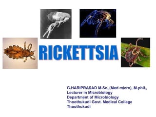
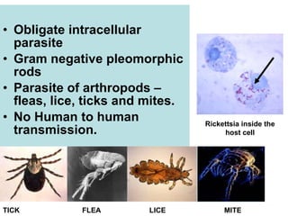
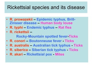
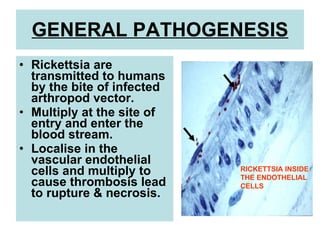
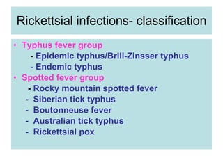
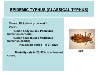
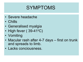
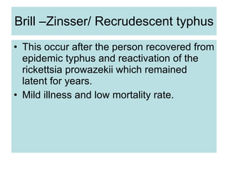
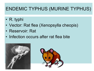
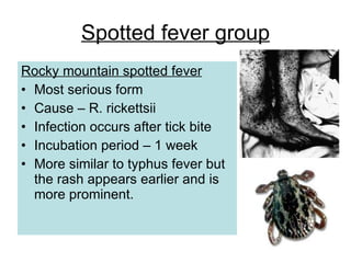
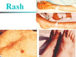
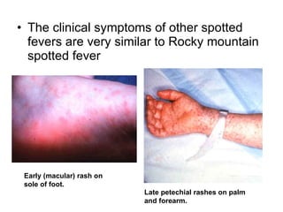
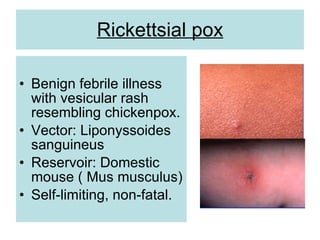
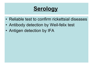
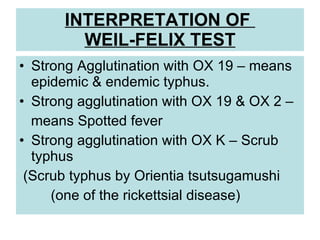
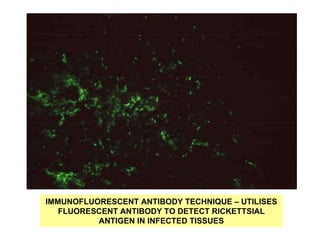
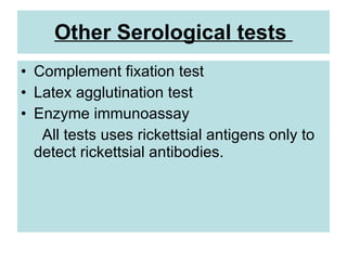
G.HARIPRASAD M.Sc.,(Med micro), M.phil.,
Lecturer in Microbiology
Department of Microbiology
Thoothukudi Govt. Medical College
Thoothukudi
• Obligate intracellular
parasite
• Gram negative pleomorphic
rods
• Parasite of arthropods –
fleas, lice, ticks and mites.
• No Human to human
transmission.
Rickettsia inside the
host cell
TICK FLEA LICE MITE
Rickettsial species and its disease
• R. prowazekii – Epidemic typhus, Brill-
Zinsser disease – Human body louse
• R. typhi – Endemic typhus – Rat flea
• R. rickettsii –
Rocky-Mountain spotted fever-Ticks
• R. conori – Boutonneuse fever - Ticks
• R. australis – Australian tick typhus - Ticks
• R. siberica – Siberian tick typhus - Ticks
• R. akari – Rickettsial pox - Mites
GENERAL PATHOGENESIS
• Rickettsia are
transmitted to humans
by the bite of infected
arthropod vector.
• Multiply at the site of
entry and enter the
blood stream.
• Localise in the
vascular endothelial
cells and multiply to
cause thrombosis lead
to rupture & necrosis.
RICKETTSIA INSIDE
THE ENDOTHELIAL
CELLS
Rickettsial infections- classification
• Typhus fever group
- Epidemic typhus/Brill-Zinsser typhus
- Endemic typhus
• Spotted fever group
- Rocky mountain spotted fever
- Siberian tick typhus
- Boutonneuse fever
- Australian tick typhus
- Rickettsial pox
EPIDEMIC TYPHUS (CLASSICAL TYPHUS)
Cause: Rickettsia prowazekii
Vector:
Human body louse ( Pediculus
humanus corporis)
Human head louse ( Pediculus
humanus capitis)
Incubation period – 5-21 days
Mortality rate is 20-30% in untreated
cases.
LICE
SYMPTOMS
• Severe headache
• Chills
• Generalised myalgia
• High fever ( 39-410
C)
• Vomiting
• Macular rash after 4-7 days – first on trunk
and spreads to limb.
• Lacks conciousness.
Brill –Zinsser/ Recrudescent typhus
• This occur after the person recovered from
epidemic typhus and reactivation of the
rickettsia prowazekii which remained
latent for years.
• Mild illness and low mortality rate.
ENDEMIC TYPHUS (MURINE TYPHUS)
• R. typhi
• Vector: Rat flea (Xenopsylla cheopis)
• Reservoir: Rat
• Infection occurs after rat flea bite
Spotted fever group
Rocky mountain spotted fever
• Most serious form
• Cause – R. rickettsii
• Infection occurs after tick bite
• Incubation period – 1 week
• More similar to typhus fever but
the rash appears earlier and is
more prominent.
• The clinical symptoms of other spotted
fevers are very similar to Rocky mountain
spotted fever
Late petechial rashes on palm
and forearm.
Early (macular) rash on
sole of foot.
Rickettsial pox
• Benign febrile illness
with vesicular rash
resembling chickenpox.
• Vector: Liponyssoides
sanguineus
• Reservoir: Domestic
mouse ( Mus musculus)
• Self-limiting, non-fatal.
Complications of rickettsial
diseases
• Bronchopneumonia
• Congestive heart failure
• Multi-organ failure
• Deafness
• Disseminated intravascular coagulopathy (DIC)
• Myocarditis (inflammation of heart muscle)
• Endocarditis (inflammation of heart lining)
• Glomerulonephritis (inflammation of kidney)
LABORATORY DIAGNOSIS
• Isolation from experimental animals
• Serology
Specimens:
Blood – collected in febrile illness
Note: Rickettsia is highly infectious so
specimens should be handled very carefully.
ISOLATION
• Blood is inoculated in guinea pigs/mice.
• Observed on 3rd
– 4th
week.
• Animal responds to different rickettsial
species can vary
Symptoms:
• Rise in temperature – all species.
• Scrotal inflammation,swelling,necrosis –
R.typhi, R.conori, R.akari ( except
R.prowazekii)
Serology
• Reliable test to confirm rickettsial diseases
• Antibody detection by Weil-felix test
• Antigen detection by IFA
• Heterophile agglutination test using
non motile proteus strains (OX 19, OX 2, OX K)
to find rickettsial antibodies in patient’s serum.
Procedure:
• Serum is diluted in three separate series of tubes
followed by the addition of equal amount of
OX19,OX2,OXK in 3 separate series of tubes.
• Incubation at 370
C for overnight.
• Observe for agglutination.
WEIL-FELIX TEST
INTERPRETATION OF
WEIL-FELIX TEST
• Strong Agglutination with OX 19 – means
epidemic & endemic typhus.
• Strong agglutination with OX 19 & OX 2 –
means Spotted fever
• Strong agglutination with OX K – Scrub
typhus
(Scrub typhus by Orientia tsutsugamushi
(one of the rickettsial disease)
IMMUNOFLUORESCENT ANTIBODY TECHNIQUE – UTILISES
FLUORESCENT ANTIBODY TO DETECT RICKETTSIAL
ANTIGEN IN INFECTED TISSUES
Other Serological tests
• Complement fixation test
• Latex agglutination test
• Enzyme immunoassay
All tests uses rickettsial antigens only to
detect rickettsial antibodies.
Treatment
• Treatment should be started early in the
first week of illness.
• Doxycycline (first choice)
• Tetracycline (alternate)
PROPHYLAXIS
• Vector control
• Live vaccine & killed vaccine are available
but not much effective

rickettsia-111014075555-phpapp01 (1).pdf

  • 1.
    G.HARIPRASAD M.Sc.,(Med micro),M.phil., Lecturer in Microbiology Department of Microbiology Thoothukudi Govt. Medical College Thoothukudi
  • 2.
    • Obligate intracellular parasite •Gram negative pleomorphic rods • Parasite of arthropods – fleas, lice, ticks and mites. • No Human to human transmission. Rickettsia inside the host cell TICK FLEA LICE MITE
  • 3.
    Rickettsial species andits disease • R. prowazekii – Epidemic typhus, Brill- Zinsser disease – Human body louse • R. typhi – Endemic typhus – Rat flea • R. rickettsii – Rocky-Mountain spotted fever-Ticks • R. conori – Boutonneuse fever - Ticks • R. australis – Australian tick typhus - Ticks • R. siberica – Siberian tick typhus - Ticks • R. akari – Rickettsial pox - Mites
  • 4.
    GENERAL PATHOGENESIS • Rickettsiaare transmitted to humans by the bite of infected arthropod vector. • Multiply at the site of entry and enter the blood stream. • Localise in the vascular endothelial cells and multiply to cause thrombosis lead to rupture & necrosis. RICKETTSIA INSIDE THE ENDOTHELIAL CELLS
  • 5.
    Rickettsial infections- classification •Typhus fever group - Epidemic typhus/Brill-Zinsser typhus - Endemic typhus • Spotted fever group - Rocky mountain spotted fever - Siberian tick typhus - Boutonneuse fever - Australian tick typhus - Rickettsial pox
  • 6.
    EPIDEMIC TYPHUS (CLASSICALTYPHUS) Cause: Rickettsia prowazekii Vector: Human body louse ( Pediculus humanus corporis) Human head louse ( Pediculus humanus capitis) Incubation period – 5-21 days Mortality rate is 20-30% in untreated cases. LICE
  • 7.
    SYMPTOMS • Severe headache •Chills • Generalised myalgia • High fever ( 39-410 C) • Vomiting • Macular rash after 4-7 days – first on trunk and spreads to limb. • Lacks conciousness.
  • 8.
    Brill –Zinsser/ Recrudescenttyphus • This occur after the person recovered from epidemic typhus and reactivation of the rickettsia prowazekii which remained latent for years. • Mild illness and low mortality rate.
  • 9.
    ENDEMIC TYPHUS (MURINETYPHUS) • R. typhi • Vector: Rat flea (Xenopsylla cheopis) • Reservoir: Rat • Infection occurs after rat flea bite
  • 10.
    Spotted fever group Rockymountain spotted fever • Most serious form • Cause – R. rickettsii • Infection occurs after tick bite • Incubation period – 1 week • More similar to typhus fever but the rash appears earlier and is more prominent.
  • 12.
    • The clinicalsymptoms of other spotted fevers are very similar to Rocky mountain spotted fever Late petechial rashes on palm and forearm. Early (macular) rash on sole of foot.
  • 13.
    Rickettsial pox • Benignfebrile illness with vesicular rash resembling chickenpox. • Vector: Liponyssoides sanguineus • Reservoir: Domestic mouse ( Mus musculus) • Self-limiting, non-fatal.
  • 14.
    Complications of rickettsial diseases •Bronchopneumonia • Congestive heart failure • Multi-organ failure • Deafness • Disseminated intravascular coagulopathy (DIC) • Myocarditis (inflammation of heart muscle) • Endocarditis (inflammation of heart lining) • Glomerulonephritis (inflammation of kidney)
  • 15.
    LABORATORY DIAGNOSIS • Isolationfrom experimental animals • Serology Specimens: Blood – collected in febrile illness Note: Rickettsia is highly infectious so specimens should be handled very carefully.
  • 16.
    ISOLATION • Blood isinoculated in guinea pigs/mice. • Observed on 3rd – 4th week. • Animal responds to different rickettsial species can vary Symptoms: • Rise in temperature – all species. • Scrotal inflammation,swelling,necrosis – R.typhi, R.conori, R.akari ( except R.prowazekii)
  • 17.
    Serology • Reliable testto confirm rickettsial diseases • Antibody detection by Weil-felix test • Antigen detection by IFA
  • 18.
    • Heterophile agglutinationtest using non motile proteus strains (OX 19, OX 2, OX K) to find rickettsial antibodies in patient’s serum. Procedure: • Serum is diluted in three separate series of tubes followed by the addition of equal amount of OX19,OX2,OXK in 3 separate series of tubes. • Incubation at 370 C for overnight. • Observe for agglutination. WEIL-FELIX TEST
  • 19.
    INTERPRETATION OF WEIL-FELIX TEST •Strong Agglutination with OX 19 – means epidemic & endemic typhus. • Strong agglutination with OX 19 & OX 2 – means Spotted fever • Strong agglutination with OX K – Scrub typhus (Scrub typhus by Orientia tsutsugamushi (one of the rickettsial disease)
  • 20.
    IMMUNOFLUORESCENT ANTIBODY TECHNIQUE– UTILISES FLUORESCENT ANTIBODY TO DETECT RICKETTSIAL ANTIGEN IN INFECTED TISSUES
  • 21.
    Other Serological tests •Complement fixation test • Latex agglutination test • Enzyme immunoassay All tests uses rickettsial antigens only to detect rickettsial antibodies.
  • 22.
    Treatment • Treatment shouldbe started early in the first week of illness. • Doxycycline (first choice) • Tetracycline (alternate)
  • 23.
    PROPHYLAXIS • Vector control •Live vaccine & killed vaccine are available but not much effective