
Downloaded 268 times
![STAGES OF DIABETIC RETINOPATHY
1. Nonproliferative Diabetic Retinopathy
[NPDR]: Early and advanced.
2. Proliferative Diabetic Retinopathy [PDR].](https://image.slidesharecdn.com/retinaanddiabeticretinopathy-131122053212-phpapp02/85/Diabetic-Retinopathy-13-320.jpg)
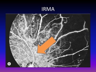
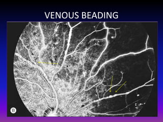
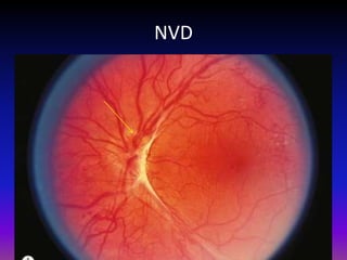
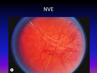
![PDR
• It is characterized by neovascularization [new
blood vessel formation], which is of 2 types:
1. Neovascularization of the disc [NVD],
2. Neovascularization elsewhere [NVE].
• NVD: New vessels arise within ≤1 disc
diameter of optic nerve.
• NVE: New vessels arise within >1 disc
diameter of optic nerve.](https://image.slidesharecdn.com/retinaanddiabeticretinopathy-131122053212-phpapp02/85/Diabetic-Retinopathy-27-320.jpg)
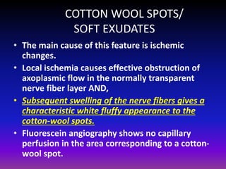
![VITREOUS TRACTION AND RETINAL
DETACHMENT
• The new vessels usually progress through a stage
of further proliferation, with associated
connective tissue formation.
• As PDR progresses, the fibrous component
becomes more prominent.
• Vitreous traction is transmitted to the retina
along these proliferations and may lead to
traction retinal detachment.
*. [Davis et al. have stressed the role of the
contracting vitreous in the production of vitreous
hemorrhage, retinal breaks, and retinal
detachment.]](https://image.slidesharecdn.com/retinaanddiabeticretinopathy-131122053212-phpapp02/85/Diabetic-Retinopathy-30-320.jpg)
![ANTI VEGF AGENTS
• Anti-VEGF drugs are available for the
treatment of macular degeneration.
• Recently, a protein kinase C inhibitor [PKCI]
has been shown to reduce diabetes-induced
hemodynamic abnormalities in patients with
diabetic retinopathy and reduce the risk of
vision loss in patients with macular edema.](https://image.slidesharecdn.com/retinaanddiabeticretinopathy-131122053212-phpapp02/85/Diabetic-Retinopathy-37-320.jpg)
![PAN RETINAL PHOTOCOAGULATION [PRP]
Eyes with High Risk Characteristics [HRC]
HRC is defined as presence of any of the following:
1. NVD> 1/4th -1/3rd of the disc area,
2. NVD and vitreous hemorrhage.
3. NVE> ½ of the disc area.
4. Vitreous/ Preretinal hemorrhage.
•
The ETDRS found that PRP significantly retards
the development of HRC in eyes with very severe
NPDR and macular edema.](https://image.slidesharecdn.com/retinaanddiabeticretinopathy-131122053212-phpapp02/85/Diabetic-Retinopathy-38-320.jpg)
![PRP
Mechanism of PRP [Proposed explanations]:
1. PRP decreases the production of vasoproliferative
factors by eliminating some of the hypoxic retina.
2. PRP stimulates the release of antiangiogenic
factors from the retinal pigment epithelium by
thinning the retina.
3. PRP increases oxygenation of the remaining
retina by allowing increased diffusion of oxygen
from the choroid.
4. PRP leads to an increase in vasoinhibitors by
directly stimulating the retinal pigment
epithelium to produce inhibitors of
vasoproliferation.](https://image.slidesharecdn.com/retinaanddiabeticretinopathy-131122053212-phpapp02/85/Diabetic-Retinopathy-39-320.jpg)
![Continued….
• The surgical objectives are:
1. To clear the media,
2. To release all anterior-posterior traction,
3. To release tangential traction via delamination or
segmentation (cutting the fibrotic bridges between
areas of tractional detachment), and
4. To perform endophotocoagulation.
•
A possible cause of failure following an otherwise
successful vitrectomy is NEOVASCULARIZATION OF
IRIS [NVI] resulting in neovascular glaucoma.](https://image.slidesharecdn.com/retinaanddiabeticretinopathy-131122053212-phpapp02/85/Diabetic-Retinopathy-42-320.jpg)
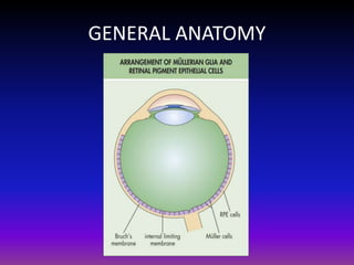
This document discusses the anatomy of the retina and stages of diabetic retinopathy. It begins with the general anatomy of the retina and then explains the pathogenesis of diabetic retinopathy, including the role of aldose reductase pathway and VEGF. It describes the stages of diabetic retinopathy from non-proliferative to proliferative and the signs associated with each stage such as microaneurysms, hemorrhages, neovascularization. The document concludes with discussing the diagnosis and treatments for diabetic retinopathy including pan-retinal photocoagulation, anti-VEGF agents, and vitrectomy.


![PERI-PROSTHETIC FRACTURE NAIL-PLATE CONSTRUCT [NPC].pptx](https://cdn.slidesharecdn.com/ss_thumbnails/drarunkumardrmohamedashrafperiprostheticfrasturenail-plateconstructnpc-260209164459-7e9d15a1-thumbnail.jpg?width=640&height=640&fit=bounds)
![ONFH[AVN HIP] -TRIPLE REGIME -A NOVAL SURGICAL CONCEPT .pptx](https://cdn.slidesharecdn.com/ss_thumbnails/onfhavnhip2026koaconcalicutdrgokuldevdrmashraf-260210064517-213ec005-thumbnail.jpg?width=640&height=640&fit=bounds)

