
Download as PDF, PPTX
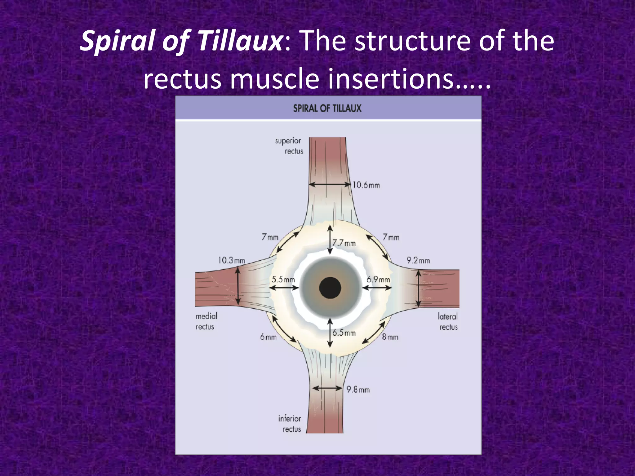
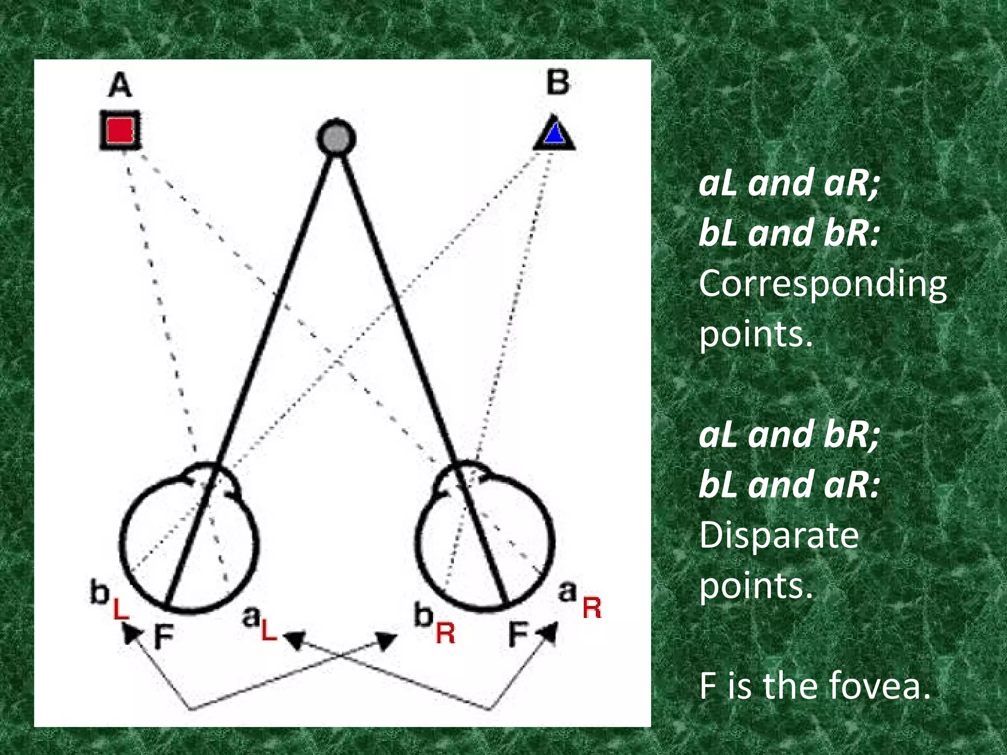
![Insertion of Recti Muscles
• The rectus muscles insert into the sclera just
anterior to the equator of the globe.
• The spatial formation created by connecting
their insertion is called the spiral of Tillaux.
• Note that the medial rectus inserts closest to
the limbus, followed by the inferior, lateral,
and superior recti in that order. [MILS]](https://image.slidesharecdn.com/diseasesofocularmotilitywithanemphasisonsquint-131205031257-phpapp02/75/Diseases-of-ocular-motility-with-an-emphasis-on-squint-9-2048.jpg)
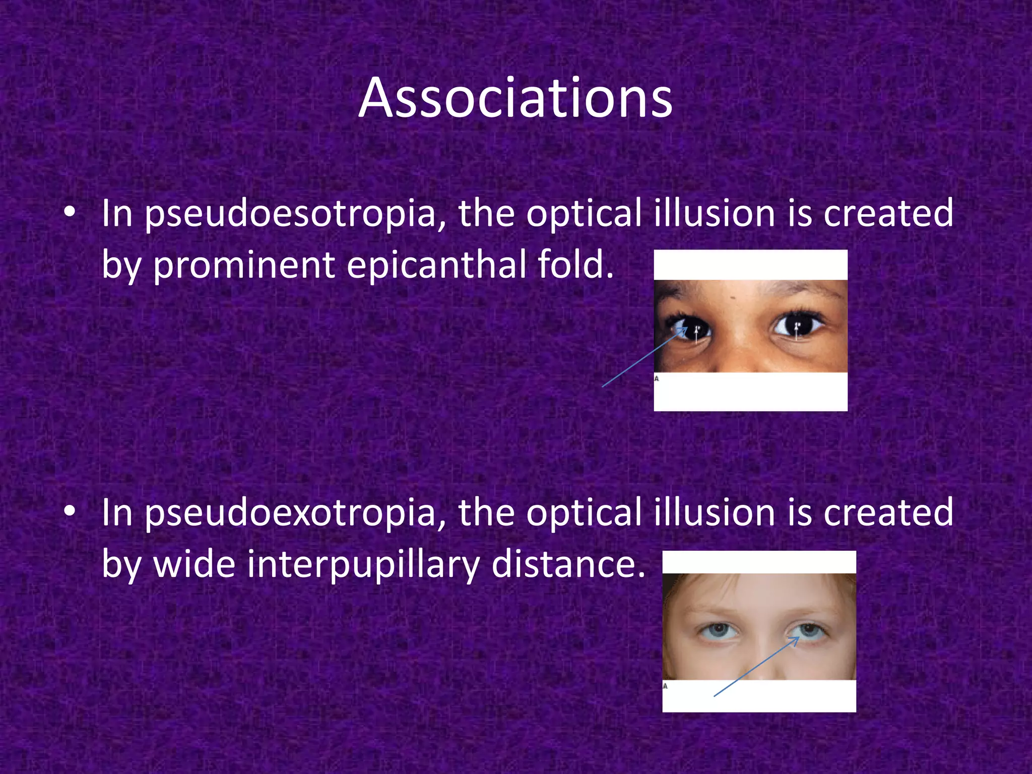
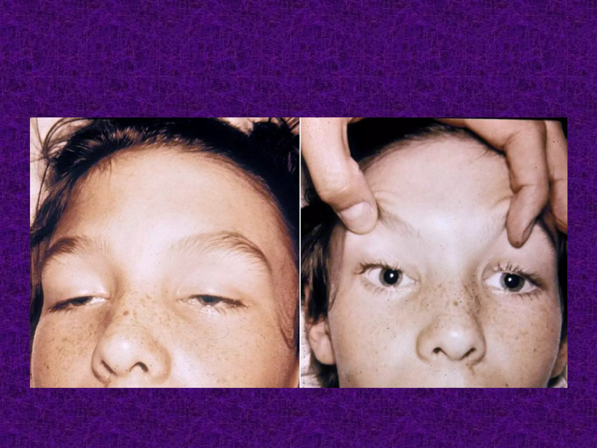
![Apparent/ Pseudo-strabismus
• It is also known as “Pseudostrabismus”.
• This is nothing but an optical illusion caused
by prominent epicanthal folds/ wide
interpupillary distances (IPD). [See next page]
• It is of 2 types:
1. Pseudo-esotropia/ Apparent convergent
squint.
2. Pseudo-exotropia/ Apparent divergent
squint.](https://image.slidesharecdn.com/diseasesofocularmotilitywithanemphasisonsquint-131205031257-phpapp02/75/Diseases-of-ocular-motility-with-an-emphasis-on-squint-40-2048.jpg)

![Secondary Esotropia
It occurs due to monoocular lesions that prevents the
development of binocular vision/ maintenance of it.
Ex.:
Cataract,
Aphakia,
Anisometropia,
Severe congenital ptosis,
Retinoblastoma etc.
[* Note that in Retinoblastoma, strabismus is the second
most common manifestation after leukocoria.]](https://image.slidesharecdn.com/diseasesofocularmotilitywithanemphasisonsquint-131205031257-phpapp02/75/Diseases-of-ocular-motility-with-an-emphasis-on-squint-60-2048.jpg)

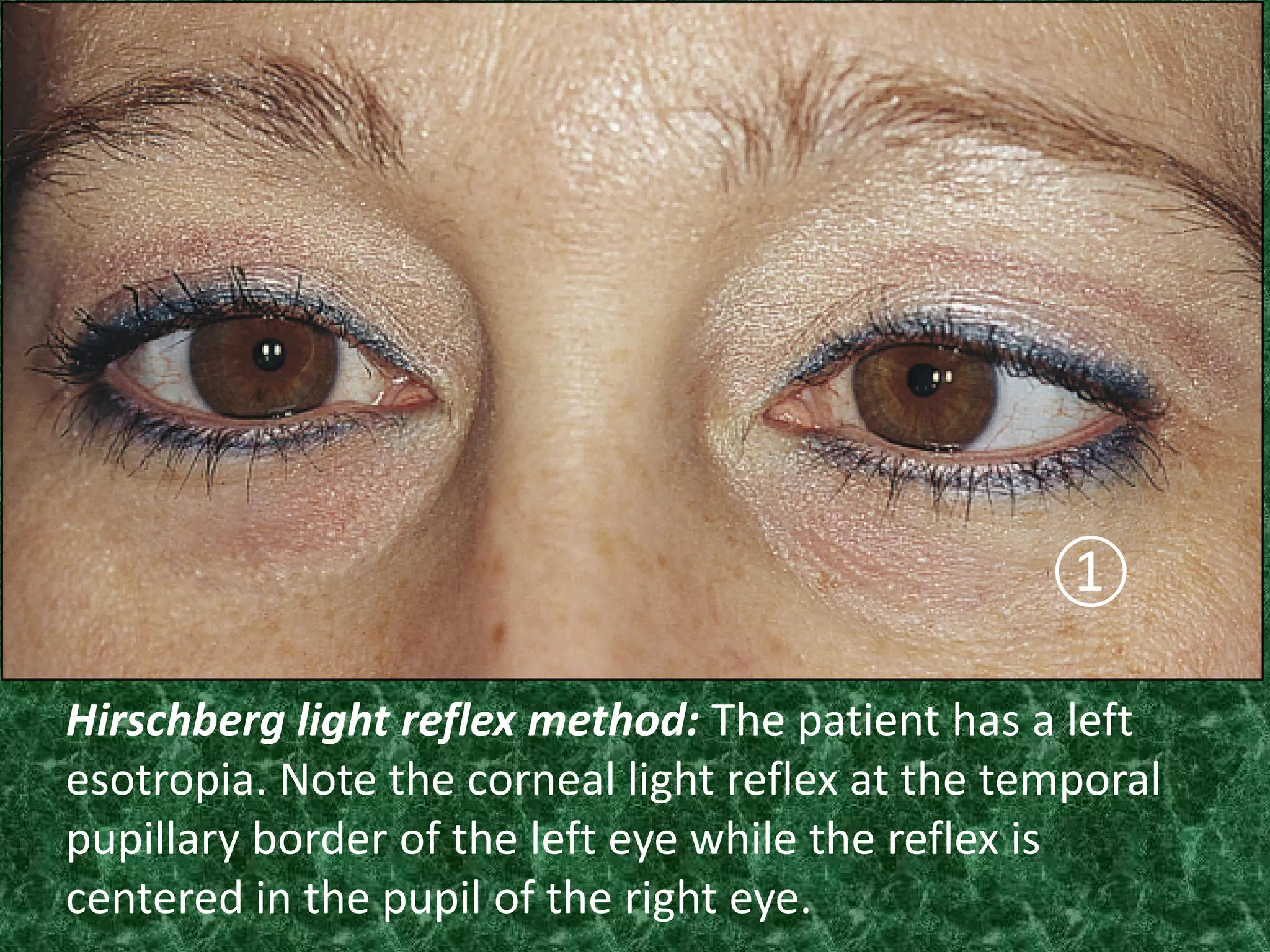
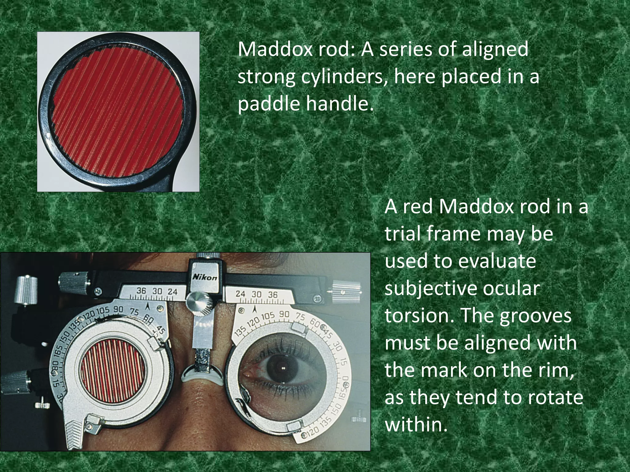
![Krimsky light reflex method [same patient as in
previous photograph ①]. The strength of a baseout prism over the fixing right eye sufficient to
center the pupillary light reflex in the esotropic
left eye is defined as the amount of left esotropia.](https://image.slidesharecdn.com/diseasesofocularmotilitywithanemphasisonsquint-131205031257-phpapp02/75/Diseases-of-ocular-motility-with-an-emphasis-on-squint-107-2048.jpg)
The document discusses disorders of ocular motility, covering the anatomy and functions of extraocular muscles, mechanisms of eye movement, strabismus types, and their clinical implications. It details various forms of strabismus, including apparent, latent, comitant, and incomitant, along with their causes, symptoms, and treatments. The document serves as a comprehensive lecture on ocular motility, addressing both theoretical and practical aspects relevant to eye movement disorders.


