This document discusses diabetic retinopathy and its stages: background diabetic retinopathy, pre-proliferative diabetic retinopathy, proliferative diabetic retinopathy, and advanced diabetic eye disease. It covers the signs, symptoms, management, and treatment for each stage. The document also discusses other common diabetic eye complications like retinal occlusive diseases, optic disc issues, glaucoma, and cranial nerve palsies. The role of optometrists in screening, referring, and low vision care for patients with diabetic eye disease is highlighted.
This lecture is part of the yearly Basic Course Lectures in Ophthalmology given by the Dept of Ophthalmology and Visual Sciences at the Philippine General Hospital.
Originally given by Dr Pearl Tamesis-Villalon, it is a 1:30:00 hour lecture on the pathologic lesions seen in the vitreous, retina and choroid. It is meant for the general physician and the beginning ophthalmology resident who is interested in the basics of retinal pathology.
It includes pathologic changes seen in hypertension, diabetes, vaso occlusive disease, vitreous, membranes, choroid, retinal pigment epithelium, retinal detachments, etc. Lesions such as hemorrhages, cotton wool spots, hard exudates and their location in the retinal layers are explained. Fluorescein angiogram and OCT images are also incorporated.
Some images were grabbed from the internet, apologies for not making the necessary acknowledgements.
About disease of Conjunctiva
1. inflammatory conditions of conjunctiva
2.Symptomatic conditions of conjunctiva
3. degenerative conditions of conjunctiva
4. tumors of conjunctiva
5. cyst of conjunctiva
Dry Eye and Ocular surface diseases in diabetes mellitusDhwanit Khetwani
RELATION OF DIABETES WITH DRY EYE AND OTHER OCULAR SURFACE DISEASES, MADE FOR THE PURPOSE PROTOCOL PRESENTATION. MADE BY DR DHWANIT KHETWANI OPHTHALMOLOGY RESIDENT
Retinal vein occlusion (RVO) is an obstruction of the retinal venous system by thrombus formation and may involve the central, hemi-central or branch retinal vein.
The most common aetiological factor is compression by adjacent atherosclerotic retinal arteries.
Other possible causes are external compression or disease of the vein wall e.g. vasculitis.
Coats' disease, (also known as exudative retinitis or retinal telangiectasis, sometimes spelled Coates' disease), is a rare congenital, nonhereditary eye disorder, causing full or partial blindness, characterized by abnormal development of blood vessels behind the retina.
Central Retinal Artery Occlusion (CRAO) for undergraduate MBBS Students.
Covers the basics of Aetiology, pathophysiology, clinical features, types, associated conditions and management of CRAO.
Also encompasses salient points for PGMEE
WHAT WE SHOULD DO FOR PROGRESSIVE COMPLICATIONS OF PDR INSPITE OF “ADEQUATE” ...DrAbdelLatifsiam
PURPOSE
To draw attention to severe cases of Proliferative Diabetic Vitreo-Retinopathy which continue to progress, in spite of what was thought to be adequate laser treatment
Vitreous hemorrhage is the extravasation, or leakage, of blood into the areas in and around the vitreous humor of the eye.[1] The vitreous humor is the clear gel that fills the space between the lens and the retina of the eye. A variety of conditions can result in blood leaking into the vitreous humor, which can cause impaired vision, floaters, and photopsia.
It's an indepth presentation by Dr. Shah-Noor Hassan.
Diabetic retinopathy is a complication of diabetes, caused by high blood sugar levels damaging the back of the eye (retina). It can cause blindness if left undiagnosed and untreated. However, it usually takes several years for diabetic retinopathy to reach a stage where it could threaten your sight.
The retina is the light-sensitive layer of cells at the back of the eye that converts light into electrical signals. The signals are sent to the brain which turns them into the images you see.
The retina needs a constant supply of blood, which it receives through a network of tiny blood vessels. Over time, a persistently high blood sugar level can damage these blood vessels in 3 main stages:
background retinopathy – tiny bulges develop in the blood vessels, which may bleed slightly but don't usually affect your vision
pre-proliferative retinopathy – more severe and widespread changes affect the blood vessels, including more significant bleeding into the eye
proliferative retinopathy – scar tissue and new blood vessels, which are weak and bleed easily, develop on the retina, this can result in some loss of vision
In this case-based presentation, Dr. Lori Myers unscrambles the alphabet soup of Diabetic Retinopathy, providing clear explanations and outstanding images to describe the diagnosis, risk stratification, and treatment of diabetic retinopathy.
Ozempic: Preoperative Management of Patients on GLP-1 Receptor Agonists Saeid Safari
Preoperative Management of Patients on GLP-1 Receptor Agonists like Ozempic and Semiglutide
ASA GUIDELINE
NYSORA Guideline
2 Case Reports of Gastric Ultrasound
Explore natural remedies for syphilis treatment in Singapore. Discover alternative therapies, herbal remedies, and lifestyle changes that may complement conventional treatments. Learn about holistic approaches to managing syphilis symptoms and supporting overall health.
Prix Galien International 2024 Forum ProgramLevi Shapiro
June 20, 2024, Prix Galien International and Jerusalem Ethics Forum in ROME. Detailed agenda including panels:
- ADVANCES IN CARDIOLOGY: A NEW PARADIGM IS COMING
- WOMEN’S HEALTH: FERTILITY PRESERVATION
- WHAT’S NEW IN THE TREATMENT OF INFECTIOUS,
ONCOLOGICAL AND INFLAMMATORY SKIN DISEASES?
- ARTIFICIAL INTELLIGENCE AND ETHICS
- GENE THERAPY
- BEYOND BORDERS: GLOBAL INITIATIVES FOR DEMOCRATIZING LIFE SCIENCE TECHNOLOGIES AND PROMOTING ACCESS TO HEALTHCARE
- ETHICAL CHALLENGES IN LIFE SCIENCES
- Prix Galien International Awards Ceremony
These simplified slides by Dr. Sidra Arshad present an overview of the non-respiratory functions of the respiratory tract.
Learning objectives:
1. Enlist the non-respiratory functions of the respiratory tract
2. Briefly explain how these functions are carried out
3. Discuss the significance of dead space
4. Differentiate between minute ventilation and alveolar ventilation
5. Describe the cough and sneeze reflexes
Study Resources:
1. Chapter 39, Guyton and Hall Textbook of Medical Physiology, 14th edition
2. Chapter 34, Ganong’s Review of Medical Physiology, 26th edition
3. Chapter 17, Human Physiology by Lauralee Sherwood, 9th edition
4. Non-respiratory functions of the lungs https://academic.oup.com/bjaed/article/13/3/98/278874
Couples presenting to the infertility clinic- Do they really have infertility...Sujoy Dasgupta
Dr Sujoy Dasgupta presented the study on "Couples presenting to the infertility clinic- Do they really have infertility? – The unexplored stories of non-consummation" in the 13th Congress of the Asia Pacific Initiative on Reproduction (ASPIRE 2024) at Manila on 24 May, 2024.
MANAGEMENT OF ATRIOVENTRICULAR CONDUCTION BLOCK.pdfJim Jacob Roy
Cardiac conduction defects can occur due to various causes.
Atrioventricular conduction blocks ( AV blocks ) are classified into 3 types.
This document describes the acute management of AV block.
Pulmonary Thromboembolism - etilogy, types, medical- Surgical and nursing man...VarunMahajani
Disruption of blood supply to lung alveoli due to blockage of one or more pulmonary blood vessels is called as Pulmonary thromboembolism. In this presentation we will discuss its causes, types and its management in depth.
Report Back from SGO 2024: What’s the Latest in Cervical Cancer?bkling
Are you curious about what’s new in cervical cancer research or unsure what the findings mean? Join Dr. Emily Ko, a gynecologic oncologist at Penn Medicine, to learn about the latest updates from the Society of Gynecologic Oncology (SGO) 2024 Annual Meeting on Women’s Cancer. Dr. Ko will discuss what the research presented at the conference means for you and answer your questions about the new developments.
ARTIFICIAL INTELLIGENCE IN HEALTHCARE.pdfAnujkumaranit
Artificial intelligence (AI) refers to the simulation of human intelligence processes by machines, especially computer systems. It encompasses tasks such as learning, reasoning, problem-solving, perception, and language understanding. AI technologies are revolutionizing various fields, from healthcare to finance, by enabling machines to perform tasks that typically require human intelligence.
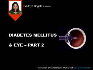
Diabetes melitis & eye part 2 presentation at www.eyenirvaan.com
1. DIABETES MELLITUS
& EYE – PART 2
Pradnya Gogate B. Optom,
To view more presentations and articles, visit www.eyenirvaan.com
2. Diabetic retinopathy has four
stages:
Background diabetic retinopathy
Pre-proliferative diabetic retinopathy
Proliferative diabetic retinopathy
Advanced diabetic eye disease
To view more presentations and articles, visit www.eyenirvaan.com
3. Background diabetic retinopathy
Microaneurysms-
In inner nuclear layer
Appear as small, round, red dots
Hard exudates-
In outer plexiform and inner nuclear layer
Distributed in circinate pattern
To view more presentations and articles, visit www.eyenirvaan.com
4. Background diabetic retinopathy
Flame shaped
haemorrhages- follow
the course of retinal
nerve fiber layer
Dot-blot haemorrhages
– within compact
middle layers
To view more presentations and articles, visit www.eyenirvaan.com
6. Management of background
diabetic retinopathy
Proper control of sugar level
Regular follow up
To view more presentations and articles, visit www.eyenirvaan.com
7. Preproliferative diabetic
retinopathy
Cotton wool spots (Soft exudates)
due to capillary occlusion in retinal nerve fiber layer and
the subsequent build-up of transported material within
the nerve axons causes white and opaque appearance
IRMA’S(Intra Retinal Microvascular
Abnormalities)
Venous changes like dilatation, beading, looping
and sausage-like segmentation
Arteriolar narrowing and may cause central
retinal artery occlusion(CRAO)
large dark blot hemorrhages
To view more presentations and articles, visit www.eyenirvaan.com
9. Management Preproliferative
diabetic retinopathy
Close follow up
Photocoagulation is usually unnecessary
unless FFA shows extensive areas of
peripheral capillary non-perfusion
To view more presentations and articles, visit www.eyenirvaan.com
10. Proliferative diabetic
retinopathy
Neovascularisation is hallmark of PDR
NVD(new vessels at disc)
More than one quarter of retina has to be non-
perfused for NVD
NVE (new vessels elsewhere)
Starts as endothelial proliferations arising from
veins
They pass through the defects in the ILM to lie in
potential vitro-retinal space
Forms fibrovascular epiretinal membrane
To view more presentations and articles, visit www.eyenirvaan.com
11. Proliferative diabetic
retinopathy
Recurrent vitreous haemorrhages
Fibrovascular component becomes adherent
to posterior vitreous and leaks plasma
constituents
Contraction of vitreous results in elevation of
blood vessels above the plane of retina
New vessels may regress if vitreous detaches
completely at this stage
Pulling from Partially detached vitreous
causes vitreous haemorrhage
To view more presentations and articles, visit www.eyenirvaan.com
14. Management proliferative
diabetic retinopathy
Pan retinal photocoagulation(PRP)
Thousands(2000-3000) of spots are burned around the
peripheral retina.
Destroys the ischemic retina, decreasing the angiogenic
stimulus, leads to regression and even the complete
disappearance of the new vessels.
side effects,
peripheral vision loss
decreased night vision (from the rod photoreceptor
loss)
To view more presentations and articles, visit www.eyenirvaan.com
16. Early Treatment Diabetic
Retinopathy Study Group (EDTRS)
Non-Proliferative Diabetic Retinopathy
Minimal NPDR
Mild NPDR
Moderate NPDR
Severe NPDR
Very Severe NPDR
Proliferative Diabetic Retinopathy (PDR)
Early PDR
High Risk (HR) PDR
To view more presentations and articles, visit www.eyenirvaan.com
17. Early Treatment Diabetic
Retinopathy Study Group (EDTRS)
Minimal NPDR
Presence of microaneurysms only
Mild NPDR
Microaneurysms plus one or more of the
following:
Intra-retinal hemorrhages
Hard exudates away from the macula
CWS
To view more presentations and articles, visit www.eyenirvaan.com
18. Early Treatment Diabetic
Retinopathy Study Group (EDTRS)
Moderate NPDR
Microaneurysms/ hemorrhages in at least one
quadrant plus one or more of the following:
CWS
IRMA
Venous beading
To view more presentations and articles, visit www.eyenirvaan.com
19. Early Treatment Diabetic
Retinopathy Study Group (EDTRS)
Severe NPDR
Any one of the following (4-2-1 rule):
Intra-retinal hemorrhages - severe, in 4 quadrants
Venous beading in 2 quadrants
Moderately severe IRMA in 1 quadrant
To view more presentations and articles, visit www.eyenirvaan.com
20. Early Treatment Diabetic
Retinopathy Study Group (EDTRS)
Very Severe NPDR
Any two of the following:
Intra-retinal hemorrhages - severe, in 4 quadrants
Venous beading in 2 quadrants
Moderately severe IRMA in 1 quadrant
To view more presentations and articles, visit www.eyenirvaan.com
21. Early Treatment Diabetic
Retinopathy Study Group (EDTRS)
Early PDR
One or more of the following:
NVD < ¼ DD
NVE without hemorrhage
Pre-retinal or vitreous hemorrhage and NVE < ½
DD without NVD
To view more presentations and articles, visit www.eyenirvaan.com
22. Early Treatment Diabetic
Retinopathy Study Group (EDTRS)
High Risk PDR
One or more of the following:
NVD > ¼ DD
NVD with hemorrhage
NVE > ½ DD with hemorrhage
To view more presentations and articles, visit www.eyenirvaan.com
23. Advanced diabetic eye disease
Pre-retinal
haemorrhage(boat-shaped
haemorrhage)
Tractional retinal
detachment
Pulling away of
neurosensory retina from
RPE by avascular or
fibrovascular vitreous
membranes.
Nonvascular glaucoma
(90 days glaucoma)
To view more presentations and articles, visit www.eyenirvaan.com
24. Management of Advanced
diabetic eye disease
Pan Retinal Photocoagulation(PRP)
Three port pars plana vitrectomy
Involves removing the vitreous humor
replacing it with saline.
removes hemorrhaged blood, inflammatory cells, and other
debris
removes any fine strands of vitreous attached to the retina
to relieve traction
Endo-laser is done
To view more presentations and articles, visit www.eyenirvaan.com
25. Three port pars plana
vitrectomy
To view more presentations and articles, visit www.eyenirvaan.com
26. Retinal Occlusive Diseases
Central Retinal Vein Occlusion (CRVO)
Branch Retinal Vein Occlusion (BRVO)
Central Retinal Artery Occlusion (CRAO)
Branch Retinal Artery Occlusion (BRAO)
To view more presentations and articles, visit www.eyenirvaan.com
27. Optic Disc
Anterior Ischemic Optic Neuropathy
Diabetic Papillitis
To view more presentations and articles, visit www.eyenirvaan.com
28. Anterior Ischemic Optic
Neuropathy(AION)
Interference with the
posterior cilliary artery supply
to anterior part of optic disc
Sudden painless loss of
vision
Altitudinal field defect
Swelling of optic disc
Flame shaped hemorrhages
To view more presentations and articles, visit www.eyenirvaan.com
29. Diabetic Papillopathy
Mild to moderate visual loss
Ranges from mild disc swelling without
haemorrhages to
Florid swelling with capillary telangeiectesis,
nerve fiber haemorrhages, exudates, CME
with or without macular star
Treatment
Good diabetic control
To view more presentations and articles, visit www.eyenirvaan.com
31. Glaucoma
Primary open angle glaucoma(POAG)
Neovascular glaucoma(NVG)
To view more presentations and articles, visit www.eyenirvaan.com
32. Cranial nerves
III, IV,VI,VII
III is the commonest affected
To view more presentations and articles, visit www.eyenirvaan.com
33. Role of optometrists
Screening and counseling of patients
Referring the patient to ophthalmologist for
investigations and treatment
Referring the patient to diabetologist for
diabetes control
Prescribing the appropriate low vision
devices,if required
To view more presentations and articles, visit www.eyenirvaan.com