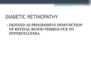
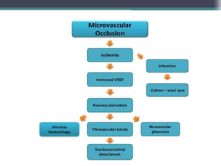
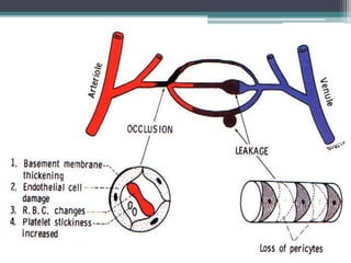
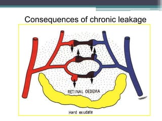
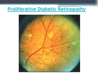
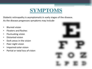
Diabetic Retinopathy
AZIZUDDIN
MBBS-III
JMDC
DIABETIC RETINOPATHY
• DEFINED AS PROGRESSIVE DYSFUNCTION
OF RETINAL BLOOD VESSELS DUE TO
HYPERGYLCEMIA.
Retinal Topography
Retina: Schematic Diagram
Diabetic Retinopathy
• The best predictor of diabetic retinopathy is the
duration of the disease.
• Type I diabetes The first 5 years of type 1 diabetes has a
very low risk of retinopathy. All patients develop
retinopathy in 15 years.
• Type II diabetes: Risk of retinopathy increases with
duration of diabetes , hypertension and smoking, and
renal pathology.
Diabetic Retinopathy
Risk factors:
• Diabetic age and age of the patient
• Glycemic control
• Hypertension
• Poor renal status
• Smoking
• Pregnancy, obesity, hyperlipidemia, anemia
Consequences of chronic leakage
Clinical Features
• Asymptomatic in the beginning
• Symptoms depend upon the retinal changes.
• Painless diminution of vision
• Dilated fundus examination shows typical
features.
Fundus Findings
Features of NPDR
● Microaneurysms
● Dot and blot hemorrhage
● Retinal edema and exudates
● Dilatation and beading of
retinal veins
● Intra retinal Microvascular
Abnormalities (IRMA)
Features of PDR
Neovascularization disc (NVD)
Neovascularization elsewhere(NVE)
In addition to other changes of NPDR
Early NPDR
Macular Edema
Proliferative Diabetic Retinopathy
Extra retinal fibrovascular proliferation
extends beyond the internal limiting
membrane (ILM).
• It’s the commonest cause of spontaneous
vitreous hemorrhage in adults.
• About 2/3rd of Type I diabetics are likely to
develop PDR over 3 decades.
Proliferative Diabetic Retinopathy
Causes of Visual Impairment
NPDR
● Macular edema (capillary
leakage)
● Macular ischemia (capillary
occlusion)
● Sequelae from ischemia
related neovascularization
PDR
• Vitreous hemorrhage, retinal
detachment.
• Diabetic macular edema
• Ischemic macular changes
Diagnosis of Diabetic Retinopathy
Clinical Exam
• Direct ophthalmoscopy under mydriasis
• Slit lamp biomicroscopy using +90 D lens
• Fluorescein angiography
Screening Schedule
• : Normal fundus , rare microaneurysms:
Annual review
• Mild NPDR: Every 9 months
• Moderate NPDR: Every 6 months
• Severe NPDR, CSME: Every 2-4 months
• PDR: Every 2-4 months
Treatment of
Diabetic Macular Edema (DME)
Laser Photocoagulation
• Grid laser :For diffuse retinal thickening outside FAZ.
• Focal laser : microaneurysms in centre of hard exudates.
Medical Treatment:
-intravitreal injection: Triamcinolome acetonide / anti-
VEGF
- posterior sub-tenon injection of corticosteroid
Surgery: Parsplana vitrectomy
Treatment of PDR
• Panretinal Laser photocoagulation (PRP) : For
NVD And NVE
• Vitreoretinal surgery and PRP :
- Severe persistent vitreous hemorrhage
- Dense, persistent premacular hemorrhage
- Progressive proliferation despite laser therapy
- Retinal detachment involving macula
• Spot size (200-500 m)
• Follow-up 4 to 8 weeks
• Area covered by complete PRP
• Initial treatment is 1200
+ burns
Laser Panretinal Photocoagulation(PRP)
Prevention Visual Loss
• Patient education and good diabetic control
• Control of hypertension, hyperlipidemia, renal
disease, anaemia, and avoidance of smoking
• Periodic dilated fundus examination
• Early treatment of macular edema and PDR
• Treatment of complications

Diabetic Retinopathy

  • 1.
  • 2.
    DIABETIC RETINOPATHY • DEFINEDAS PROGRESSIVE DYSFUNCTION OF RETINAL BLOOD VESSELS DUE TO HYPERGYLCEMIA.
  • 3.
  • 4.
  • 5.
    Diabetic Retinopathy • Thebest predictor of diabetic retinopathy is the duration of the disease. • Type I diabetes The first 5 years of type 1 diabetes has a very low risk of retinopathy. All patients develop retinopathy in 15 years. • Type II diabetes: Risk of retinopathy increases with duration of diabetes , hypertension and smoking, and renal pathology.
  • 6.
    Diabetic Retinopathy Risk factors: •Diabetic age and age of the patient • Glycemic control • Hypertension • Poor renal status • Smoking • Pregnancy, obesity, hyperlipidemia, anemia
  • 14.
  • 17.
    Clinical Features • Asymptomaticin the beginning • Symptoms depend upon the retinal changes. • Painless diminution of vision • Dilated fundus examination shows typical features.
  • 18.
    Fundus Findings Features ofNPDR ● Microaneurysms ● Dot and blot hemorrhage ● Retinal edema and exudates ● Dilatation and beading of retinal veins ● Intra retinal Microvascular Abnormalities (IRMA) Features of PDR Neovascularization disc (NVD) Neovascularization elsewhere(NVE) In addition to other changes of NPDR
  • 19.
  • 20.
  • 21.
    Proliferative Diabetic Retinopathy Extraretinal fibrovascular proliferation extends beyond the internal limiting membrane (ILM). • It’s the commonest cause of spontaneous vitreous hemorrhage in adults. • About 2/3rd of Type I diabetics are likely to develop PDR over 3 decades.
  • 22.
  • 24.
    Causes of VisualImpairment NPDR ● Macular edema (capillary leakage) ● Macular ischemia (capillary occlusion) ● Sequelae from ischemia related neovascularization PDR • Vitreous hemorrhage, retinal detachment. • Diabetic macular edema • Ischemic macular changes
  • 25.
    Diagnosis of DiabeticRetinopathy Clinical Exam • Direct ophthalmoscopy under mydriasis • Slit lamp biomicroscopy using +90 D lens • Fluorescein angiography
  • 26.
    Screening Schedule • :Normal fundus , rare microaneurysms: Annual review • Mild NPDR: Every 9 months • Moderate NPDR: Every 6 months • Severe NPDR, CSME: Every 2-4 months • PDR: Every 2-4 months
  • 27.
    Treatment of Diabetic MacularEdema (DME) Laser Photocoagulation • Grid laser :For diffuse retinal thickening outside FAZ. • Focal laser : microaneurysms in centre of hard exudates. Medical Treatment: -intravitreal injection: Triamcinolome acetonide / anti- VEGF - posterior sub-tenon injection of corticosteroid Surgery: Parsplana vitrectomy
  • 28.
    Treatment of PDR •Panretinal Laser photocoagulation (PRP) : For NVD And NVE • Vitreoretinal surgery and PRP : - Severe persistent vitreous hemorrhage - Dense, persistent premacular hemorrhage - Progressive proliferation despite laser therapy - Retinal detachment involving macula
  • 29.
    • Spot size(200-500 m) • Follow-up 4 to 8 weeks • Area covered by complete PRP • Initial treatment is 1200 + burns Laser Panretinal Photocoagulation(PRP)
  • 30.
    Prevention Visual Loss •Patient education and good diabetic control • Control of hypertension, hyperlipidemia, renal disease, anaemia, and avoidance of smoking • Periodic dilated fundus examination • Early treatment of macular edema and PDR • Treatment of complications