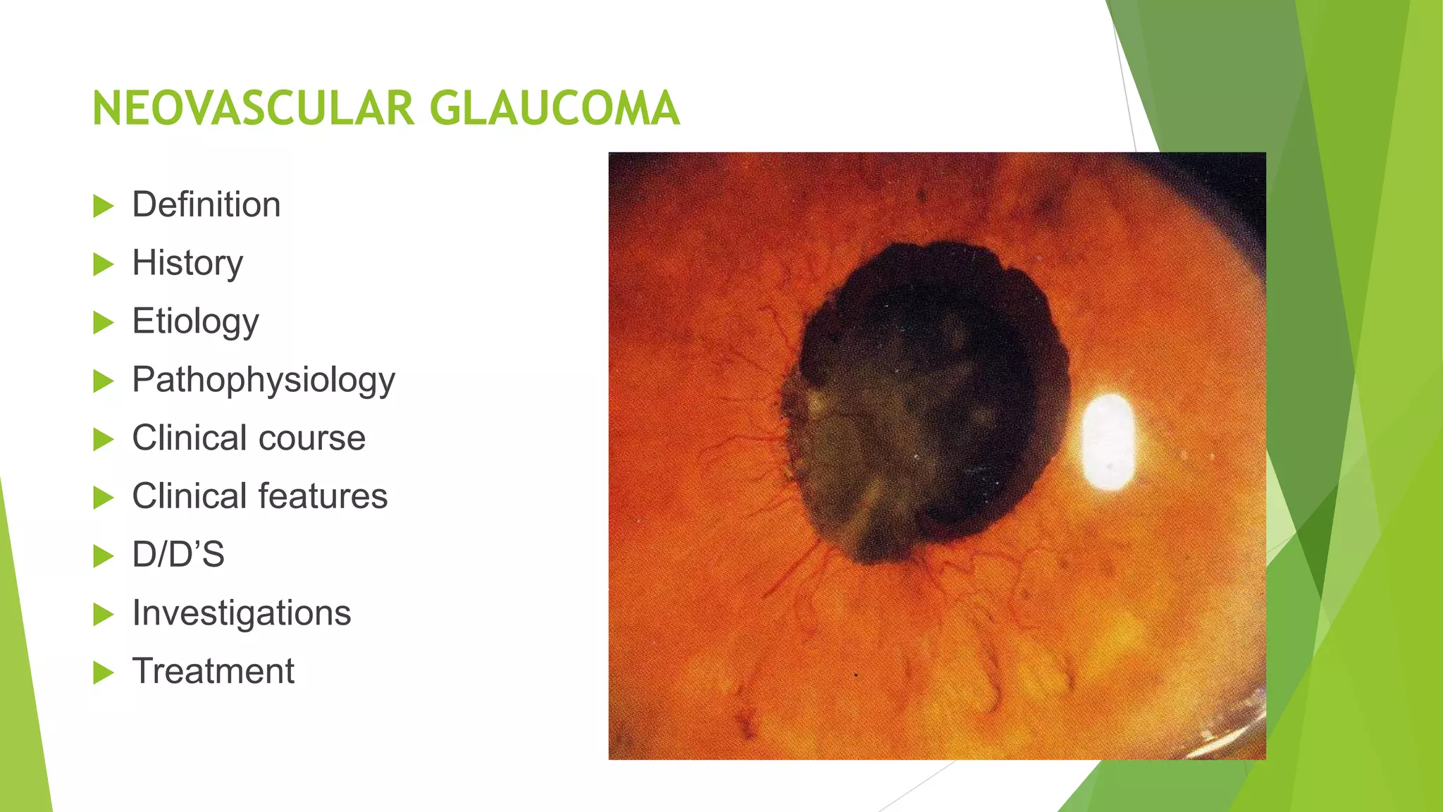
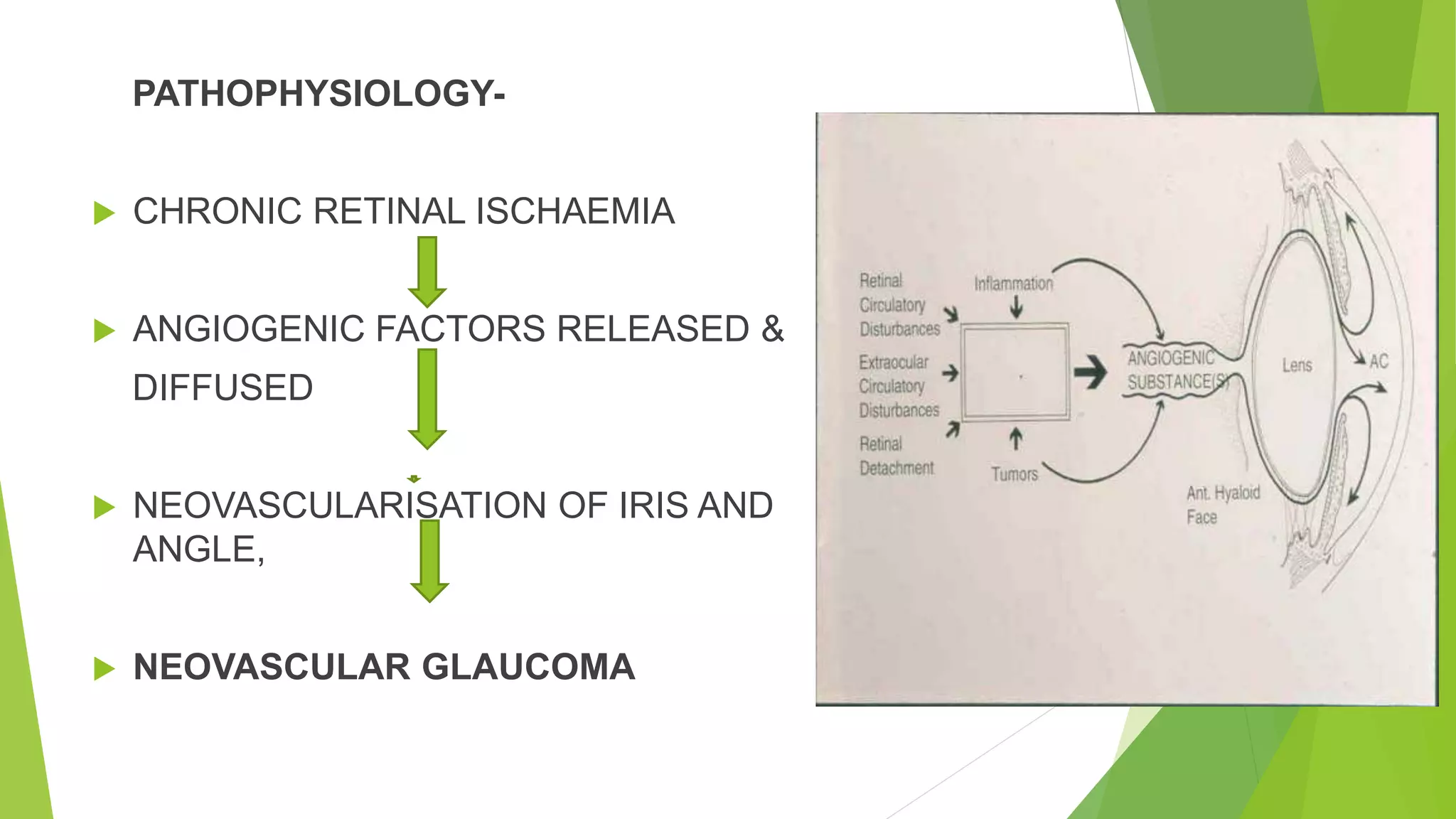
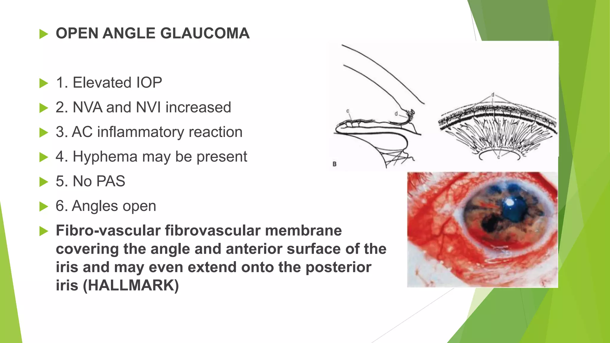
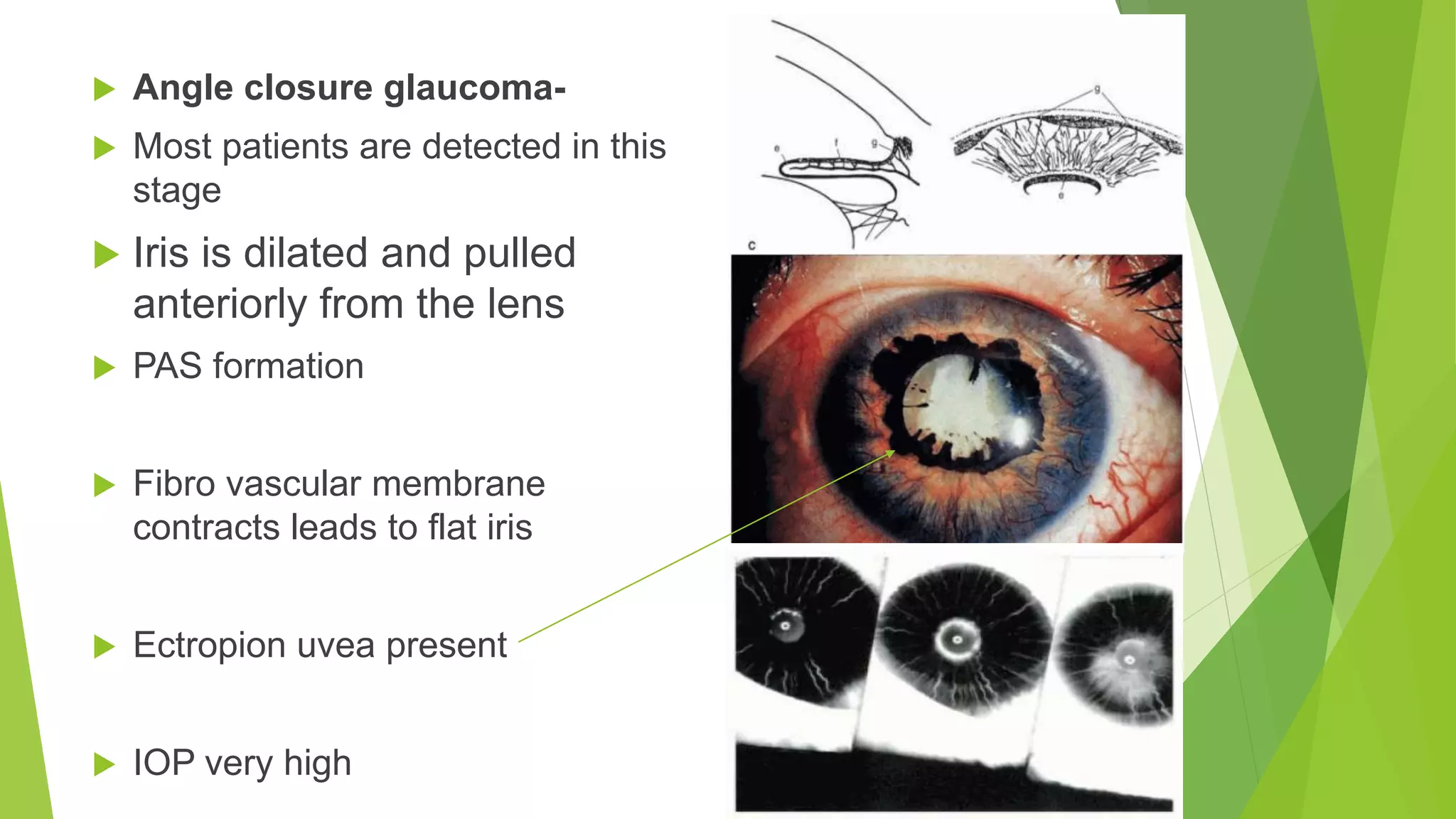
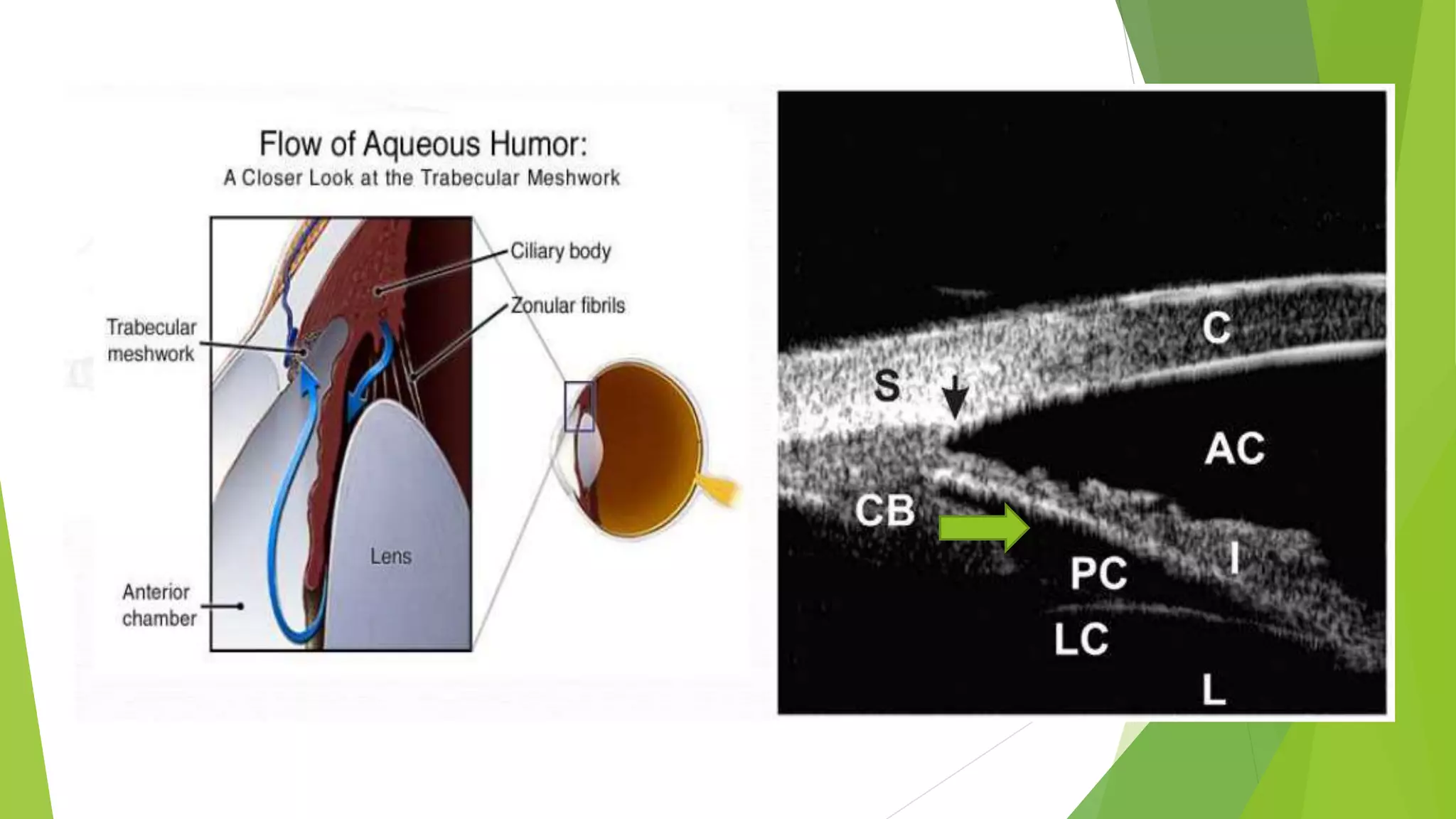
NEOVASCULAR GLAUCOMA /
AQUEOUS MISDIRECTION /
UVEITIC GLAUCOMA
PRESENTER - DR. GAURAV SHUKLA
NEOVASCULAR GLAUCOMA
 Definition
 History
 Etiology
 Pathophysiology
 Clinical course
 Clinical features
 D/D’S
 Investigations
 Treatment
 DEFINITION-
 Severe form of secondary glaucoma characterised by fibro
vascular proliferation in the anterior chamber angle
 ALSO KNOWN AS-
 1. Thrombotic glaucoma
 2. Rubeotic glaucoma
 3. Congestive glaucoma
 4.Hemorrhagic glaucoma
 HISTORY-
 1906 - Coats , NVI in CRVO termed as RUBEOSIS IRIDIS
 1937 - Kurtz , NVA leading to PAS formation
 1963 - Weiss et al, coined the term NEOVASCULAR GLAUCOMA
 ETIOLOGY-
 Proliferative diabetic retinopathy (M.C.C)- approx. 33%
 CRVO - 28% (CRVO > CRAO)
 Ocular ischemic disease d/t carotid artery obstruction-13%
 Others-
PATHOPHYSIOLOGY-
 CHRONIC RETINAL ISCHAEMIA
 ANGIOGENIC FACTORS RELEASED &
DIFFUSED
 NEOVASCULARISATION OF IRIS AND
ANGLE,
 NEOVASCULAR GLAUCOMA
 Lens and vitreous act as mechanical barriers and also releases
vaso-inhibitory factors
 So any complicated cataract surgery with PCR, aphakia more
predisposition
 VEGF conc 50-100 times more in aqueous humor in NVG
 STAGES-
 PRE RUBEOSIS STAGE
 PRE GLAUCOMA (RUBEOSIS IRIDIS )
 OPEN ANGLE GLAUCOMA
 ANGLE CLOSURE GLAUCOMA
 PRE RUBEOSIS STAGE-
 In patients with predisposing risk factors such as PDR, CRVO, etc
 It is important to understand the risk of developing rubeosis irides
and the chances for progression to NVG
 2. Look carefully for NVI and NVA under high magnification
 PRE GLAUCOMA STAGE : RUBEOSIS
IRIDIS
 a. NVI +/- NVA
 b. IOP normal
 d. dilated tufts of preexisting capillaries
and fine, randomly oriented vessels on
the surface of the iris
A: Angle neovascularization in a patient with a central retinal vein occlusion. Note vessels
are superficial and found on the ciliary body band and trabecular meshwork.
B: Neovascular glaucoma has progressed to angle closure in this patient.
C: A patient with open-angle neovascular glaucoma, with heavy neovascularization of the open
angle.
 OPEN ANGLE GLAUCOMA
 1. Elevated IOP
 2. NVA and NVI increased
 3. AC inflammatory reaction
 4. Hyphema may be present
 5. No PAS
 6. Angles open
 Fibro-vascular fibrovascular membrane
covering the angle and anterior surface of the
iris and may even extend onto the posterior
iris (HALLMARK)
 Angle closure glaucoma-
 Most patients are detected in this
stage
 Iris is dilated and pulled
anteriorly from the lens
 PAS formation
 Fibro vascular membrane
contracts leads to flat iris
 Ectropion uvea present
 IOP very high
CLINICAL FEATURES
 SYMPTOMS
 Severe pain
 Headache ,vomiting
 Redness
 Watering
 Defective vision
 Photophobia
 SIGNS
 Reduced vision
 Ciliary injection
 Corneal edema
 AC with flare
 Hyphema
 Fixed dilated pupil
 NVI, NVA
 Raised IOP
 DIFFERENTIAL DIAGONOSIS
 1. PACG - no NVI and NVA
 2. UVEITIC GLAUCOMA - KP’S + ,Complicated cataract, band shaped
keratopathy
 3. Fuch Heterochromic Iritis - stellate KP’S, NVA+ ,NVI and NVG are rare
 4. ICE syndrome - corneal decompensation, iris atrophy
 5. Old trauma - angle recession, iris pigment clumps, no NVI
 6. Lens induced glaucoma
 INVESTIGATIONS
 OCULAR :-
 Fundus examination-
 Fundus Fluorescein Angiogram- to assess retinal ischemia
 Iris angiography- in cases of doubtful NVI, to confirm the diagnosis
 B scan ultrasound- if retina is not visible
 Electroretinogram – to assess for retinal ischemia
 SYSTEMIC :-
 - BP, FBS PPBS, Carotid Doppler, lipid profile, renal profile
 TREATMENT-
 A. Identifying the underlying etiology and start adequate
treatment to prevent the development and progression of NVG
 B. Once NVG develops and IOP is high, the major point of
management is control of high IOP to prevent optic nerve
damage
 Management-
 Intravitreal bevacizumab- injecting 1.25-mg bevacizumab in the
vitreous cavity or 1.0- to 1.25-mg bevacizumab in the anterior chamber
before or concomitant with pan retinal photocoagulation PRP
 Pan retinal photocoagulation (PRP)- Ist line treatment
 DM - In established cases of PDR, PRP +/- IVB done to prevent NVG
 CRVO -PRP indicated only after 2 clock hours of NVA/NVI (CVOS)
 Ocular Ischemic Syndrome -PRP indicated for cases with retinal
ischemia on FFA & refer for neurological and cardiology assessment
 Pan retinal photocoagulation-
 Make ischemic retina anoxic by reducing O2 demand
 Decreased angiogenic factor
 Decreased new vessels
 Reduces neo vascularization
 Transscleral Pan-retinal cryotherapy+ cyclo-cryotherapy
 Gonio-photocoagulation-
 a. Adjunct to PRP
 b. LASER therapy aimed at directly treating the NVA before development
of NVG
 c. No role once glaucoma is established
 d. Low-energy argon laser shots (0.2 seconds, 50-100 um, 100 - 200
mW) are applied to the neovascular tufts
 Medical management-
 Aqueous suppressants- beta blockers, carbonic anhydrase
inhibitors, alpha2 agonists
 Topical prostaglandin analogues can be tried though they may
increase ocular inflammation
 Miotics are c/I- as they can increase inflammation and discomfort
 Surgical management-
 Trabeculectomy
 Tube shunts
 Cycloablation
 Surgical management :-
 1. Medical management of glaucoma with intravitreal anti-VEGF along
with retinal ablation
 It may be sufficient to control the IOP in the open angle stage of NVG
 2. Advanced stage with synechial angle closure surgical intervention
for IOP lowering is often required.
 Trabeculectomy :-
 a. Intraoperative use of anti-fibrotic agents -to reduce the risk of
bleb failure due to subconjunctival scarring
 b. The success rate of trabeculectomy with MMC in NVG at 1 year
has been reported to be around 62.6% and reduced to 51.7% at 5
years *
 c. With the use of preoperative Bevacizumab, success rate may
improve up to 95%**
 * Takihara Y, Inatani M, Fukushima M, Iwao K, Iwao M, Tanihara H. Trabeculectomy with mitomycin C for
neovascular glaucoma: prognostic factors for surgical failure.Am J Ophthalmol 2009; 147:912–8.
 ** Saito Y, Higashide T, Takeda H, Ohkubo S, Sugiyama K.Beneficial effects of preoperative intravitreal bevacizumab
on trabeculectomy outcomes in neovascular glaucoma. Acta Ophthalmol 2010; 88:96–102.
 Tube shunts-
 I. Glaucoma drainage devices - considered as a primary
surgical procedure especially NVG, high risk for failure of
conventional filtering surgery
 II. Scarred conjunctiva, active inflammation, vigorous new
vessel growth and prior failure of trabeculectomy -
indications to consider tube shunt surgery in NVG
 Cycloablation :-
 For refractive NVG, no PL eye to relieve pain-
 Cyclocryotherapy
 TSCPC, other contact and non contact trans scleral cyclo destructive
procedures
 Cyclo photocoagulation- 12-24 burn spots ,posterior to limbus over
360 degrees , 1500-2000 MW, 1.5-2 secs
AQUEOUS – MISDIRECTION /
MALIGNANT GLAUCOMA
 Malignant Glaucoma / Aqueous Misdirection Syndrome
 Definition-
 Von Graefe 1869
 A shallow or flat anterior chamber with an inappropriately high
intraocular pressure despite a patent iridectomy
 European Glaucoma Society; II edition
 Secondary angle closure glaucoma with ‘’posterior’’ pushing
mechanism, without pupillary block, caused by the ciliary body and
iris rotating forward
 Malignant Glaucoma – Etiology
 Surgery for angle-closure glaucoma
 Cessation of topical cycloplegic therapy
 Initiation of topical miotic therapy
 Laser iridotomy
 Laser capsulotomy
 Laser cyclophotocoagulation
 Cataract extraction
 Central retinal vein occlusion
 Argon laser suture lysis
 Hyperopia
 Short axial lengths, or nanophthalmos
 Spontaneously
 Pathogenesis-
 Posterior misdirection of aqueous flow
 Through hyaloid membrane into or behind the
vitreous body
 Increase in vitreous volume
 Anterior rotation of the ciliary body, forward
movement of the lens-iris diaphragm
 Shallower anterior chamber
 Increase in intraocular pressure
Malignant glaucoma: Cilio-lenticular
block
Malignant glaucoma: Cilio-vitreal block
 Clinical Presentation-
 Shallow or flat anterior chamber both centrally and peripherally
 No iris bombe
 IOP is elevated
 Great posterior resistance may be noted, during reformation of
anterior chamber postoperatively through the paracentesis site with
viscoelastic substance
 Anterior chamber may not deepen
 IOP may rise substantially
 Patent pi may be there
 Lens iris diaphragm ant placed
 Trigger factors-
 Small, crowded anterior segment
 Angle closure
 Swelling and inflammation of the ciliary processes
 Anterior rotation of the ciliary body
 Forward movement of the lens-iris diaphragm
Differential diagnosis-
Pupillary block v/s Malignant Glaucoma
 Investigations-
 Ultrasound biomicroscoscopy (UBM)
 Ultrasound B scan: exclude other pathologies
 Ultrasound biomicroscopy-
 Confirm the diagnosis by the
visualization of the anterior segment
structures:-
 Irido-corneal touch
 Appositional angle closure
 Anterior rotation of the ciliary body
 Apposition to the iris
 MANAGEMENT-
 Medical therapy
 Laser therapy
 Pars plana vitrectomy
 Medical treatment-
 First step (good results in 50% of cases)
 Cycloplegia with atropin 1%x 4-6/d
 Mydriasis with phenilephrin 2,5%x 4-6/d
 Anti glaucoma medications
 Steroids
 Mechanism of action-
 posterior push of the iris lens diaphragm
 Cilliary muscles relaxation
 Long time treatment with atropin required to prevent recurrence
 β blockers, ι agonists
 Hyperosmotics agents: Glycerol (po), Manitol (2g/kg iv)
 Laser Therapy
 The second line of treatment
 Neodymium : yttrium-aluminum-garnet (Nd:YAG) laser
 Anterior hyaloid rupture to release the trapped
aqueous from the vitreous
 Several openings are made peripherally
 Placement of the iridectomies should be peripheral
to enable anterior migration of the aqueous
 Pars plana vitrectomy
 Mechanism-
 To debulk the vitreous
 To disrupt the anterior hyaloid face
 Ac reformation
 +/- lensectomy / phacoemulsification
 Needed if-
 Medical or laser therapy fails
 Phakic eyes for which laser treatment is not
possible
 Large pi through in phakic eye a good
option
 Pars plana vitrectomy-
 Pseudophakic
 vitrectomy + anterior hyaloidotomy
 Phakic-
 Pars plana vitrectomy ¹ lensectomy
 Fellow eye-
 Narrow angle is present
The laser peripheral iridotomy is performed before
 Risk of aqueous misdirection may be reduced after iridotomy if the
angle remains open and the IOP is normal
 Failure to provide prompt therapy to the fellow - bilateral blindness
UVEITIC GLAUCOMA
 Uveitis is one of the most common of the inflammatory processes
 Acute cases (< 3 months duration) involves the anterior uvea (iritis
or iridocyclitis)
 Chronic forms are seen having frequencies:-
 Anterior (45%), intermediate (15%), posterior (14%) and panuveitis
(24%)
 • Iridocyclitis most common form causing increased IOP
 • 10% of pts with uveitis will develop OHT/OAG
Uveitic glaucoma
 Multifactorial
 Hypertensive uveitis ( trabeculitis)
 Open angle sec glaucoma - steroid induced , trabeculitis,
chronic inflammatory glaucoma – trab scarring, neovascular
glaucoma
 Closed angle sec glaucoma- acute pupil block , chronic
angle closure , NVG
 Hypotony (acute with active acute inflammation or chronic
due to ciliary shut down )
 Pathophysiology-
 Secondary OAG
 Obstruction of TM by inflammatory debris, RBCs, WBCs, fibrin,
viscous inflammatory aqueous
 Direct inflammation and swelling of the TM endothelial cells
(trabeculitis)
 Steroid responsiveness
 Formation of vascular or cuticular membranes overlying the TM as
a result of chronic recurrent Inflammation
 Pathophysiology-
 Secondary ACG
 Pupillary block secondary to
posterior synechiae
 Extensive peripheral anterior
synechiae
 Choroidal/ciliary effusions with
anterior rotation of the ciliary body
and resultant angle closure
 Neovascular glaucoma
 General Principles
 It is uncommon for acute anterior uveitis of short duration (< 3 months)
to cause persistent IOP elevation
 Main aim of treatment - suppressing inflammation
 Full physical, CXR, SI joint films, ACE levels, HLA-B27, RPR, FTA-ABS,
ANA, RF, HIV
 HLA B27 positive accounts for 50% of cases in Caucasians,
 Signs and symptoms
 Pain, photophobia,
tearing, decreased VA
 Ciliary flush, miosis, cells
& flare, KPs, vitreous
cells,
 Ant/post synechiae, iris
atrophy, corneal edema
and IOP changes.
 Treatment for Uveitic Glaucoma-
 Depending upon the degree of inflammation present
 Aimed to reduce the acute inflammation and controlling the IOP
 A long term treatment goal is to prevent any permanent structural
damage such as cataract, corneal decompensation and glaucoma
 Specific Tx required in certain cases – eg: ABx for STD, TB,
Toxoplasmosis
 Treatment Principles for Uveitic Glaucoma
 Identification of the mechanism of IOP elevation and treatment of the
underlying cause
 Cycloplegics and corticosteroids - mainstays of treatment to control
inflammation and prevent synechiae
 Steroid potency:-
 Difluprednate (Durezol)>Dexamethasone>Prednisolone>
 Loteprednol (Lotemax)>FML Steroids must be tapered off
 Topical NSAIDS as adjunct in known steroid responders
 Systemic immunosuppresive Tx successful in 70% of patients
unresponsive to other Tx
 Treatment Principles for Uveitic Glaucoma
 Select target IOP based on duration of IOP elevation
 Elevated IOP initially treated with B blockers and CAIs, both topical
and systemic
 Alpha agonists effective, although granulomatous anterior uveitis in
patients taking brominidine 0.2%
 Miotics are C/I
 Laser trabeculoplasty generally ineffective in uveitic glaucoma and
may result in IOP spikes.
 Laser PI is the treatment of choice although higher rate of secondary
closure - only in pupillary block
 Gonio-synechiolysis, both laser and surgical- effective in reversing PAS (
combined with cataract surgery )
 Tube shunt surgery
 Slightly higher success rate than Trabeculectomy but often need
medications
 Cyclo-destructive surgery an option in eyes with poor visual potential
 Common Uveitic Entities Associated with
Glaucoma-
 Fuchs Heterochromic Iridocyclitis (FHI)-
 Chronic low grade inflammatory condition
 Heterochromia, KPs, vitreous opacities, cataract
and glaucoma
 M=F
 U/L in 95% of cases.
 Pts usually present with a white, quiet eye
complaining of decreased VA due to cataract
 Minimal flare and cell, fine stellate KPs
 Stromal iris atrophy d/t iris Transillumination
defects.
 PAS, posterior synechiae are unusual
 Light NV of the iris and AC angle
 Fine iris nodules occur in 20% of
patients
 FA shows delayed filling, sector
ischemia and NV of the iris
 Iris transillumination may be present.
 Management of FHI
 Cataract surgery in these patients is generally associated with good
visual outcomes
 Higher incidence of post op CME, IOP spikes, hyphema and Post op
uveitis
 Glaucoma Incidence is greater with longer term follow up
 Does not respond to aggressive steroid therapy and may develop
steroid induced glaucoma if used inappropriately.
 Standard glaucoma medications are useful but often ineffective in the
long term
 Avoid pilocarpine
 Trabeculectomy with MMC as well as tube shunt sx =
good success rates if medical treatment fail
 Glaucomatocyclitic Crisis (Posner-Schlossman Syndrome)
 Recurrent unilateral attacks of mild anterior uveitis with marked
elevations of IOP
 Young to middle aged adults, age 20-50 yrs.
 Symptoms, slight in relation to the level of IOP, such as slight
ocular discomfort, blurred vision and
 Recurrences = monthly to yearly.
 Mild ciliary flush, epithelial edema, faint flare and scant KPs
 Angle is open, no synechiae
 A/W subsequent POAG.
 Etiology unknown
 IOP range = 40-60 mmHg
 IOP return to normal between attacks
 Self limiting condition
 Corticosteroids = controlling the inflammatory process (Unlike FHI)
 CAIs, B-blockers and Alpha agonists are all effective in controlling IOP
during acute attacks
 Prophylactic antiglaucoma meds and corticosteroids are not necessary
between attacks
 NSAIDS
 Glaucoma surgical procedures may be indicated if severe
or prolonged attacks lead to progressive ON damage.
 Common Uveitis Entities Associated
with Glaucoma
 Sarcoidosis-
 A multi system inflammatory disorder
 Young adults and African Americans
 Non caseating granulomas involving the lungs,
liver, spleen, skin, eyes and CNS
 Glaucoma occurred in 10% of pts c Sarcoid
related iridocyclitis
 Ocular manifestations occur in 38-50% of
sarcoidosis patients
 MC involves -anterior uveitis,
chorioretinitis, retinal periphlebitis, optic
neuritis and lacrimal gland involvement
 Diagnosis is made by Chest Xray = (hilar
lymphadenopathy),
 ACE levels and lymph node or skin
biopsy
 Both an acute form and chronic relapsing form
 Initially u/l with an insidious onset
 Frequently develops into a chronic phase often
becoming b/l
 Characterized by mutton fat KPs, iris nodules
(Busacca and Koeppe), nodules in the AC angle
and synechiae.
 Glaucoma is mc associated with chronic relapsing form of sarcoid
anterior uveitis
 Corticosteroids are usefull in the treatment of systemic and ocular
sarcoidosis
 May require prolonged course and taper of corticosteroids
 Cycloplegics
 CAIs, B-Blockers and Alpha agonists effective
 As always avoid miotics
 Trabeculectomy c MMC, tube surgery.
THANK YOU

Neovascular glaucoma (NVG)

  • 1.
    NEOVASCULAR GLAUCOMA / AQUEOUSMISDIRECTION / UVEITIC GLAUCOMA PRESENTER - DR. GAURAV SHUKLA
  • 2.
    NEOVASCULAR GLAUCOMA  Definition History  Etiology  Pathophysiology  Clinical course  Clinical features  D/D’S  Investigations  Treatment
  • 3.
     DEFINITION-  Severeform of secondary glaucoma characterised by fibro vascular proliferation in the anterior chamber angle  ALSO KNOWN AS-  1. Thrombotic glaucoma  2. Rubeotic glaucoma  3. Congestive glaucoma  4.Hemorrhagic glaucoma
  • 4.
     HISTORY-  1906- Coats , NVI in CRVO termed as RUBEOSIS IRIDIS  1937 - Kurtz , NVA leading to PAS formation  1963 - Weiss et al, coined the term NEOVASCULAR GLAUCOMA
  • 5.
     ETIOLOGY-  Proliferativediabetic retinopathy (M.C.C)- approx. 33%  CRVO - 28% (CRVO > CRAO)  Ocular ischemic disease d/t carotid artery obstruction-13%  Others-
  • 7.
    PATHOPHYSIOLOGY-  CHRONIC RETINALISCHAEMIA  ANGIOGENIC FACTORS RELEASED & DIFFUSED  NEOVASCULARISATION OF IRIS AND ANGLE,  NEOVASCULAR GLAUCOMA
  • 8.
     Lens andvitreous act as mechanical barriers and also releases vaso-inhibitory factors  So any complicated cataract surgery with PCR, aphakia more predisposition  VEGF conc 50-100 times more in aqueous humor in NVG
  • 9.
     STAGES-  PRERUBEOSIS STAGE  PRE GLAUCOMA (RUBEOSIS IRIDIS )  OPEN ANGLE GLAUCOMA  ANGLE CLOSURE GLAUCOMA
  • 10.
     PRE RUBEOSISSTAGE-  In patients with predisposing risk factors such as PDR, CRVO, etc  It is important to understand the risk of developing rubeosis irides and the chances for progression to NVG  2. Look carefully for NVI and NVA under high magnification
  • 11.
     PRE GLAUCOMASTAGE : RUBEOSIS IRIDIS  a. NVI +/- NVA  b. IOP normal  d. dilated tufts of preexisting capillaries and fine, randomly oriented vessels on the surface of the iris
  • 12.
    A: Angle neovascularizationin a patient with a central retinal vein occlusion. Note vessels are superficial and found on the ciliary body band and trabecular meshwork. B: Neovascular glaucoma has progressed to angle closure in this patient. C: A patient with open-angle neovascular glaucoma, with heavy neovascularization of the open angle.
  • 13.
     OPEN ANGLEGLAUCOMA  1. Elevated IOP  2. NVA and NVI increased  3. AC inflammatory reaction  4. Hyphema may be present  5. No PAS  6. Angles open  Fibro-vascular fibrovascular membrane covering the angle and anterior surface of the iris and may even extend onto the posterior iris (HALLMARK)
  • 14.
     Angle closureglaucoma-  Most patients are detected in this stage  Iris is dilated and pulled anteriorly from the lens  PAS formation  Fibro vascular membrane contracts leads to flat iris  Ectropion uvea present  IOP very high
  • 15.
    CLINICAL FEATURES  SYMPTOMS Severe pain  Headache ,vomiting  Redness  Watering  Defective vision  Photophobia  SIGNS  Reduced vision  Ciliary injection  Corneal edema  AC with flare  Hyphema  Fixed dilated pupil  NVI, NVA  Raised IOP
  • 17.
     DIFFERENTIAL DIAGONOSIS 1. PACG - no NVI and NVA  2. UVEITIC GLAUCOMA - KP’S + ,Complicated cataract, band shaped keratopathy  3. Fuch Heterochromic Iritis - stellate KP’S, NVA+ ,NVI and NVG are rare  4. ICE syndrome - corneal decompensation, iris atrophy  5. Old trauma - angle recession, iris pigment clumps, no NVI  6. Lens induced glaucoma
  • 18.
     INVESTIGATIONS  OCULAR:-  Fundus examination-  Fundus Fluorescein Angiogram- to assess retinal ischemia  Iris angiography- in cases of doubtful NVI, to confirm the diagnosis  B scan ultrasound- if retina is not visible  Electroretinogram – to assess for retinal ischemia  SYSTEMIC :-  - BP, FBS PPBS, Carotid Doppler, lipid profile, renal profile
  • 19.
     TREATMENT-  A.Identifying the underlying etiology and start adequate treatment to prevent the development and progression of NVG  B. Once NVG develops and IOP is high, the major point of management is control of high IOP to prevent optic nerve damage
  • 20.
     Management-  Intravitrealbevacizumab- injecting 1.25-mg bevacizumab in the vitreous cavity or 1.0- to 1.25-mg bevacizumab in the anterior chamber before or concomitant with pan retinal photocoagulation PRP  Pan retinal photocoagulation (PRP)- Ist line treatment  DM - In established cases of PDR, PRP +/- IVB done to prevent NVG  CRVO -PRP indicated only after 2 clock hours of NVA/NVI (CVOS)  Ocular Ischemic Syndrome -PRP indicated for cases with retinal ischemia on FFA & refer for neurological and cardiology assessment
  • 21.
     Pan retinalphotocoagulation-  Make ischemic retina anoxic by reducing O2 demand  Decreased angiogenic factor  Decreased new vessels  Reduces neo vascularization
  • 22.
     Transscleral Pan-retinalcryotherapy+ cyclo-cryotherapy  Gonio-photocoagulation-  a. Adjunct to PRP  b. LASER therapy aimed at directly treating the NVA before development of NVG  c. No role once glaucoma is established  d. Low-energy argon laser shots (0.2 seconds, 50-100 um, 100 - 200 mW) are applied to the neovascular tufts
  • 23.
     Medical management- Aqueous suppressants- beta blockers, carbonic anhydrase inhibitors, alpha2 agonists  Topical prostaglandin analogues can be tried though they may increase ocular inflammation  Miotics are c/I- as they can increase inflammation and discomfort
  • 24.
     Surgical management- Trabeculectomy  Tube shunts  Cycloablation
  • 25.
     Surgical management:-  1. Medical management of glaucoma with intravitreal anti-VEGF along with retinal ablation  It may be sufficient to control the IOP in the open angle stage of NVG  2. Advanced stage with synechial angle closure surgical intervention for IOP lowering is often required.
  • 26.
     Trabeculectomy :- a. Intraoperative use of anti-fibrotic agents -to reduce the risk of bleb failure due to subconjunctival scarring  b. The success rate of trabeculectomy with MMC in NVG at 1 year has been reported to be around 62.6% and reduced to 51.7% at 5 years *  c. With the use of preoperative Bevacizumab, success rate may improve up to 95%**  * Takihara Y, Inatani M, Fukushima M, Iwao K, Iwao M, Tanihara H. Trabeculectomy with mitomycin C for neovascular glaucoma: prognostic factors for surgical failure.Am J Ophthalmol 2009; 147:912–8.  ** Saito Y, Higashide T, Takeda H, Ohkubo S, Sugiyama K.Beneficial effects of preoperative intravitreal bevacizumab on trabeculectomy outcomes in neovascular glaucoma. Acta Ophthalmol 2010; 88:96–102.
  • 27.
     Tube shunts- I. Glaucoma drainage devices - considered as a primary surgical procedure especially NVG, high risk for failure of conventional filtering surgery  II. Scarred conjunctiva, active inflammation, vigorous new vessel growth and prior failure of trabeculectomy - indications to consider tube shunt surgery in NVG
  • 28.
     Cycloablation :- For refractive NVG, no PL eye to relieve pain-  Cyclocryotherapy  TSCPC, other contact and non contact trans scleral cyclo destructive procedures  Cyclo photocoagulation- 12-24 burn spots ,posterior to limbus over 360 degrees , 1500-2000 MW, 1.5-2 secs
  • 30.
    AQUEOUS – MISDIRECTION/ MALIGNANT GLAUCOMA
  • 32.
     Malignant Glaucoma/ Aqueous Misdirection Syndrome  Definition-  Von Graefe 1869  A shallow or flat anterior chamber with an inappropriately high intraocular pressure despite a patent iridectomy  European Glaucoma Society; II edition  Secondary angle closure glaucoma with ‘’posterior’’ pushing mechanism, without pupillary block, caused by the ciliary body and iris rotating forward
  • 33.
     Malignant Glaucoma– Etiology  Surgery for angle-closure glaucoma  Cessation of topical cycloplegic therapy  Initiation of topical miotic therapy  Laser iridotomy  Laser capsulotomy  Laser cyclophotocoagulation  Cataract extraction  Central retinal vein occlusion  Argon laser suture lysis  Hyperopia  Short axial lengths, or nanophthalmos  Spontaneously
  • 34.
     Pathogenesis-  Posteriormisdirection of aqueous flow  Through hyaloid membrane into or behind the vitreous body  Increase in vitreous volume  Anterior rotation of the ciliary body, forward movement of the lens-iris diaphragm  Shallower anterior chamber  Increase in intraocular pressure
  • 35.
  • 36.
  • 37.
     Clinical Presentation- Shallow or flat anterior chamber both centrally and peripherally  No iris bombe  IOP is elevated  Great posterior resistance may be noted, during reformation of anterior chamber postoperatively through the paracentesis site with viscoelastic substance  Anterior chamber may not deepen  IOP may rise substantially  Patent pi may be there  Lens iris diaphragm ant placed
  • 38.
     Trigger factors- Small, crowded anterior segment  Angle closure  Swelling and inflammation of the ciliary processes  Anterior rotation of the ciliary body  Forward movement of the lens-iris diaphragm
  • 39.
  • 41.
    Pupillary block v/sMalignant Glaucoma
  • 42.
     Investigations-  Ultrasoundbiomicroscoscopy (UBM)  Ultrasound B scan: exclude other pathologies
  • 43.
     Ultrasound biomicroscopy- Confirm the diagnosis by the visualization of the anterior segment structures:-  Irido-corneal touch  Appositional angle closure  Anterior rotation of the ciliary body  Apposition to the iris
  • 44.
     MANAGEMENT-  Medicaltherapy  Laser therapy  Pars plana vitrectomy
  • 45.
     Medical treatment- First step (good results in 50% of cases)  Cycloplegia with atropin 1%x 4-6/d  Mydriasis with phenilephrin 2,5%x 4-6/d  Anti glaucoma medications  Steroids  Mechanism of action-  posterior push of the iris lens diaphragm  Cilliary muscles relaxation  Long time treatment with atropin required to prevent recurrence  β blockers, ι agonists  Hyperosmotics agents: Glycerol (po), Manitol (2g/kg iv)
  • 46.
     Laser Therapy The second line of treatment  Neodymium : yttrium-aluminum-garnet (Nd:YAG) laser  Anterior hyaloid rupture to release the trapped aqueous from the vitreous  Several openings are made peripherally  Placement of the iridectomies should be peripheral to enable anterior migration of the aqueous
  • 47.
     Pars planavitrectomy  Mechanism-  To debulk the vitreous  To disrupt the anterior hyaloid face  Ac reformation  +/- lensectomy / phacoemulsification  Needed if-  Medical or laser therapy fails  Phakic eyes for which laser treatment is not possible  Large pi through in phakic eye a good option
  • 48.
     Pars planavitrectomy-  Pseudophakic  vitrectomy + anterior hyaloidotomy  Phakic-  Pars plana vitrectomy ¹ lensectomy
  • 49.
     Fellow eye- Narrow angle is present The laser peripheral iridotomy is performed before  Risk of aqueous misdirection may be reduced after iridotomy if the angle remains open and the IOP is normal  Failure to provide prompt therapy to the fellow - bilateral blindness
  • 50.
  • 51.
     Uveitis isone of the most common of the inflammatory processes  Acute cases (< 3 months duration) involves the anterior uvea (iritis or iridocyclitis)  Chronic forms are seen having frequencies:-  Anterior (45%), intermediate (15%), posterior (14%) and panuveitis (24%)  • Iridocyclitis most common form causing increased IOP  • 10% of pts with uveitis will develop OHT/OAG
  • 52.
    Uveitic glaucoma  Multifactorial Hypertensive uveitis ( trabeculitis)  Open angle sec glaucoma - steroid induced , trabeculitis, chronic inflammatory glaucoma – trab scarring, neovascular glaucoma  Closed angle sec glaucoma- acute pupil block , chronic angle closure , NVG  Hypotony (acute with active acute inflammation or chronic due to ciliary shut down )
  • 53.
     Pathophysiology-  SecondaryOAG  Obstruction of TM by inflammatory debris, RBCs, WBCs, fibrin, viscous inflammatory aqueous  Direct inflammation and swelling of the TM endothelial cells (trabeculitis)  Steroid responsiveness  Formation of vascular or cuticular membranes overlying the TM as a result of chronic recurrent Inflammation
  • 55.
     Pathophysiology-  SecondaryACG  Pupillary block secondary to posterior synechiae  Extensive peripheral anterior synechiae  Choroidal/ciliary effusions with anterior rotation of the ciliary body and resultant angle closure  Neovascular glaucoma
  • 56.
     General Principles It is uncommon for acute anterior uveitis of short duration (< 3 months) to cause persistent IOP elevation  Main aim of treatment - suppressing inflammation  Full physical, CXR, SI joint films, ACE levels, HLA-B27, RPR, FTA-ABS, ANA, RF, HIV  HLA B27 positive accounts for 50% of cases in Caucasians,
  • 57.
     Signs andsymptoms  Pain, photophobia, tearing, decreased VA  Ciliary flush, miosis, cells & flare, KPs, vitreous cells,  Ant/post synechiae, iris atrophy, corneal edema and IOP changes.
  • 58.
     Treatment forUveitic Glaucoma-  Depending upon the degree of inflammation present  Aimed to reduce the acute inflammation and controlling the IOP  A long term treatment goal is to prevent any permanent structural damage such as cataract, corneal decompensation and glaucoma  Specific Tx required in certain cases – eg: ABx for STD, TB, Toxoplasmosis
  • 59.
     Treatment Principlesfor Uveitic Glaucoma  Identification of the mechanism of IOP elevation and treatment of the underlying cause  Cycloplegics and corticosteroids - mainstays of treatment to control inflammation and prevent synechiae  Steroid potency:-  Difluprednate (Durezol)>Dexamethasone>Prednisolone>  Loteprednol (Lotemax)>FML Steroids must be tapered off  Topical NSAIDS as adjunct in known steroid responders  Systemic immunosuppresive Tx successful in 70% of patients unresponsive to other Tx
  • 60.
     Treatment Principlesfor Uveitic Glaucoma  Select target IOP based on duration of IOP elevation  Elevated IOP initially treated with B blockers and CAIs, both topical and systemic  Alpha agonists effective, although granulomatous anterior uveitis in patients taking brominidine 0.2%  Miotics are C/I  Laser trabeculoplasty generally ineffective in uveitic glaucoma and may result in IOP spikes.  Laser PI is the treatment of choice although higher rate of secondary closure - only in pupillary block
  • 61.
     Gonio-synechiolysis, bothlaser and surgical- effective in reversing PAS ( combined with cataract surgery )  Tube shunt surgery  Slightly higher success rate than Trabeculectomy but often need medications  Cyclo-destructive surgery an option in eyes with poor visual potential
  • 62.
     Common UveiticEntities Associated with Glaucoma-  Fuchs Heterochromic Iridocyclitis (FHI)-  Chronic low grade inflammatory condition  Heterochromia, KPs, vitreous opacities, cataract and glaucoma  M=F  U/L in 95% of cases.  Pts usually present with a white, quiet eye complaining of decreased VA due to cataract  Minimal flare and cell, fine stellate KPs  Stromal iris atrophy d/t iris Transillumination defects.
  • 63.
     PAS, posteriorsynechiae are unusual  Light NV of the iris and AC angle  Fine iris nodules occur in 20% of patients  FA shows delayed filling, sector ischemia and NV of the iris  Iris transillumination may be present.
  • 64.
     Management ofFHI  Cataract surgery in these patients is generally associated with good visual outcomes  Higher incidence of post op CME, IOP spikes, hyphema and Post op uveitis  Glaucoma Incidence is greater with longer term follow up  Does not respond to aggressive steroid therapy and may develop steroid induced glaucoma if used inappropriately.  Standard glaucoma medications are useful but often ineffective in the long term  Avoid pilocarpine
  • 65.
     Trabeculectomy withMMC as well as tube shunt sx = good success rates if medical treatment fail
  • 66.
     Glaucomatocyclitic Crisis(Posner-Schlossman Syndrome)  Recurrent unilateral attacks of mild anterior uveitis with marked elevations of IOP  Young to middle aged adults, age 20-50 yrs.  Symptoms, slight in relation to the level of IOP, such as slight ocular discomfort, blurred vision and  Recurrences = monthly to yearly.  Mild ciliary flush, epithelial edema, faint flare and scant KPs  Angle is open, no synechiae  A/W subsequent POAG.  Etiology unknown
  • 67.
     IOP range= 40-60 mmHg  IOP return to normal between attacks  Self limiting condition  Corticosteroids = controlling the inflammatory process (Unlike FHI)  CAIs, B-blockers and Alpha agonists are all effective in controlling IOP during acute attacks  Prophylactic antiglaucoma meds and corticosteroids are not necessary between attacks
  • 68.
     NSAIDS  Glaucomasurgical procedures may be indicated if severe or prolonged attacks lead to progressive ON damage.
  • 69.
     Common UveitisEntities Associated with Glaucoma  Sarcoidosis-  A multi system inflammatory disorder  Young adults and African Americans  Non caseating granulomas involving the lungs, liver, spleen, skin, eyes and CNS  Glaucoma occurred in 10% of pts c Sarcoid related iridocyclitis
  • 70.
     Ocular manifestationsoccur in 38-50% of sarcoidosis patients  MC involves -anterior uveitis, chorioretinitis, retinal periphlebitis, optic neuritis and lacrimal gland involvement  Diagnosis is made by Chest Xray = (hilar lymphadenopathy),  ACE levels and lymph node or skin biopsy
  • 71.
     Both anacute form and chronic relapsing form  Initially u/l with an insidious onset  Frequently develops into a chronic phase often becoming b/l  Characterized by mutton fat KPs, iris nodules (Busacca and Koeppe), nodules in the AC angle and synechiae.
  • 72.
     Glaucoma ismc associated with chronic relapsing form of sarcoid anterior uveitis  Corticosteroids are usefull in the treatment of systemic and ocular sarcoidosis  May require prolonged course and taper of corticosteroids  Cycloplegics  CAIs, B-Blockers and Alpha agonists effective  As always avoid miotics  Trabeculectomy c MMC, tube surgery.
  • 73.