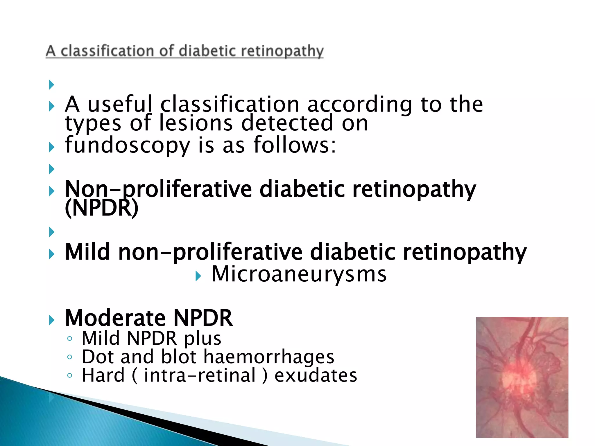
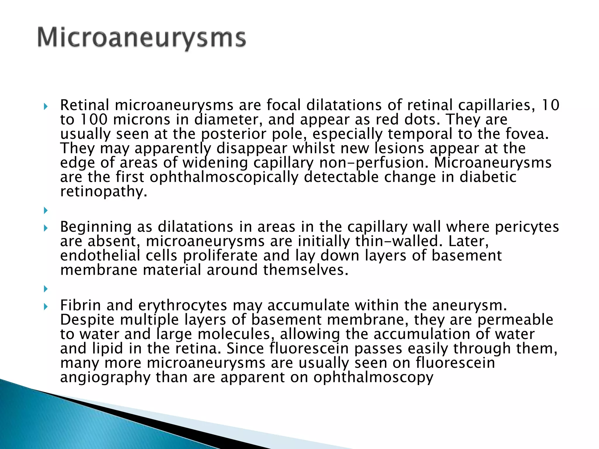
This document discusses diabetic retinopathy and its effects. It begins by noting the increasing prevalence of diabetes worldwide and that diabetes is a leading cause of blindness. It then describes the classification of diabetic retinopathy from mild non-proliferative to proliferative stages. Specific lesions associated with each stage are defined, including microaneurysms, hemorrhages, exudates, neovascularization, and retinal detachment. Risk factors for progression and visual loss from macular edema are also covered. The document emphasizes the importance of good blood sugar control in preventing and slowing the progression of diabetic retinopathy.
Retinal vein occlusion (RVO) is an obstruction of the retinal venous system by thrombus formation and may involve the central, hemi-central or branch retinal vein.
The most common aetiological factor is compression by adjacent atherosclerotic retinal arteries.
Other possible causes are external compression or disease of the vein wall e.g. vasculitis.
Comprehensive review of Ophthalmic Manifestations of Systemic Disorders for undergraduate medical students and general practionaers. Lecture was taken by Associate Professor Dr. Zia ul Mazhry at Central Park Medical College Lahore Pakistan.
Automatic detection Non-proliferative Diabetic Retinopathy using image proces...IJERA Editor
Diabetes is a chronic disease that is reaching epidemic proportions worldwide. There are currently more than
190 million people with diabetes worldwide. The World Health Organization (WHO) estimates that this will rise
to 221 million by the year 2010, largely due to population growth, ageing, urbanization and a sedentary lifestyle.
Diabetes is currently the fourth main cause of death in most developed countries. In Singapore, the prevalence
of diabetes in our population is 8.2% according to the 2004 National Health Survey. This is expected to grow as
our population age.
Diabetic Retinopathy, if not well managed and controlled, can progress steadily to devastating
complications like blindness. At present, various analyses on complicated interaction between hereditary and
environmental factors are being undertaken regarding the onset of diabetes. The development of diabetic
complication has become a major concern regarding the prognosis of diabetic patients.
Diabetes Retinopathy is one of the most common diseases that people get affected by over the years. By doing
this paper, we hope to detect the stages of Diabetic Retinopathy as early as possible so as to prevent and cure
more Singaporeans from falling prey to this disease
Diabetic retinopathy is a complication of diabetes, caused by high blood sugar levels damaging the back of the eye (retina). It can cause blindness if left undiagnosed and untreated. However, it usually takes several years for diabetic retinopathy to reach a stage where it could threaten your sight.
The retina is the light-sensitive layer of cells at the back of the eye that converts light into electrical signals. The signals are sent to the brain which turns them into the images you see.
The retina needs a constant supply of blood, which it receives through a network of tiny blood vessels. Over time, a persistently high blood sugar level can damage these blood vessels in 3 main stages:
background retinopathy – tiny bulges develop in the blood vessels, which may bleed slightly but don't usually affect your vision
pre-proliferative retinopathy – more severe and widespread changes affect the blood vessels, including more significant bleeding into the eye
proliferative retinopathy – scar tissue and new blood vessels, which are weak and bleed easily, develop on the retina, this can result in some loss of vision
Normal vision means attaining 20/20 on a routine eye exam ie, one can read 3/8-inch letters at 20 feet. Approximately 285 million people worldwide cannot pass this test without correcting their vision. Sight problems range from normal to moderate or severe visual impairment. Thirty-nine million people are blind and ~90% of visually impaired people live in low-income settings. This presentation digs into the details and current treatments. This information is for educational purposes only and all medical cases should be discussed with licensed healthcare providers.
ARTIFICIAL INTELLIGENCE IN HEALTHCARE.pdfAnujkumaranit
Artificial intelligence (AI) refers to the simulation of human intelligence processes by machines, especially computer systems. It encompasses tasks such as learning, reasoning, problem-solving, perception, and language understanding. AI technologies are revolutionizing various fields, from healthcare to finance, by enabling machines to perform tasks that typically require human intelligence.
Recomendações da OMS sobre cuidados maternos e neonatais para uma experiência pós-natal positiva.
Em consonância com os ODS – Objetivos do Desenvolvimento Sustentável e a Estratégia Global para a Saúde das Mulheres, Crianças e Adolescentes, e aplicando uma abordagem baseada nos direitos humanos, os esforços de cuidados pós-natais devem expandir-se para além da cobertura e da simples sobrevivência, de modo a incluir cuidados de qualidade.
Estas diretrizes visam melhorar a qualidade dos cuidados pós-natais essenciais e de rotina prestados às mulheres e aos recém-nascidos, com o objetivo final de melhorar a saúde e o bem-estar materno e neonatal.
Uma “experiência pós-natal positiva” é um resultado importante para todas as mulheres que dão à luz e para os seus recém-nascidos, estabelecendo as bases para a melhoria da saúde e do bem-estar a curto e longo prazo. Uma experiência pós-natal positiva é definida como aquela em que as mulheres, pessoas que gestam, os recém-nascidos, os casais, os pais, os cuidadores e as famílias recebem informação consistente, garantia e apoio de profissionais de saúde motivados; e onde um sistema de saúde flexível e com recursos reconheça as necessidades das mulheres e dos bebês e respeite o seu contexto cultural.
Estas diretrizes consolidadas apresentam algumas recomendações novas e já bem fundamentadas sobre cuidados pós-natais de rotina para mulheres e neonatos que recebem cuidados no pós-parto em unidades de saúde ou na comunidade, independentemente dos recursos disponíveis.
É fornecido um conjunto abrangente de recomendações para cuidados durante o período puerperal, com ênfase nos cuidados essenciais que todas as mulheres e recém-nascidos devem receber, e com a devida atenção à qualidade dos cuidados; isto é, a entrega e a experiência do cuidado recebido. Estas diretrizes atualizam e ampliam as recomendações da OMS de 2014 sobre cuidados pós-natais da mãe e do recém-nascido e complementam as atuais diretrizes da OMS sobre a gestão de complicações pós-natais.
O estabelecimento da amamentação e o manejo das principais intercorrências é contemplada.
Recomendamos muito.
Vamos discutir essas recomendações no nosso curso de pós-graduação em Aleitamento no Instituto Ciclos.
Esta publicação só está disponível em inglês até o momento.
Prof. Marcus Renato de Carvalho
www.agostodourado.com
Ozempic: Preoperative Management of Patients on GLP-1 Receptor Agonists Saeid Safari
Preoperative Management of Patients on GLP-1 Receptor Agonists like Ozempic and Semiglutide
ASA GUIDELINE
NYSORA Guideline
2 Case Reports of Gastric Ultrasound
Title: Sense of Smell
Presenter: Dr. Faiza, Assistant Professor of Physiology
Qualifications:
MBBS (Best Graduate, AIMC Lahore)
FCPS Physiology
ICMT, CHPE, DHPE (STMU)
MPH (GC University, Faisalabad)
MBA (Virtual University of Pakistan)
Learning Objectives:
Describe the primary categories of smells and the concept of odor blindness.
Explain the structure and location of the olfactory membrane and mucosa, including the types and roles of cells involved in olfaction.
Describe the pathway and mechanisms of olfactory signal transmission from the olfactory receptors to the brain.
Illustrate the biochemical cascade triggered by odorant binding to olfactory receptors, including the role of G-proteins and second messengers in generating an action potential.
Identify different types of olfactory disorders such as anosmia, hyposmia, hyperosmia, and dysosmia, including their potential causes.
Key Topics:
Olfactory Genes:
3% of the human genome accounts for olfactory genes.
400 genes for odorant receptors.
Olfactory Membrane:
Located in the superior part of the nasal cavity.
Medially: Folds downward along the superior septum.
Laterally: Folds over the superior turbinate and upper surface of the middle turbinate.
Total surface area: 5-10 square centimeters.
Olfactory Mucosa:
Olfactory Cells: Bipolar nerve cells derived from the CNS (100 million), with 4-25 olfactory cilia per cell.
Sustentacular Cells: Produce mucus and maintain ionic and molecular environment.
Basal Cells: Replace worn-out olfactory cells with an average lifespan of 1-2 months.
Bowman’s Gland: Secretes mucus.
Stimulation of Olfactory Cells:
Odorant dissolves in mucus and attaches to receptors on olfactory cilia.
Involves a cascade effect through G-proteins and second messengers, leading to depolarization and action potential generation in the olfactory nerve.
Quality of a Good Odorant:
Small (3-20 Carbon atoms), volatile, water-soluble, and lipid-soluble.
Facilitated by odorant-binding proteins in mucus.
Membrane Potential and Action Potential:
Resting membrane potential: -55mV.
Action potential frequency in the olfactory nerve increases with odorant strength.
Adaptation Towards the Sense of Smell:
Rapid adaptation within the first second, with further slow adaptation.
Psychological adaptation greater than receptor adaptation, involving feedback inhibition from the central nervous system.
Primary Sensations of Smell:
Camphoraceous, Musky, Floral, Pepperminty, Ethereal, Pungent, Putrid.
Odor Detection Threshold:
Examples: Hydrogen sulfide (0.0005 ppm), Methyl-mercaptan (0.002 ppm).
Some toxic substances are odorless at lethal concentrations.
Characteristics of Smell:
Odor blindness for single substances due to lack of appropriate receptor protein.
Behavioral and emotional influences of smell.
Transmission of Olfactory Signals:
From olfactory cells to glomeruli in the olfactory bulb, involving lateral inhibition.
Primitive, less old, and new olfactory systems with different path
micro teaching on communication m.sc nursing.pdfAnurag Sharma
Microteaching is a unique model of practice teaching. It is a viable instrument for the. desired change in the teaching behavior or the behavior potential which, in specified types of real. classroom situations, tends to facilitate the achievement of specified types of objectives.
Pulmonary Thromboembolism - etilogy, types, medical- Surgical and nursing man...VarunMahajani
Disruption of blood supply to lung alveoli due to blockage of one or more pulmonary blood vessels is called as Pulmonary thromboembolism. In this presentation we will discuss its causes, types and its management in depth.
These lecture slides, by Dr Sidra Arshad, offer a quick overview of physiological basis of a normal electrocardiogram.
Learning objectives:
1. Define an electrocardiogram (ECG) and electrocardiography
2. Describe how dipoles generated by the heart produce the waveforms of the ECG
3. Describe the components of a normal electrocardiogram of a typical bipolar leads (limb II)
4. Differentiate between intervals and segments
5. Enlist some common indications for obtaining an ECG
Study Resources:
1. Chapter 11, Guyton and Hall Textbook of Medical Physiology, 14th edition
2. Chapter 9, Human Physiology - From Cells to Systems, Lauralee Sherwood, 9th edition
3. Chapter 29, Ganong’s Review of Medical Physiology, 26th edition
4. Electrocardiogram, StatPearls - https://www.ncbi.nlm.nih.gov/books/NBK549803/
5. ECG in Medical Practice by ABM Abdullah, 4th edition
6. ECG Basics, http://www.nataliescasebook.com/tag/e-c-g-basics
NVBDCP.pptx Nation vector borne disease control programSapna Thakur
NVBDCP was launched in 2003-2004 . Vector-Borne Disease: Disease that results from an infection transmitted to humans and other animals by blood-feeding arthropods, such as mosquitoes, ticks, and fleas. Examples of vector-borne diseases include Dengue fever, West Nile Virus, Lyme disease, and malaria.
These simplified slides by Dr. Sidra Arshad present an overview of the non-respiratory functions of the respiratory tract.
Learning objectives:
1. Enlist the non-respiratory functions of the respiratory tract
2. Briefly explain how these functions are carried out
3. Discuss the significance of dead space
4. Differentiate between minute ventilation and alveolar ventilation
5. Describe the cough and sneeze reflexes
Study Resources:
1. Chapter 39, Guyton and Hall Textbook of Medical Physiology, 14th edition
2. Chapter 34, Ganong’s Review of Medical Physiology, 26th edition
3. Chapter 17, Human Physiology by Lauralee Sherwood, 9th edition
4. Non-respiratory functions of the lungs https://academic.oup.com/bjaed/article/13/3/98/278874
2. ( The World Health Organisation (1992) definition of
blindness is vision less than 3/60 in the better eye with best
available spectacle correction. )
Diabetes is therefore one of the most serious challenges to
health care world-wide. According to recent projections it will
affect 239 million people by 2010- doubling in prevalence
since 1994. Diabetes will affect 28 million in western Europe,
18.9 million in North America 138.2 million in Asia, 1.3
million in Australasia.
Diabetes mellitus is the most common cause of blindness
amongst individuals of working-age ( 20-65 years). The
prevalence of blindness due to DR in Western Communities is
estimated as between 1.6-1.9/ 100,000
3. About 2% of type 2 diabetics have CSME at
diagnosis and 10.2% have other signs of DR
already present when their diabetes is
discovered.
Mitchell and co- workers found that 15.8 %
of undiagnosed diabetics in an elderly
Australian population had signs of DR,
according to the recent Blue Mountains Eye
Study. Indeed it may often take from 9-12
years for type 2 diabetes to be diagnosed
4. Hyperglycaemia causes-
BM thickening
non enzymaitc glycosylation
increased free radical activity
increased flux through the polyol pathway
osmotic damage
Haemostatic abnormalities of the microcirculation-
It has also been postulated that platelet abnormalities in diabetics may
contribute to diabetic retinopathy. There are three steps in platelet
coagulation: initial adhesion, secretion, and further aggregation. It has been
shown that the platelets in diabetic patients are "stickier" than platelets of
non-diabetics They secrete prostaglandins that cause other platelets to
adhere to them (aggregation) and blockage of the vessel and endothelial
damage.
5.
6.
A useful classification according to the
types of lesions detected on
fundoscopy is as follows:
Non-proliferative diabetic retinopathy
(NPDR)
Mild non-proliferative diabetic retinopathy
Microaneurysms
Moderate NPDR
◦ Mild NPDR plus
◦ Dot and blot haemorrhages
◦ Hard ( intra-retinal ) exudates
7. Moderate-to-severe non-proliferative
diabetic retinopathy
The above lesions, usually with exacerbation,
plus:
1. Cotton-wool spots -4
2. Venous beading and loops-2
3. Intraretinal microvascular abnormalities ( IRMA )-1
8. Proliferative diabetic retinopathy
Neovascularization of the retina, optic disc or
iris
Fibrous tissue adherent to vitreous face of
retina
Retinal detachment
Vitreous haemorrhage
Pre retinal haemorrhage
10. Retinal microaneurysms are focal dilatations of retinal capillaries, 10
to 100 microns in diameter, and appear as red dots. They are
usually seen at the posterior pole, especially temporal to the fovea.
They may apparently disappear whilst new lesions appear at the
edge of areas of widening capillary non-perfusion. Microaneurysms
are the first ophthalmoscopically detectable change in diabetic
retinopathy.
Beginning as dilatations in areas in the capillary wall where pericytes
are absent, microaneurysms are initially thin-walled. Later,
endothelial cells proliferate and lay down layers of basement
membrane material around themselves.
Fibrin and erythrocytes may accumulate within the aneurysm.
Despite multiple layers of basement membrane, they are permeable
to water and large molecules, allowing the accumulation of water
and lipid in the retina. Since fluorescein passes easily through them,
many more microaneurysms are usually seen on fluorescein
angiography than are apparent on ophthalmoscopy
11. When the wall of a capillary or microaneurysm is sufficiently weakened, it
may rupture, giving rise to an intraretinal haemorrhage. If the hemorrhage is
deep (i.e., in the inner nuclear layer or outer plexiform layer), it usually is
round or oval ("dot or blot")
Dot haemorrhages appear as bright red dots and are the same size as large
microaneurysms. Blot haemorrhages are larger lesions they are located
within the mid retina and often within or surrounding areas of ischaemia.
(1,4,)
If the hemorrhage is more superficial and in the nerve fiber layer, it takes a
flame or splinter shape, which is indistinguishable from a hemorrhage seen in
hypertensive retinopathy. They often absorb slowly after several weeks. Their
presence strongly suggests the co-existence of systemic hypertension.
Diabetics with normal blood pressure may have multiple splinter
haemorrhages. Nevertheless, when an ophthalmologist sees numerous
splinter haemorrhages in a diabetic patient, the patient's blood pressure
must be checked because a frequent complication of diabetes is systemic
hypertension.
12.
13.
14. Cotton wool spots result from occlusion of
retinal pre-capillary arterioles supplying the
nerve fibre layer with concomitant swelling
of local nerve fibre axons. Also called "soft
exudates" or "nerve fibre layer infarctions"
they are white, fluffy lesions in the nerve
fibre layer. Fluorescein angiography shows
no capillary perfusion in the area of the soft
exudate. They are very common in DR,
especially if the patient is also hypertensive.
15.
16. Hard exudates ( Intra-retinal lipid exudates )
are yellow deposits of lipid and protein within
the sensory retina. Accumulations of lipids
leak from surrounding capillaries and
microaneuryisms, they may form a circinate
pattern. Hyperlipidaemia may correlate with
the development of hard exudates.
17. Accumulations of
lipids leak from
surrounding
capillaries and
microaneuryisms,
they may form a
circinate pattern.
18. Intra-retinal microvascular abnormalities ( IRMA)
are abnormal, dilated retinal capillaries or may
represent intraretinal neovacularization which
has not breached the internal limiting
membrane of the retina.
They indicate severe non-proliferative diabetic
retinopathy that may rapidly progress to
proliferative retinopathy.
Venous beading has an appearance resembling
sausage-shaped dilatation of the retinal veins.
It is another sign of severe non proliferative
diabetic retinopathy.
19.
20. Macular oedema is thus an important
manifestation of DR because it is now the
leading cause of legal blindness in
diabetics. The intercellular fluid comes from
leaking microaneurysms or from diffuse
capillary leakage .It should be stressed
however that current regimes now lay
emphasis on the treatment of retinal
thickening by grid laser than direct
treatment of microaneuyrisns and other
discreet lesions.
21. The leading cause of visual loss amongst diabetics. Diagnosed by stereoscopic
assessment of retinal thickening, usually by slit lamp biomicroscopy.
Defined as the presence of one or more of the following, ( Modified Airlie -House
Criteria )
Retinal oedema within 500 microns of the centre fovea.
Hard exudates within 500 microns of fovea if associated with adjacent retinal
thickening
Retinal oedema that is one disc diameter or larger, any part of which is within one
disc diameter of the centre of the fovea.
Laser grid photocoagulation reduces the risk of visual loss by 50% at 2 years
22.
23.
24. Maculopathy in type 1 diabetics is often due
to drop out of the perifoveal capillaries with
non perfusion and the consequent
development of an ischaemic maculopathy.
Enlargement of the foveal avascular zone
(FAZ) is frequently seen on fluorescein
angiography. Ischaemic maculopathy is not
uncommon in type 2 diabetics, maculopathy
in this group may show both changes due to
ischaemia but also retinal thickening.
25.
26.
Retinal ischaemia due to widespread capillary non perfusion results
in the production of vasoproliferative substances and to the
development of neovascularization. Neovascularization can involve
the retina, optic disc or the iris( rubeosis iridis).
Rubeosis iridis is a sign of severe proliferative disease, it may cause
intractable glaucoma.
Bleeding from fragile new vessels involving the retina or optic disc
can result in vitreous or retinal haemorrhage. Retinal damage can
result from persistent vitreous haemorrhage.
Pre-retinal haemorrhages are often associated with retinal
neovascularization, they may dramatically reduce vision within a
few minutes.
27.
28.
29.
30. Contraction of associated fibrous tissue
formed by proliferative disease tissue can
result in deformation of the retina and
tractional retinal detachment
31.
32. There are two types of diabetic retinal
detachments: those caused by traction alone
(nonrhegmatogenous) and those caused by
traction and retinal break formation
(rhegmatogenous)
Characteristics of nonrhegmatogenous
detachment in PDR include the following: (1)
the detached retina is usually confined to the
posterior fundus and infrequently extends
more than two thirds of the distance to the
equator; (2) it has a taut and shiny surface; (3)
it is concave toward the pupil; and (4) there is
no shifting of subretinal fluid.
33. The history of any visual symptoms or changes in vision
2. Measurement of visual acuity (unaided, with spectacles / pinhole
as necessary)
3. Iris examination by slit lamp biomicroscopy prior to pupil
mydriasis.
4. Pupil mydriasis. ( tropicamide 0.5 % ) -the risk of precipitating
angle closure glaucoma is actually very small. Patients should be
accompanied by a relative and instructed not to drive home.
5. Examination of the crystalline lens by slit lamp biomicroscopy.
6. Fundus examination by slit lamp biomicroscopy using diagnostic
contact lens or slit lamp indirect ophthalmoscopy.
34. It is now proven that good diabetic control may slow
the development and progression of diabetic
retinopathy in both type 1 and type 2 diabetes.
For example, the United Kingdom Prospective
Diabetes Study 1998 (UKPDS) followed 5,102 newly
diagnosed type 2 diabetics prospectively since 1977.
Those diabetics who were intensively treated and
achieved tight control with either insulin or
suphonylurea had diabetic endpoints 12% lower than
less well controlled diabetics.
Overall there was a 25% reduction in microvascular
end points in the group exhibiting good glycaemic
control.
35. Recent literature indicates that there is a
striking correlation between the presence of
systemic hypertension and progression of
diabetic retinopathy. Recent studies have
delineated the role of treating associated
hypertension and the slowing of the
progress of DR. It is important to note that
many type 2 diabetics will need a
combination of anti-hypertensive agents to
lower their blood pressure.
36.
37. The hypertension in diabetes study was launched within the original
UKPDS study in 1987.
The study compared diabetics whose blood pressure was tightly
controlled ( BP < 150/85)with ACE inhibitors and beta blockers with
a cohort whose blood pressure was less tightly controlled. (BP
<180/ 95 ) Median follow up was 8.4 years.
The reduction of macrovascular events was significant with a 32%
reduction in diabetes related deaths. There was a 44% reduction in
stroke and a 34% reduction in overall macrovascular disease.
UKPDS is a unique study in that it also looked at microvascular end
points in type 2 diabetics. Overall the tight control group had a 37%
reduction in microvascular disease, this was a more striking
reduction than tight glycaemic control.
This effect was manifested as a reduction of the risk of having to
undergo laser photocoagulation by 34%.
38. The risk of reduction of visual acuity was
lowered by 47%.
Atenolol and Captopril were equally
effective in reducing the risk of progression
of retinopathy in type 2 diabetics.
The Hypertension Optimal Treatment ( HOT
) study indicates that the lowest incidents of
cardiac events occurs when blood pressure
is lowered to 82.6 mmHg diastolic and 136
mmHg systolic.
39. The EUCLID study is currently investigating the
prophylactic treatment of type 1 diabetics with the
Angiotensin Converting Enzyme (ACE) Inhibitor
Lisinopril and the progression of nephropathy and
other microvascular disease including DR .
Preliminary reports are of a specific benefit are
encouraging, with a claimed 50% reduction in
progression of DR in type 1 diabetics.
The study did not look at maculopathy- so that
implications are unclear for type 2 diabetics,
although no specific advantage of ACE inhibitors
(Captopril) over Atenolol was seen in UKPDS.(31)
40. There is evidence in the literature that
diabetics who have exudative maculopathy
with extensive lipid exudes benefit from
active treatment of hyperlipidaemia
41. Diabetic nephropathy accelerates the
progression of retinopathy, especially
macular oedema, inter alia via increased
levels of fibrinogen and lipoprotein and
associated hypertension.
The visual prognosis is often better if the
nephropathy is treated by renal
transplantation rather than by dialysis
Any anaemia resulting from renal disease
must be aggressively treated.
Diabetic retinopathy is a common prelude to
the development of renal disease.
42. Pregnancy may accelerate the progression of diabetic retinopathy.
Frequency of monitoring NPDR should therefore be
increased.Women who begin a pregnancy with no retinopathy, the
risk of developing diabetic retinopathy is about 10%.
Those with DR at the onset of pregnancy may show progression,
with increased haemorrhages, soft exudates, and macular edema.
There is no doubt that women who maintain good metabolic control
during pregnancy have fewer spontaneous abortions and fewer
children with birth defects.
Those with untreated PDR at the onset frequently do poorly unless
they are treated with panretinal photocoagulation. Finally, patients
with previously treated PDR often do not worsen during the
pregnancy.
Women who begin pregnancy with poorly controlled diabetes and
who are suddenly brought under strict control frequently have
severe deterioration of their retinopathy and do not always recover
after delivery
43. Cataract surgery may lead to progression of
pre-existing macular oedema and
proliferative diabetic retinopathy. However,
cataracts may impede fundoscopy and
therefore interfere with the treatment of
diabetic retinopathy. If possible, diabetic
retinopathy should be treated prior to
cataract surgery
44. Tightening of glycaemic control may initially
produce worsening of retinopathy. The postulated
mechanism includes lowering of retinal blood low
or overproduction of IGF-1 by the liver.
It is therefore recommended that monitoring of
retinopathy is increased if major changes to
glycaemic control are made particularly in
previously poorly controlled diabetics. Ideally
glycated haemoglobin ( HbA1c) should be
maintained below 7%.
45. The mainstay of treatment of diabetic retinopathy is
retinal laser photocoagulation, an ablative treatment.
Laser therapy is highly effective; the rate of severe
visual loss at 2 years due to proliferative disease can
be reduced by 60%.
Laser photocoagulation causes a retinal burn which
is visible on fundoscopy. Retinal and optic disc
neovascularization can regress with the use of
retinal laser photocoagulation.
Rubeosis iridis requires urgent panretinal
photocoagulation to prevent ocular pain and
blindness from glaucoma.
46.
47.
48.
49. The indications for laser therapy now
include CSME which is treated with a
macular laser grid or treatment of focal
lesions such as microaneuryisms. Early
referral and detection of disease is
important as treatment of maculopathy is far
more successful if undertaken at an early
stage of the disease process.
There is a reduction in the rate of loss of
vision by 50% at 2 years with macular grid
therapy.
Pregnant patients should undergo laser
therapy if the usual indications are met.
50. The technique of laser photocoagulation
delivery involves the application of
eyedrops
( for pupil dilatation and corneal
anaesthesia ) and the application of an
optical contact lens. Mild proliferative
retinopathy is usually treated with at least
600 burns placed between the retinal
equator and the retinal vascular arcades. A
complete panretinal photocoagulation
treatment requires at least 1500 burns.
51. Although laser therapy can be highly effective in preventing blindness, it is
associated with numerous complications.
Retinal vein occlusion can follow inadvertent photocoagulation of a retinal
vein. Rarely, there may be loss of central acuity from inadvertent
photocoagulation of the fovea.
Vitreous haemorrhage can follow photocoagulation of retinal or choroidal
vessels.
There may be visual field restriction, decreased contrast sensitivity, impaired
night vision or impaired colour vision.
Visual field constriction may impair fitness to drive although
ophthalmologists increasingly strive to avoid this most undesirable problem,
for example by avoiding confluent laser burns.
A recent study indicates that 88% of diabetics who have undergone laser
photocoagulation would pass the Esterman binocular field test which is the
legal criterion for fitness to drive in the United Kingdom, even if both eyes
were treated. 42% of uniocular fields failed to make the criterion of a 120
degree horizontal field. Patients who have already lost the sight in one eye
therefore have a significant chance of failing to meet legal parameters for
fitness to drive in the United Kingdom.
Headache can sometimes follow laser therapy. The headache is usually
relieved with rest and simple analgesia. Glaucoma must be excluded if the
headache is severe or persistent.
52. Vitrectomy, plays a vital role in the
management of severe complications of
diabetic retinopathy.
The major indications are nonclearing
vitreous hemorrhage, traction retinal
detachment, and combined
traction/rhegmatogenous retinal
detachment. Less common indications are
macular edema with a thickened and taut
posterior hyaloid, macular heterotopia, and
tight preretinal macular hemorrhage.