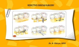
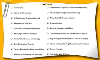
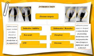
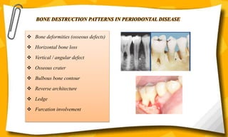
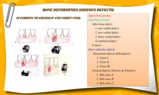
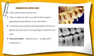
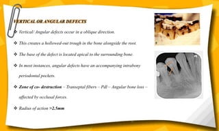
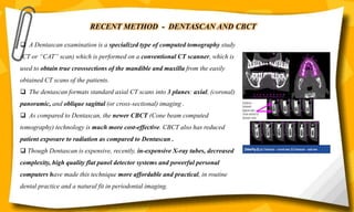
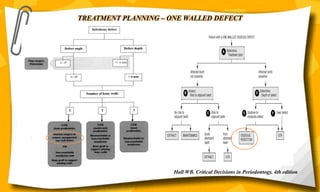
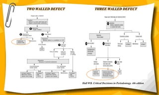
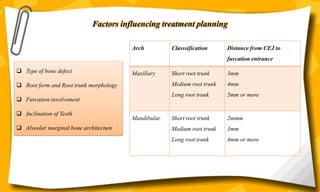
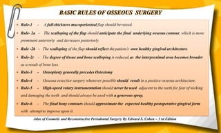
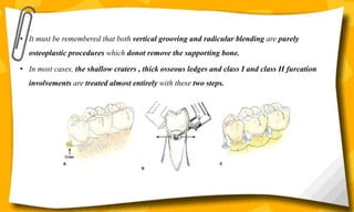
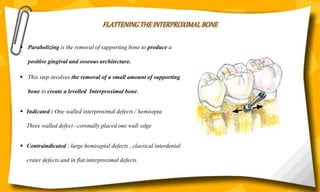
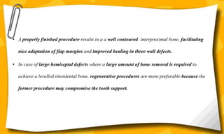
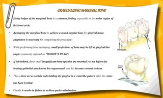
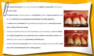
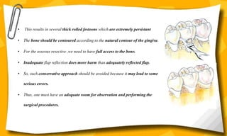
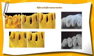
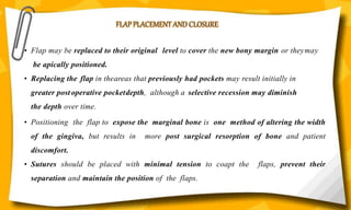
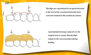
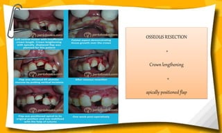
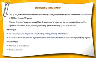
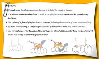
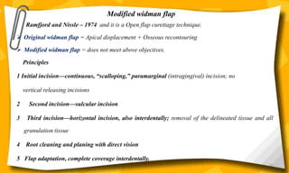
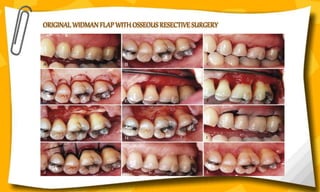
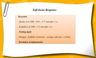
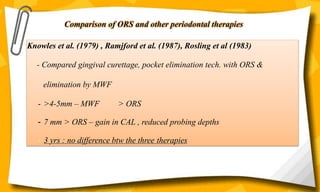
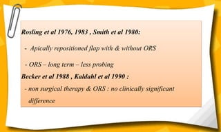
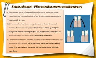
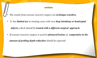
SlideShare a Scribd company logo
RESECTIVE OSSEOUS SURGERY
Dr. R. Dhivya.,MDS
CONTENT
 Examination, Diagnosis and Treatment Planning
 Factors influencing treatment planning
 Basic Rules Of Osseous Surgery
 Armamentarium
 Surgical procedure and Videos
 Correction of various soft and hard tissue defects
 Flap management after Osseous resective surgery
 Wound healing following Osseous resective surgery
 Recent Concepts
 Conclusion and Reference
 Introduction
 Historical Background
 Definitions and Terminologies used
 Rationale and objectives
 Indications and Contraindications
 Advantages and disadvantages
 Normal alveolar bone morphology
 Bone Destruction Patterns
 Factors Determining Bone Morphology
 Concept of osteoplasty and ostectomy
Osseous surgery
Inductive /Additive Subtractive / Resective
Bone grafts
GTR
Osteoplasty
Ostectomy
INTRODUCTION
Additive osseous
surgery includes
procedures
directed at
restoring the
alveolar bone to
its original level
subtractive osseous
surgery is designed
to restore the form
of preexisting
alveolar bone to the
level present at the
time of surgery or
slightly more apical
to this level
• Schluger (1949) - “Osseous Resection — A Basic Principle in Periodontal Surgery"
• Goldman (1950) - "The Development of Physiologic Gingival Contours by Gingivoplasty"
• Friedman (1955) - "Periodontal Osseous Surgery : Osteoplasty and Osteotomy"
• Prichard (1957) - "The Infrabony Technique as a Predictable Procedure"
• Goldman and Cohen (1958) - "The Infrabony Pocket: Classification and Treatment”
• Ochsenbein (1958) - "Osseous Resection in Periodontal Surgery"
• Prichard (1961) – Gingivoplasty , gingivectomy and osseous surgery.
• Ochsenbein (1986) - "A Primer for Osseous Surgery"
HISTORICAL BACKGROUND
Father of osseous surgery
DEFINITIONS AND TERMINOLOGY
OSTEOPLASTY : reshaping of the alveolar process to
achieve a more physiological form without removal of
supporting bone .
OSTECTOMY : bone that is part of the attachment
apparatus is removed to eliminate a periodontal
pocket and establish gingival contours that will be
maintained
DEFINITIVE OSSEOUS SURGERY : establishes a positive
or normal parabolic osseous form
COMPROMISE OSSEOUS SURGERY: indicates an osseous
topography requiring extensive osseous removal that would
be detrimental to the long-term prognosis of the tooth
- Aspect of periodontal surgery which deals with
the modification of the bony support of the
teeth - World workshop - 1989
- Surgical removal of the gingiva & reshaping of
the bone to eliminate the pocket and correct
unphysiologic bone architecture.
- ( Friedman et al – 1992)
- Procedure by which changes in the alveolar bone can be
accomplished to rid it of deformities induced by
periodontal disease process or other related factors –
exostosis & tooth supra eruption - Sims and Carranza
(1996)
Elimination of
periodontal
pocket and
creation of
physiological
parabolic
contour.
This contour
will maintain
physiologic
gingival
architecture
Regeneration
of periodontal
apparatus
destroyed by
periodontal
disease.
Create
environment
suitable to
restorative and
prosthodontic
treatment
RATIONALE AND OBJECTIVES
Periodontal disease
Discrepancies in level &
shape of bone
Pocket recurrence
Easley - 1967
INDICATIONS
• Residual osseous defect remaining after regenerative procedures.
• Class I or class II furcation involvement.
• Facilitates certain restorative/prosthetic dental procedures
• Fractured roots for removal.
• Bony exostoses, interdental craters, shelf like bone, bony protruberance.
• Short anatomic crowns can be lengthened by a combination of orthodontic tooth extrusion.
• Optimal crown length for cosmetic purpose.
CONTRAINDICATIONS
• Position of the external oblique line in the mandibular molar area and maxillary sinus,
which is very close to the osseous defect and root proximity.
• A periodontal pocket of more than 8mm exists after initial therapy.
• The bottom of osseous defect extends apically against multiple tooth–root trunks.
• The deep intrabony defect is more than 3-4mm or the bottom of the osseous defect is more
than one half of the root length from the cemento enamel junction.
• Extended tooth mobility.
ADVANTAGES AND DISADVANTAGES
• Reliable
• Short term (8-12 weeks)
• Obtain gingiva-alveolar bone
morphology that facilitates easy
maintenance
• Attachment loss
• Root exposure
• Compromising esthetics
• Strong possibility of hypersensitivity
• Strong possibility of root surface caries
• Possibility of phonetic impediment
• Architecture - interproximal bone coronal to labial/lingual/palatal - pyramidal
• This architecture more prominent in Anterior Max and mand areas and In posterior –
interdental bone – closer to labial/lingual bony margins.
• Form of the interdental bone – tooth form, narrower embrasure : more pyramidal - wider
embrassure : flat
• Bony margins - CEJ – marginal bone – scalloping : more in anteriors than posteriors
NORMAL ALVEOLAR BONE MORPHOLOGY
MORPHOLOGICALLY DESCRIPTIVE TERMS
Positive Architecture Negative Architecture
Flat Architecture
BONE DESTRUCTION PATTERNS IN PERIODONTAL DISEASE
 Bone deformities (osseous defects)
 Horizontal bone loss
 Vertical / angular defect
 Osseous crater
 Bulbous bone contour
 Reverse architecture
 Ledge
 Furcation involvement
ACCORDING TO GOLDMAN AND COHEN (1958)
Supra bony pocket
Infra bony pocket
Infra bony defect
1. one walled defect
2. two walled defect
3. three walled defect
4.combined defect
Craters
Inter radicular defects
Horizontal defects (Glickman’s)
1. Class I
2. Class II
3. Class III
Vertical defects (Tarnow & Fletcher)
1. Sub-class A
2. Sub-class B
3. Sub-class C
BONE DEFORMITIES (OSSEOUS DEFECTS)
HORIZONTAL BONE LOSS
 Most common pattern of bone loss
 Bone is reduced in such a way that the bone margin is
approximately perpendicular to the teeth surface
 Interdental septa and facial and lingual plates of bone are
affected, but necessarily to an equal degree around the same
tooth.
 Zone of Irritation – Gingival tissues - not affected by
occlusal forces.
14
VERTICAL OR ANGULAR DEFECTS
 Vertical/ Angular defects occur in a oblique direction.
 This creates a hollowed-out trough in the bone alongside the root.
 The base of the defect is located apical to the surrounding bone.
 In most instances, angular defects have an accompanying intrabony
periodontal pockets.
 Zone of co- destruction – Transeptal fibers – Pdl – Angular bone loss –
affected by occlusal forces.
 Radius of action >2.5mm
15
OSSEOUS CRATERS
 Osseous craters are concavities in the crest of the interdental bone
confined within the facial and lingual walls
 Craters have been found to make up about one-third of all defects
and about two-thirds of all mandibular defects.
 They occur twice as often in posterior segments as in anterior
segments.
Ochsenbein divided bony craters into three basic types :
Crater type Dimension
Shallow crater 1 -2 mm
Medium crater 3 -4 mm
Deep crater 5 mm or more
16
Heights of facial and lingual crests of the crater
- Equal in 85% cases
- Facial > Lingual in 6.5%
- Lingual > Facial in 6.5%
Several authors have also used descriptive terms to define special morphological
characteristics: funnel shaped defects, moat-like defects, trenches
The high frequency of interdental craters have been attributed to
 The interdental area collects plaque and is difficult to clean
 The normal flat or even slightly concave faciolingual shape of the interdental septum in
lower molars may favor crater formation.
 Vascular patterns from the gingiva to the centre of the crest may provide a pathway for
inflammation.
BULBOUS BONE CONTOURS
 Bulbous bone contours are bony enlargements caused by
exostoses, adaptation to function, or buttressing bone
formation.
 They are found more frequently in the maxilla than in the
mandible.
REVERSED ARCHITECTURE
 They are produced by loss of interdental bone, including
the facial plates, without concomitant loss of radicular
bone.
 The normal architecture of bone is thus reversed
 These defects are more commonly seen in the mandible
LEDGES
- Ledges are plateau like bone margins caused by resorption of
thickened bony plates
FURCATION INVOLVEMENT
 The term Furcation involvement refers to the invasion of the
bifurcation and trifurcation of multi-rooted teeth by periodontal
disease.
 Denuded furcation may be visible clinically or covered by the
wall of pocket.
 Extend of involvement is determined by exploration with a
blunt probe, along with a simultaneous blast of warm air to
facilitate visualization.
19
FACTORS DETERMINING BONE MORPHOLOGY IN PERIODONTAL DISEASE
 Normal variations in alveolar bone
 Exostoses
 Trauma from occlusion
 Buttressing bone formation
 Food impaction
 Aggressive periodontitis
20
NORMAL VARIATION OF ALVEOLAR BONE
The thickness, width
and crestal
angulation of the
interdental septa .
The thickness of the
facial and lingual
alveolar plates.
The presence of
fenestrations and
dehiscence.
The alignment of the
teeth.
Root and root trunk
anatomy.
Root position within
the alveolar process.
Proximity with
another tooth
surface.
21
EXOSTOSES
 Overgrowths of bone
 They can occur as small or large nodules, sharp ridges, spike-like projections.
 Buccal exostoses : These are seen in about 25% of all teeth, and 77% of all individuals.
 Lingual Exostoses : This may be seen in 11% of all teeth, and in 50% of all individuals.
BUTTRESSING BONE FORMATION
 Buttressing bone formation has been described as the development of
thickened or exostotic buccal alveolar bone in response to heavy occlusal
forces.
 Bone formation sometimes occurs in an attempt to buttress the bony
trabeculae weakened by resorption.
 When it occurs within the jaw, it is termed “central buttressing bone”
 When it occurs on the external surface, it is referred to as “peripheral
buttressing bone” formation.
 This may cause bulging of bone contours, termed as “Lipping”
23
FOOD IMPACTION :
a) interdental bone defects occur when there is
abnormal or absence of proximal contact.
b) food impaction here , results in inverted bone
architecture.
AGGRESSIVE PERIODONTITIS :
a) vertical or angular bone defects.
CONCEPT OF OSTEOPLASTY AND OSTECTOMY
OSTEOPLASTY : Reshaping of the alveolar process to achieve a more physiological form without removal of
supporting bone .
INDICATIONS :
• Pocket elimination
• Tori reduction,
• Intra bony defects adjacent to edentulous ridges
• Incipient furcation involvement
• Thick heavy ledges and exostoses
• Shallow osseous craters
• Small intra bony defects
Osteoplasty includes the techniques of vertical
grooving or festooning (ochsenbein,1958) and
radicular blending (carranza, 1984).
They donot necessitate removing supporting bone
Indications
• Sufficient bone remaining for establishing physiologic
contours without attachment compromise
• No esthetic or anatomic limitations
• To treat Shallow (1-2mm deep) to medium (3-4 mm
deep) intrabony and hemiseptal osseous defects
• Interdental craters
• Horizontal bone loss with irregular marginal bone
height
• Moderate to advanced furcation involvements
• Correct reversals in the osseous topography
Ostectomy includes the techniques such as flattening
interproximal bone and gradualising marginal bone
OSTECTOMY : Bone that is part of the attachment apparatus is removed to eliminate a
periodontal pocket and establish gingival contours that will be maintained.
They necessitate removal of supporting bone
Contraindications
• Areas of insufficient remaining attachment or where ostectomy might
unfavourably alter the prognosis of the adjacent teeth
• Anatomic limitations (prominent external oblique ridge , zygomatic arch)
• Esthetic limitations (anteriorly , high smile line etc)
Advantages
• Predictable pocket elimination
• Establishment of physiologic gingival and osseous architecture
• Establishment of a favorable prosthetic environment
Disadvantages
• Loss of attachment
• Esthetic compromise
• Increased root sensitivity
• The endpoint of osteoplasty procedure done in conjucation with a modified Widman flap or
an apically positioned flap , is the achievement of close tissue adaptation at the time of
suturing.
• The endpoint of ostectomy procedure done in conjucation with a apically positioned flap or a
thinned palatal flap, is the elimination of an intrabony pocket.
Examination, Diagnosis and Treatment Planning
Clinical Examination , Radiographs , Transgingival probing
Clinical Examination :
• Signs and symptoms of periodontitis, inflamed gingiva with plaque and calculus, increased flow of GCF and
bleeding on probing and exudation are commonly found in suitable patients.
• Pocket depth greater than normal gingival sulcus, presence of furcation defects should be observed.
Gingival examination:
• Physiologic gingival contour and Clinical attachment level
• Width of the attached gingiva and Thickness of the gingiva
Radiograph Examination :
• Routine dental radiographs donot accurately document the extent of bony defects.
• Well made radiographs provide useful information about interproximal bone loss, caries, and root trunk length
and root morphology
BONE SOUNDING
• Before making a surgical entry , bone sounding is done to evaluate the
bone topography.
• The buccal and lingual plates may have different thickness and
variable topography due to the reparative process going on in the
inflamed periodontal tissue.
• Bone sounding or transgingival probing with local anesthesia may
aid in the diagnosis of buccal/lingual bony defects by more accurately ,
determining the underlying bony contours.
• Greenberg et al. (1976), reported that bone sounding yielded
accurate measurements when compared to surgical entry
measurements
 The "sounding" technique for plotting the morphological outline
of the alveolar bone in mesiodistal or buccolingual sections.
 This procedure performed with local anesthesia utilized the
periodontal probe pushed through the gingival tissues as a
sounding device to determine the shape of infrabony defects until
the tip of the probe contacts the bone.
 In the case of vertical defects , bone sounding may provide
insight into the topography of the remaining walls, which many
times is not possible with radiographs due to overlapping of the
buccal and lingual/palatal plates.
RECENT METHOD - DENTASCAN AND CBCT
 A Dentascan examination is a specialized type of computed tomography study
(CT or “CAT” scan) which is performed on a conventional CT scanner, which is
used to obtain true crosssections of the mandible and maxilla from the easily
obtained CT scans of the patients.
 The dentascan formats standard axial CT scans into 3 planes: axial, (coronal)
panoramic, and oblique sagittal (or cross-sectional) imaging .
 As compared to Dentascan, the newer CBCT (Cone beam computed
tomography) technology is much more cost-effective. CBCT also has reduced
patient exposure to radiation as compared to Dentascan .
 Though Dentascan is expensive, recently, in-expensive X-ray tubes, decreased
complexity, high quality flat panel detector systems and powerful personal
computers have made this technique more affordable and practical, in routine
dental practice and a natural fit in periodontal imaging.
TREATMENT PLANNING – ONE WALLED DEFECT
Hall WB. Critical Decisions in Periodontogy. 4th edition
TWO WALLED DEFECT THREE WALLED DEFECT
Hall WB. Critical Decisions in Periodontogy. 4th edition
Factors influencing treatment planning
 Type of bone defect
 Root form and Root trunk morphology
 Furcation involvement
 Inclination of Teeth
 Alveolar marginal bone architecture
Arch Classsification Distance from CEJ to
furcation entrance
Maxillary Short root trunk
Medium root trunk
Long root trunk
3mm
4mm
5mm or more
Mandibular Short root trunk
Medium root trunk
Long root trunk
2mmm
3mm
4mm or more
BASIC RULES OF OSSEOUS SURGERY
 Rule-1 - A full-thickness mucoperiosteal flap should beraised.
 Rule- 2a - The scalloping of the flap should anticipate the final underlying osseous contour, which is more
prominent anteriorly and decreases posteriorly.
 Rule -2b - The scalloping of the flap should reflect thepatient’s own healthy gingival architecture.
 Rule-2c - The degree of tissue and bone scalloping is reduced,as the interproximal area becomes broader
as a result of bone loss.
 Rule-3 - Osteoplasty generally precedes Ostectomy
 Rule-4 - Osseous resective surgery whenever possible should result in a positive osseous architecture.
 Rule-5 - High-speed rotary instrumentation should never be used adjacent to the teeth for fear of nicking
and damaging the teeth and should always be used with a generous spray.
 Rule-6 - The final bony contours should approximate the expected healthy postoperative gingival form
with attemptto improve upon it.
Atlas of Cosmetic and Reconstructive Periodontal Surgery By Edward S. Cohen – 3 rd Edition
ARMAMENTARIUM
A number of rotary and hand instruments have been used forosseous resective surgery
Rotary for osteoplastic steps and Hand for ostectomy steps
Rongeurs - Friedman
Carbide round burs
Diamond burs
Interproximal files :
Schluger
Sugarman
Back action chisels
Ochsenbein chisels
• Bone reshaping can be done by hand or rotary instruments.
• Rotary instruments used – use of water coolant is critical.
• Overheating of bone - Necrosis of Bone.
• Thick bone such as tori and heavier ledges – rotary instruments are used.
• When reshaping the areas close to the root proximity , hand instruments are more suitable.
• As bone removal is fast with these instruments,care should be taken not to reduce bone over the
root surface to such an extent that resorption and dehiscence are inevitable.
In other words Reducing heavy bone mass - Rotary instruments are suitable
Reshaping thin bone in close proximity to the root - Hand instruments are more suitable
• Large diameter burs are more suitable in making horizontal and vertical grooving and small
diameter burs usually produce pits in the bone which are difficult to reshape.
• Small diameter burs may invade the nutrient canals in the bone which doesnot usually happen
with large diameter burs.
• In areas with thin bone, the use of hand instrument is recommended because with them there
are fewer chances of root damage.
• Thin chisels and rasps are very effective in reshaping and levelling the craters and carving the
thin bony margins.
• Many clinicains use a chisel and mallet while finishing the bone reshaping procedure because
it is more controlled as compared to rotary instruments.
SURGICAL PROCEDURES
STEPS FOR RESECTIVE OSSEOUS SURGERY
VERTICALGROOVING
RADICULAR BLENDING
FLATENNING THE INTERPROXIMAL BONE
GRADUALIZING MARGINAL BONE
Vertical Grooving
• In health, the alveolar bone and gingiva covering the roots of teeth have a
relative prominence over the interradicular counterpart, especially in the
anterior dentition.
• This creates a self – maintainable architecture of periodontal tissue.
• Another point to be remembered is that the prime objective of periodontal
surgery is to replace a pocket with the selective recession which is
maintainable.
• Vertical grooving or festooning – reduce buccal & lingual thickness of
bone interdentally till the desired depth and later on these are merged
with the bone covering the root surfaces, thereby creating smooth
elevations and depressions.
• These grooves are carried to the line angles of adjacent teeth and determine the buccolingual
width of the bone.
• Instrument : no. 6, 8 or 10 round bur + high speed handpiece+ copious irrigation.
• Indication : shallow craters, thick bony ledges.
• Contraindication : Interproximal bone between most of the upper first and second molars because of sharp
distal flare of the distobuccal root of first molar.
• I f the procedure is attempted in these areas, exposure of the root commonly results,which may complicate the
procedure.
RADICULAR BLENDING
• This is the second step which involves gradualization of the bone on th entire rot
surface, thus creating a smooth,blended surface for good flap adaptation.
• In areas with thick bony ledges this procedure results in a smooth blended
bone surface, whereas in areas with thin bone, where vertical grooving is very
minor or the radicular bone is thin or fenestrated; theis step is not necessary.
• Instrument : bur no. 6,8 or 10 – high speed handpiece.
• Back & forth motion
• Scribing : Ochsenbien chisels – 1 or 2
• Indication : shallow craters, thick ledges,
• It must be remembered that both vertical grooving and radicular blending are purely
osteoplastic procedures which donot remove the supporting bone.
• In most cases, the shallow craters , thick osseous ledges and class I and class II furcation
involvements are treated almost entirely with these two steps.
FLATTENING THE INTERPROXIMAL BONE
 Parabolizing is the removal of supporting bone to produce a
positive gingival and osseous architecture.
 This step involves the removal of a small amount of supporting
bone to create a levelled Interproximal bone.
 Indicated : One walled interproximal defects / hemisepta
Three walled defect –coronally placed one wall edge
 Contraindicated : large hemiseptal defects , classical interdental
crater defects and in flat interproximal defects.
• A properly finished procedure results in a a well contoured interproximal bone, facilitating
nice adaptation of flap margins and improved healing in three wall defects.
• In case of large hemiseptal defects where a large amount of bone removal is required to
achieve a levelled interdental bone, regenerative procedures are more preferable because the
former procedure may compromise the tooth support.
GRADUALIZING MARGINAL BONE
• Heavy ledges of the marginal bone is a common finding, especially in the molar region of
the lower arch.
• Reshaping the marginal bone to achieve a sound, regular base for gingival tissue
adaptation is necessary for completing the procedure.
• While performing bone reshaping, small projections of bone may be left at gingival line
angles commonly referred as “WIDOW’S PEAK”.
• If left behind, these small insignificant bony spicules are resorbed but not before the
healing epithelial attachment has regenerated and has become coronal to them.
• Thus , these act as curtain rods holding the gingiva in a craterlike pattern after the crater
has been levelled.
• Finally it results in failure to achieve pocket elimination.
• Use of hand instruments over rotary instruments is highly recommended in this step, if
the bone is thin.
• The final outcome of the procedure is a levelled bone, which is nicely contoured around
the teeth, following root morphology and facilitating close flap adaptation.
• A common error committed during gradualizing marginal bone is removing too much
bone, leaving a thin bone margin.
• It must be remembered that even when immediately coverd with flap, an addition 0.5mm
or so of the marginal bone is resorbed due to the continuous osteoclastic activity which
ultimately causes unnecessary fenestration and dehiscence.
• Another error is over-contouring .Over contoured bony margins are not desirable as they
may not be well maintained by the overlying gingiva.
• This results in several thick rolled festoons which are extremely persistant
• The bone should be contoured according to the natural contour of the gingiva.
• For the osseous resective ,we need to have full access to the bone.
• Inadequate flap reflection does more harm than adequately reflected flap.
• So, such conservative approach should be avoided because it may lead to some
serious errors.
• Thus, one must have an adequate room for observation and performing the
surgical procedures.
Before and after osseous resection
FLAP PLACEMENT AND CLOSURE
• Flap may be replaced to their original level to cover the new bony margin or theymay
be apically positioned.
• Replacing the flap in theareas that previously had pockets may result initially in
greater postoperative pocketdepth, although a selective recession may diminish
the depth over time.
• Positioning the flap to expose the marginal bone is one method of altering the width
of the gingiva, but results in more post surgical resorption of bone and patient
discomfort.
• Sutures should be placed with minimal tension to coapt the flaps, prevent their
separation and maintain the position of the flaps.
POSTOPERATIVE MAINTENANCE
 Nonresorbable suturing materials should be removed one week after healing. Newer synthetic material can be left
for upto of 3 weeks. Resorbable sutures will get resorbed by 1- 3 weeks .
 Suture removal should be accomplished without dragging contaminated portions of the suture through the
periodontal tissues.
 This is done by lightly compressing the soft tissues immediately adjacent to the suture. Suture is then cut at the
gingival surface.
 Chlorhexidine digluconate is a valuable adjunct to postsurgical maintenance.
 Professional prophylaxis should be done every two weeks until healing is complete or the patient is maintaing
appropriate levels of plaque control. It is usually advisable to wait minimum period of six weeks before beginning
dental restorations.
SPECIFIC OSSEOUS RESHAPING SITUATIONS
 One wall hemiseptal defects-Bone to be reduced to the level of most apical portion of the defect and One wall defects next to
edentulous spaces-Edentulous ridge is reduced to the level of osseous defects.
 Dilacerated roots, root proximity, and furcations- Compromised by osseous surgery and The walls of the crater may be
reduced at the expense of the buccal , lingual or both walls. The reduction should be made to remove the least amount of
alveolar bone required to - produce a satisfactory form,
- prevent the therapeutic invasion of furcations
- blend the contours with the adjacent teeth.
 If a tooth in a surgical field has one-walled defects on both its mesial and distal surfaces, the severely affected, tooth
may be extruded by orthodontic therapy to eliminate the need for resection of bone from the adjacent teeth.
• Heavy ledges-Osteoplasty first to eliminate any exostoses or reduce the buccal/lingual bulk of the bone.
MANAGEMENT OF DEEPCRATERS
24
MANAGEMENT OFEDENTULOUS RIDGE
The selective reduction of bony defects by “ramping” the bone to the palatal or lingual to
avoid involvement of furcations has been advocated by Ochsenbein and Bohnnan(1964).
• Restoration of fractured , severely decayed , partially erupted , worn or
poorly restored teeth is often difficult.
• Periodontal exposure or prophylactic lengthening of these teeth must adhere
to certain biologic principles and an adequate biologic width must be
maintained.
• Biological width is the term applied to the dimensional width of the
dentogingival junction (epithelial attachment and underlying connective
tissue ) .
• Garguilo et al 1961 quantified this as almost a constant 2.04mm (epithelial
attachment is 0.97mm and connective tissue is 1.07mm) with a sulcus depth
of 0.69mm.
• Biological width is defined as the sum of the combined supracrestal fibers
the junctional epithelium and the sulcus (Nevins & Skurow 1984).
Crown lengthening
• Biological width should be 3mm when measured from the crest of bone.
• Tooth lengthening procedures often employ some combination of tissue
removal, osseous surgery and orthodontics.
• The amount of tooth structure exposed (4mm) must be enough to permit
proper tooth preparation and account for an adequate marginal placement.
• Impingement of the restoration on this zone results in bone resorption.
• In crown lengthening there are two methods , coronal extension and apical
extension.
• Apical extension of the crown is achieved by surgery such as gingivectomy
and apically positioned flap surgery with and without osseous resection.
• Coronal extension is achieved by surgical or orthodontic extrusion
an post and core.
Apically repositioned flap
 In 1950s and 1960s new surgical techniques for the removal of soft tissue were described.
 The importance of maintaining an adequate zone of attached gingival after surgery was
emphasized.
 Apically positioned flap surgery, in which flaps are reflected with an internal bevel incision and
sutured apical to pre-operative position.
 Norberg (1926) first advocated this technique for mucogingival problems in periodontal disease.
 Nabers (1954) described this technique for the preservation of the gingiva following surgery.
59
Following a vertical releasing incision, the reverse bevel incision
is made through the gingiva and the periosteum to separate the
inflamed tissue adjacent to the tooth from the flap.
A mucoperiosteal flap is raised and the tissue collar remaining
around the teeth, including the pocket epithelium and the
inflamed connective tissue, is removed with a currette.
Osseous surgery is
performed with the
use of a rotating
bur
60
The flaps are repositioned in an apical direction
to the level of the recontoured alveolar bone
crest and retained in this position by sutures.
A periodontal dressing is placed over the
surgical area to ensure that the flaps
remain in the correct position during
healing.
61
OSSEOUS RESECTION
+
Crown lengthening
+
apically positioned flap
THE ORIGINAL WIDMAN FLAP
 One of the first detailed descriptions of the use of a flap procedure for pocket elimination was published
in 1916 by Leonard Widman.
 Widman described a mucoperiosteal flap design aimed at removing the pocket epithelium and the
inflamed connective tissue, thereby facilitating optimal cleaning of the root surfaces
Advantages
 Less discomfort for the patient, since healing was by primary intention and
 It was possible to re-establish a proper contour of the alveolar bone in sites with angular bony defects.
Disadvantages
 Exposure of root surfaces
 Vertical incisions
63
Procedure :
 Two releasing incisions demarcate the area scheduled for surgical therapy.
 A scalloped reverse bevel incision is made in the gingival margin to connect the two releasing
incisions.
 The collar of inflamed gingival tissue is removed following the elevation of a mucoperiosteal flap.
 By bone recontouring, a "physiologic" contour of the alveolar bone may be reestablished.
 The coronal ends of the buccal and lingual flaps are placed at the alveolar bone crest and secured
in this position by interdentally placed sutures.
64
Modified widman flap
Ramfjord and Nissle – 1974 and it is a Open flap curettage technique.
 Original widman flap = Apical displacement + Osseous recontouring
 Modified widman flap = does not meet above objectives.
Principles
1 Initial incision—continuous, “scalloping,” paramarginal (intragingival) incision; no
vertical releasing incisions
2 Second incision—sulcular incision
3 Third incision—horizontal incision, also interdentally; removal of the delineated tissue and all
granulation tissue
4 Root cleaning and planing with direct vision
5 Flap adaptation, complete coverage interdentally.
65
66
Step 1: The initial incision is an internal bevel incision
to the alveolar crest starting 0.5 to 1 mm away from the
gingival margin
Step 2: The gingiva is reflected with a periosteal
elevator .
Step 3: A crevicular incision is made from the bottom
of the pocket to the bone,
67
Step 4: third incision is made in the interdental spaces
coronal to the bone with a curette or an interproximal
knife, and the gingival collar is removed .
Step 5:Tissue tags and granulation tissue are
removed with a curette.
Step 6: adapt the facial and lingual interproximal
tissue adjacent to each other in such a way that no
interproximal bone remains exposed at the time of
suturing . Interrupted direct sutures are placed .
Differences between Modified Widman and Original Widman flaps
MODIFIED WIDMAN FLAP ORIGINAL WIDMAN FLAP
Main aim is access for root debridement with
pocket reduction
Main aim is pocket elimination.
Flaps are elevated to a much lesser extent Flaps are elevated to a larger extent.
Flaps are placed at alveolar crest margins Flaps are placed apically
Less postoperative pain and swelling More post operative pain and swellling
68
ORIGINAL WIDMAN FLAP WITH OSSEOUS RESECTIVE SURGERY
IN FURCATION DEFECTS
FURCATIONPLASTY
 Odontoplasty i.e. removal of tooth substance in the furcation area in
order to widen a narrow entrance of the furca and to reduce the
horizontal depth of the involvement.
 Osteoplasty - recontouring of bony defects in the furcation area, if
indicated.
 Repositioning and suturing of the flap
Furcation Plasty—Odontoplasty and Osteoplasty
• Hamp and colleagues (1975) described furcation plasty as raising a mucoperiosteal
flap to provide access to the furcation area and combining scaling and root planing,
osteoplasty , odontoplasty to remove local irritants and open the furcation to allow
the patient access to clean and maintain the area.
• The result should be a firm, well-contoured papilla to cover the interradicular space.
• This procedure is recommended for grade I and early grade II lesions (Glickman)
• The purpose of the procedure is to
establish a condition in the dentogingival
region which facilitates self performed
plaque control.
• It results in the establishment of a soft
tissue papilla which covers the entrance
to the inter-radicular periodontal tissues.
TUNNELPROCEDURE
• Intentional creation of a Class III furcation – entrance accessible for oral hygiene procedure.
•Bone is reshaped to obtain a scalloped morphology and the soft tissues are apically
positioned.
• This procedure results in the exposure of tooth furcation into the oral environment which can
be easily cleaned by the patient
• Very conservative approach.
• Objective - cleaning the furcation area by the patient using an interdentaltooth brush.
• Main advantage - avoidance of prosthetic reconstruction and endodontictherapy.
RESECTIVE PERIODONTAL THERAPY FOR FURCATION INVOLVED TEETH
Root amputation / Root resection : involves the removal of one or more roots of a
multirooted tooth, at the same time permitting retention of the remaining tooth portion.
Hemisection is defined as removal or separation of the root with its accompanying
crown portion.
Radisection is a newer terminology for removal of roots of maxillary molars.
Bisection / Bicuspidization is the separation of mesial and distal roots of mandibular molars
along with its crown portion, where both segments are then retained individually.
Healing after osseous surgery
Caffesse et al (1968)
Conclusion : 0.06mm – 1.2mm
• Selipsky 1976
• Aeschlimann 1979
• Moghaddas &
Stahl 1980
• Carnevale et al 1994
Amount of bone lost during ORS:
Authors Surgery
• ORS
• ORS
• ORS
• ORS
Bone removed
• 0.6mm
• 0.22mm
• Avg.0.06-0.22mm
• 0.62- 1.04mm
Crestal Bone Loss From Resorption After Ors:
 Aeschlimann et al (1979) : 0.28mm
 Moghaddas & Stahl (1980) : 0.23mm to 0.88mm
 Smith et al (1980) : 0.2mm -0.3mm
 Pennel (1967) & Wilderman ( 1970) : 0.8mm
Bone loss and remodeling after flap elevation
without osseous resective surgery
- Donnenfeld et al 1964, 1970 : 0.6 – 1mm
- Wood et al 1973 : 0.62mm , 0.98mm
- Felts & McKenzie 1964 : minimal
- Pfeifer 1967, Wood 1973 – no clear clinical advantage
• Recession
- Becker et al 1988 : 0.95 – 2.77 mm after 1 yr
- Kaldahl et al 1988 : 1.72 mm after 1 yr
• Probing depth
- Bragger , Kaldahl, Carnevale : average reduction –1.23mm
Resolution of inflammation
Soft tissue Response
Knowles et al. (1979) , Ramjford et al. (1987), Rosling et al (1983)
- Compared gingival curettage, pocket elimination tech. with ORS &
elimination by MWF
- >4-5mm – MWF > ORS
- 7 mm > ORS – gain in CAL , reduced probing depths
- 3 yrs : no difference btw the three therapies
Comparison of ORS and other periodontal therapies
Rosling et al 1976, 1983 , Smith et al 1980:
- Apically repositioned flap with & without ORS
- ORS – long term – less probing
Becker et al 1988 , Kaldahl et al 1990 :
- non surgical therapy & ORS : no clinically significant
difference
Recent Advances - Fibre retention osseous resective surgery
(a) Inter-proximal and buccal view of a lower molar with an inter-dental osseous
crater. Transeptal gingival fibres inserted into the root cementum are designed as
red dots inside the crater.
(b) Inter-proximal and buccal ostectomy performed according to the classical
technique of osseous resective surgery (ORS) where the bottom of the defect is
changed into the most coronal part of the new inter-proximal bone surface. The
buccal ostectomy is executed to create a positive bony architecture
(c) Inter-proximal and buccal ostectomy performed according to the technique of ORS
with gingival fibre retention. The coronal part of the fibres is considered as the
bottom of the defect and the inter-dental and buccal ostectomy is performed
accordingly.
Fibre retention osseous resective surgery
Carnevale G. J Clin Periodontol 2007; 34: 182–187
• This novel approach shifts the bottom of the infrabony defect to the level of the
supracrestal connective tissue fibre attachment, resulting in a more conservative
bone resection.
• Practical implications: ORS with gingival fibre retention may lead to pocket
elimination and limitation of attachment loss and may thus strengthen the
classical indications for infrabony defect elimination.
COMBINATION THERAPY - Resection and Regeneration
conclusion
• The results from osseous resective surgery are technique sensitive.
• It has limited use in treating cases with very deep intrabony or hemiseptal
defects, which should be treated with a different surgical approach.
• If osseous resective surgery is used in advanced lesions, a compromise in the
amount of probing depth reduction should be expected.
REFERENCE
 Textbook of periodontology – Carranza - 10th ed.
 Textbook of Periodontal Diseases: Basic Phenomena, Clinical Management, and Occlusal
and Restorative Interrelationships - Page and Schluger - 2nd ed.
 Cohen – Atlas of Cosmetic & Reconstructive periodontal Surgery – 2nd ed.
 Hall WB. Critical Decisions in Periodontogy - 4th edition.
 Tetbook of Periobasics: A textbook of periodontics and implantology – 1st Edition
 Kronfcld, R. "Condition of Alveolar Bone Underlying Periodontal Pockets," J. Periodontology,
6:22, 1935.
 Friedman, N. "Periodontal Osseous Surgery,“ Osteoplasty, Osteoectomy, J. Periodontology,
26:257,1955.
 The role of resective periodontal surgery in the treatment of furcation defects. Massimo
Desanctis , Perio 2000 Vol 22, 2000
 Osseous Resective Surgery – Carnavale & Kaldahl, Perio 2000, vol.22 ,2000
 Osseous resective surgery: Long-term case report , Checchi et al , IJPRD 2008.
• Osseous Resection in Periodontal Surgery, Ochsenbein - 1958
• Soft tissue regrowth following Fiber Retention Osseous Resective Surgery or Osseous Resective
Surgery. A multilevel analysis, Francesco Cairo, JCP 2015.
• Osseous resective surgery with and without fibre retention technique in the treatment of shallow
intrabony defects: a splitmouth randomized clinical trial, Mario Aimetti, JCP 2015.
• The Use of a Disclosing Agent During Resective Periodontal Surgery for Improved Removal of
Biofilm, open dental journal, 2012.
• Carnevale, G. & Kaldahl, W. (2000) Osseouresective surgery. Periodontology 2000 22, 59–87.
• Carnevale, G., Sterrantino, S. & Di Febo, G. (1983) Soft and hard tissue wound healing
• following tooth preparation to the alveolar crest. International Journal of Periodontics and
Restorative Dentistry 3, 36–53.
• Caton, J., Nyman, S. & Zander, H. (1980) Histometric evaluation of periodontal surgery.II.
Connective tissue attachment levels after four regenerative procedures. Journal of Clinical
Periodontology 7, 224–231.
Resective osseous surgery

More Related Content

What's hot

What's hot (20)

Nonsurgical Periodontal Therapy
Nonsurgical Periodontal TherapyNonsurgical Periodontal Therapy
Nonsurgical Periodontal Therapy
 
Periodontal bone defects
Periodontal bone defectsPeriodontal bone defects
Periodontal bone defects
 
Periodontal plastic surgery
Periodontal plastic surgeryPeriodontal plastic surgery
Periodontal plastic surgery
 
039.splints in periodontal therapy
039.splints in periodontal therapy039.splints in periodontal therapy
039.splints in periodontal therapy
 
Peri implant Diseases and its management
Peri implant Diseases and its managementPeri implant Diseases and its management
Peri implant Diseases and its management
 
Resective osseous surgery
Resective osseous surgeryResective osseous surgery
Resective osseous surgery
 
Gingival Recession
Gingival RecessionGingival Recession
Gingival Recession
 
RESECTIVE OSSEOUS SURGERY
RESECTIVE OSSEOUS SURGERYRESECTIVE OSSEOUS SURGERY
RESECTIVE OSSEOUS SURGERY
 
Mechanism of bone loss and patterns of bone loss
Mechanism of bone loss and patterns of bone loss  Mechanism of bone loss and patterns of bone loss
Mechanism of bone loss and patterns of bone loss
 
Bone loss and patterns of bone destruction
Bone loss and patterns of bone destructionBone loss and patterns of bone destruction
Bone loss and patterns of bone destruction
 
Host modulation therapy
Host modulation therapyHost modulation therapy
Host modulation therapy
 
Coronal advanced flap in combination with a connective tissue graft. Is the t...
Coronal advanced flap in combination with a connective tissue graft. Is the t...Coronal advanced flap in combination with a connective tissue graft. Is the t...
Coronal advanced flap in combination with a connective tissue graft. Is the t...
 
Principles of flap surgery copy
Principles of flap surgery   copyPrinciples of flap surgery   copy
Principles of flap surgery copy
 
Ridge Augmentation Procedures
Ridge Augmentation Procedures Ridge Augmentation Procedures
Ridge Augmentation Procedures
 
Interdisciplinary periodontics
Interdisciplinary periodonticsInterdisciplinary periodontics
Interdisciplinary periodontics
 
"GUIDED TISSUE REGENERATION"
"GUIDED TISSUE REGENERATION""GUIDED TISSUE REGENERATION"
"GUIDED TISSUE REGENERATION"
 
Periodontal plastic and esthetic surgery
Periodontal plastic and esthetic surgeryPeriodontal plastic and esthetic surgery
Periodontal plastic and esthetic surgery
 
Periodontal regeneration
Periodontal  regenerationPeriodontal  regeneration
Periodontal regeneration
 
local drug delivery in periodontics
local drug delivery in periodonticslocal drug delivery in periodontics
local drug delivery in periodontics
 
Resective osseous surgery
Resective osseous surgeryResective osseous surgery
Resective osseous surgery
 

Similar to Resective osseous surgery

7. Remodeling Resective Osseous Surgery.pptx
7. Remodeling Resective Osseous Surgery.pptx7. Remodeling Resective Osseous Surgery.pptx
7. Remodeling Resective Osseous Surgery.pptx
malti19
 
Resorbed ridge seminar koto
Resorbed ridge seminar kotoResorbed ridge seminar koto
Resorbed ridge seminar koto
Kushal Singh
 
Mandibular 3rd molar impactions
Mandibular 3rd molar impactionsMandibular 3rd molar impactions
Mandibular 3rd molar impactions
Mohammad Akheel
 
Periodontal surgeries
Periodontal surgeriesPeriodontal surgeries
Periodontal surgeries
Moola Reddy
 

Similar to Resective osseous surgery (20)

Osseous surgery
Osseous surgeryOsseous surgery
Osseous surgery
 
Resective osseous surgery.pptx
Resective osseous surgery.pptxResective osseous surgery.pptx
Resective osseous surgery.pptx
 
RESECTIVE OSSEOUS SURGERY.pptx
RESECTIVE OSSEOUS SURGERY.pptxRESECTIVE OSSEOUS SURGERY.pptx
RESECTIVE OSSEOUS SURGERY.pptx
 
resective osseous surgeryyy.pptx
resective osseous surgeryyy.pptxresective osseous surgeryyy.pptx
resective osseous surgeryyy.pptx
 
7. Remodeling Resective Osseous Surgery.pptx
7. Remodeling Resective Osseous Surgery.pptx7. Remodeling Resective Osseous Surgery.pptx
7. Remodeling Resective Osseous Surgery.pptx
 
RESECTIVE OSSEOUS SURGERY.ppt
RESECTIVE OSSEOUS SURGERY.pptRESECTIVE OSSEOUS SURGERY.ppt
RESECTIVE OSSEOUS SURGERY.ppt
 
Resective osseous surgery
Resective osseous surgeryResective osseous surgery
Resective osseous surgery
 
Resorbed ridge seminar koto
Resorbed ridge seminar kotoResorbed ridge seminar koto
Resorbed ridge seminar koto
 
BONE-DESTRUCTION-PATTERNS-20208171318390.pptx
BONE-DESTRUCTION-PATTERNS-20208171318390.pptxBONE-DESTRUCTION-PATTERNS-20208171318390.pptx
BONE-DESTRUCTION-PATTERNS-20208171318390.pptx
 
Resective osseous surgery
Resective osseous surgeryResective osseous surgery
Resective osseous surgery
 
Fenestration and dehiscence
Fenestration and dehiscenceFenestration and dehiscence
Fenestration and dehiscence
 
Perio-Ortho interrelationships - Dr.Malvika
Perio-Ortho interrelationships - Dr.MalvikaPerio-Ortho interrelationships - Dr.Malvika
Perio-Ortho interrelationships - Dr.Malvika
 
implant bjc.ppt
implant bjc.pptimplant bjc.ppt
implant bjc.ppt
 
Mandibular 3rd molar impactions
Mandibular 3rd molar impactionsMandibular 3rd molar impactions
Mandibular 3rd molar impactions
 
RESIDUAL RIDGE RESORPTION
RESIDUAL RIDGE RESORPTIONRESIDUAL RIDGE RESORPTION
RESIDUAL RIDGE RESORPTION
 
PREPROSTHETIC SURGERY 2.pptx
PREPROSTHETIC SURGERY 2.pptxPREPROSTHETIC SURGERY 2.pptx
PREPROSTHETIC SURGERY 2.pptx
 
Enamel reduction techniques in orthodontics
Enamel reduction techniques in orthodonticsEnamel reduction techniques in orthodontics
Enamel reduction techniques in orthodontics
 
Patterns of bone destruction in periodontics
Patterns of bone destruction in periodontics Patterns of bone destruction in periodontics
Patterns of bone destruction in periodontics
 
Periodontal surgeries
Periodontal surgeriesPeriodontal surgeries
Periodontal surgeries
 
Satz/ oral surgery courses  
Satz/ oral surgery courses  Satz/ oral surgery courses  
Satz/ oral surgery courses  
 

More from Dr.R.Dhivya.,MDS (12)

Stem cells
Stem cellsStem cells
Stem cells
 
Toll like receptors
Toll like receptorsToll like receptors
Toll like receptors
 
Supportive periodontal therapy
Supportive periodontal therapySupportive periodontal therapy
Supportive periodontal therapy
 
Root biomodification
Root biomodificationRoot biomodification
Root biomodification
 
Periodontal flap surgery
Periodontal flap surgeryPeriodontal flap surgery
Periodontal flap surgery
 
Periodontal microsurgery
Periodontal microsurgeryPeriodontal microsurgery
Periodontal microsurgery
 
Local dug delivery
Local dug deliveryLocal dug delivery
Local dug delivery
 
Growth factors
Growth factorsGrowth factors
Growth factors
 
Gingival Crevicular Fluid
Gingival Crevicular FluidGingival Crevicular Fluid
Gingival Crevicular Fluid
 
Connective Tissue in health and disease
Connective Tissue in health and diseaseConnective Tissue in health and disease
Connective Tissue in health and disease
 
Bone destruction
Bone destructionBone destruction
Bone destruction
 
Antigen presenting cells
Antigen presenting cellsAntigen presenting cells
Antigen presenting cells
 

Recently uploaded

Recently uploaded (20)

Prix Galien International 2024 Forum Program
Prix Galien International 2024 Forum ProgramPrix Galien International 2024 Forum Program
Prix Galien International 2024 Forum Program
 
ANATOMY OF THE LOWER URINARY TRACT AND MALE [Autosaved] [Autosaved].pptx
ANATOMY OF THE LOWER URINARY TRACT AND MALE [Autosaved] [Autosaved].pptxANATOMY OF THE LOWER URINARY TRACT AND MALE [Autosaved] [Autosaved].pptx
ANATOMY OF THE LOWER URINARY TRACT AND MALE [Autosaved] [Autosaved].pptx
 
Final CAPNOCYTOPHAGA INFECTION by Gauri Gawande.pptx
Final CAPNOCYTOPHAGA INFECTION by Gauri Gawande.pptxFinal CAPNOCYTOPHAGA INFECTION by Gauri Gawande.pptx
Final CAPNOCYTOPHAGA INFECTION by Gauri Gawande.pptx
 
Effects of vaping e-cigarettes on arterial health
Effects of vaping e-cigarettes on arterial healthEffects of vaping e-cigarettes on arterial health
Effects of vaping e-cigarettes on arterial health
 
Arterial health throughout cancer treatment and exercise rehabilitation in wo...
Arterial health throughout cancer treatment and exercise rehabilitation in wo...Arterial health throughout cancer treatment and exercise rehabilitation in wo...
Arterial health throughout cancer treatment and exercise rehabilitation in wo...
 
Hemodialysis: Chapter 2, Extracorporeal Blood Circuit - Dr.Gawad
Hemodialysis: Chapter 2, Extracorporeal Blood Circuit - Dr.GawadHemodialysis: Chapter 2, Extracorporeal Blood Circuit - Dr.Gawad
Hemodialysis: Chapter 2, Extracorporeal Blood Circuit - Dr.Gawad
 
Scientificity and feasibility study of non-invasive central arterial pressure...
Scientificity and feasibility study of non-invasive central arterial pressure...Scientificity and feasibility study of non-invasive central arterial pressure...
Scientificity and feasibility study of non-invasive central arterial pressure...
 
DECIPHERING COMMON ECG FINDINGS IN ED.pptx
DECIPHERING COMMON ECG FINDINGS IN ED.pptxDECIPHERING COMMON ECG FINDINGS IN ED.pptx
DECIPHERING COMMON ECG FINDINGS IN ED.pptx
 
Relationship between vascular system disfunction, neurofluid flow and Alzheim...
Relationship between vascular system disfunction, neurofluid flow and Alzheim...Relationship between vascular system disfunction, neurofluid flow and Alzheim...
Relationship between vascular system disfunction, neurofluid flow and Alzheim...
 
Multiple sclerosis diet.230524.ppt3.pptx
Multiple sclerosis diet.230524.ppt3.pptxMultiple sclerosis diet.230524.ppt3.pptx
Multiple sclerosis diet.230524.ppt3.pptx
 
Retinal consideration in cataract surgery
Retinal consideration in cataract surgeryRetinal consideration in cataract surgery
Retinal consideration in cataract surgery
 
TEST BANK For Advanced Practice Nursing in the Care of Older Adults, 2nd Edit...
TEST BANK For Advanced Practice Nursing in the Care of Older Adults, 2nd Edit...TEST BANK For Advanced Practice Nursing in the Care of Older Adults, 2nd Edit...
TEST BANK For Advanced Practice Nursing in the Care of Older Adults, 2nd Edit...
 
Gauri Gawande(9) Constipation Final.pptx
Gauri Gawande(9) Constipation Final.pptxGauri Gawande(9) Constipation Final.pptx
Gauri Gawande(9) Constipation Final.pptx
 
Why invest into infodemic management in health emergencies
Why invest into infodemic management in health emergenciesWhy invest into infodemic management in health emergencies
Why invest into infodemic management in health emergencies
 
Compare home pulse pressure components collected directly from home
Compare home pulse pressure components collected directly from homeCompare home pulse pressure components collected directly from home
Compare home pulse pressure components collected directly from home
 
CURRENT HEALTH PROBLEMS AND ITS SOLUTION BY AYURVEDA.pptx
CURRENT HEALTH PROBLEMS AND ITS SOLUTION BY AYURVEDA.pptxCURRENT HEALTH PROBLEMS AND ITS SOLUTION BY AYURVEDA.pptx
CURRENT HEALTH PROBLEMS AND ITS SOLUTION BY AYURVEDA.pptx
 
DIGITAL RADIOGRAPHY-SABBU KHATOON .pptx
DIGITAL RADIOGRAPHY-SABBU KHATOON  .pptxDIGITAL RADIOGRAPHY-SABBU KHATOON  .pptx
DIGITAL RADIOGRAPHY-SABBU KHATOON .pptx
 
Temporal, Infratemporal & Pterygopalatine BY Dr.RIG.pptx
Temporal, Infratemporal & Pterygopalatine BY Dr.RIG.pptxTemporal, Infratemporal & Pterygopalatine BY Dr.RIG.pptx
Temporal, Infratemporal & Pterygopalatine BY Dr.RIG.pptx
 
180-hour Power Capsules For Men In Ghana
180-hour Power Capsules For Men In Ghana180-hour Power Capsules For Men In Ghana
180-hour Power Capsules For Men In Ghana
 
In-service education (Nursing Mangement)
In-service education (Nursing Mangement)In-service education (Nursing Mangement)
In-service education (Nursing Mangement)
 

Resective osseous surgery

  • 2. CONTENT  Examination, Diagnosis and Treatment Planning  Factors influencing treatment planning  Basic Rules Of Osseous Surgery  Armamentarium  Surgical procedure and Videos  Correction of various soft and hard tissue defects  Flap management after Osseous resective surgery  Wound healing following Osseous resective surgery  Recent Concepts  Conclusion and Reference  Introduction  Historical Background  Definitions and Terminologies used  Rationale and objectives  Indications and Contraindications  Advantages and disadvantages  Normal alveolar bone morphology  Bone Destruction Patterns  Factors Determining Bone Morphology  Concept of osteoplasty and ostectomy
  • 3. Osseous surgery Inductive /Additive Subtractive / Resective Bone grafts GTR Osteoplasty Ostectomy INTRODUCTION Additive osseous surgery includes procedures directed at restoring the alveolar bone to its original level subtractive osseous surgery is designed to restore the form of preexisting alveolar bone to the level present at the time of surgery or slightly more apical to this level
  • 4. • Schluger (1949) - “Osseous Resection — A Basic Principle in Periodontal Surgery" • Goldman (1950) - "The Development of Physiologic Gingival Contours by Gingivoplasty" • Friedman (1955) - "Periodontal Osseous Surgery : Osteoplasty and Osteotomy" • Prichard (1957) - "The Infrabony Technique as a Predictable Procedure" • Goldman and Cohen (1958) - "The Infrabony Pocket: Classification and Treatment” • Ochsenbein (1958) - "Osseous Resection in Periodontal Surgery" • Prichard (1961) – Gingivoplasty , gingivectomy and osseous surgery. • Ochsenbein (1986) - "A Primer for Osseous Surgery" HISTORICAL BACKGROUND Father of osseous surgery
  • 5. DEFINITIONS AND TERMINOLOGY OSTEOPLASTY : reshaping of the alveolar process to achieve a more physiological form without removal of supporting bone . OSTECTOMY : bone that is part of the attachment apparatus is removed to eliminate a periodontal pocket and establish gingival contours that will be maintained DEFINITIVE OSSEOUS SURGERY : establishes a positive or normal parabolic osseous form COMPROMISE OSSEOUS SURGERY: indicates an osseous topography requiring extensive osseous removal that would be detrimental to the long-term prognosis of the tooth - Aspect of periodontal surgery which deals with the modification of the bony support of the teeth - World workshop - 1989 - Surgical removal of the gingiva & reshaping of the bone to eliminate the pocket and correct unphysiologic bone architecture. - ( Friedman et al – 1992) - Procedure by which changes in the alveolar bone can be accomplished to rid it of deformities induced by periodontal disease process or other related factors – exostosis & tooth supra eruption - Sims and Carranza (1996)
  • 6. Elimination of periodontal pocket and creation of physiological parabolic contour. This contour will maintain physiologic gingival architecture Regeneration of periodontal apparatus destroyed by periodontal disease. Create environment suitable to restorative and prosthodontic treatment RATIONALE AND OBJECTIVES Periodontal disease Discrepancies in level & shape of bone Pocket recurrence Easley - 1967
  • 7. INDICATIONS • Residual osseous defect remaining after regenerative procedures. • Class I or class II furcation involvement. • Facilitates certain restorative/prosthetic dental procedures • Fractured roots for removal. • Bony exostoses, interdental craters, shelf like bone, bony protruberance. • Short anatomic crowns can be lengthened by a combination of orthodontic tooth extrusion. • Optimal crown length for cosmetic purpose.
  • 8. CONTRAINDICATIONS • Position of the external oblique line in the mandibular molar area and maxillary sinus, which is very close to the osseous defect and root proximity. • A periodontal pocket of more than 8mm exists after initial therapy. • The bottom of osseous defect extends apically against multiple tooth–root trunks. • The deep intrabony defect is more than 3-4mm or the bottom of the osseous defect is more than one half of the root length from the cemento enamel junction. • Extended tooth mobility.
  • 9. ADVANTAGES AND DISADVANTAGES • Reliable • Short term (8-12 weeks) • Obtain gingiva-alveolar bone morphology that facilitates easy maintenance • Attachment loss • Root exposure • Compromising esthetics • Strong possibility of hypersensitivity • Strong possibility of root surface caries • Possibility of phonetic impediment
  • 10. • Architecture - interproximal bone coronal to labial/lingual/palatal - pyramidal • This architecture more prominent in Anterior Max and mand areas and In posterior – interdental bone – closer to labial/lingual bony margins. • Form of the interdental bone – tooth form, narrower embrasure : more pyramidal - wider embrassure : flat • Bony margins - CEJ – marginal bone – scalloping : more in anteriors than posteriors NORMAL ALVEOLAR BONE MORPHOLOGY
  • 11. MORPHOLOGICALLY DESCRIPTIVE TERMS Positive Architecture Negative Architecture Flat Architecture
  • 12. BONE DESTRUCTION PATTERNS IN PERIODONTAL DISEASE  Bone deformities (osseous defects)  Horizontal bone loss  Vertical / angular defect  Osseous crater  Bulbous bone contour  Reverse architecture  Ledge  Furcation involvement
  • 13. ACCORDING TO GOLDMAN AND COHEN (1958) Supra bony pocket Infra bony pocket Infra bony defect 1. one walled defect 2. two walled defect 3. three walled defect 4.combined defect Craters Inter radicular defects Horizontal defects (Glickman’s) 1. Class I 2. Class II 3. Class III Vertical defects (Tarnow & Fletcher) 1. Sub-class A 2. Sub-class B 3. Sub-class C BONE DEFORMITIES (OSSEOUS DEFECTS)
  • 14. HORIZONTAL BONE LOSS  Most common pattern of bone loss  Bone is reduced in such a way that the bone margin is approximately perpendicular to the teeth surface  Interdental septa and facial and lingual plates of bone are affected, but necessarily to an equal degree around the same tooth.  Zone of Irritation – Gingival tissues - not affected by occlusal forces. 14
  • 15. VERTICAL OR ANGULAR DEFECTS  Vertical/ Angular defects occur in a oblique direction.  This creates a hollowed-out trough in the bone alongside the root.  The base of the defect is located apical to the surrounding bone.  In most instances, angular defects have an accompanying intrabony periodontal pockets.  Zone of co- destruction – Transeptal fibers – Pdl – Angular bone loss – affected by occlusal forces.  Radius of action >2.5mm 15
  • 16. OSSEOUS CRATERS  Osseous craters are concavities in the crest of the interdental bone confined within the facial and lingual walls  Craters have been found to make up about one-third of all defects and about two-thirds of all mandibular defects.  They occur twice as often in posterior segments as in anterior segments. Ochsenbein divided bony craters into three basic types : Crater type Dimension Shallow crater 1 -2 mm Medium crater 3 -4 mm Deep crater 5 mm or more 16
  • 17. Heights of facial and lingual crests of the crater - Equal in 85% cases - Facial > Lingual in 6.5% - Lingual > Facial in 6.5% Several authors have also used descriptive terms to define special morphological characteristics: funnel shaped defects, moat-like defects, trenches The high frequency of interdental craters have been attributed to  The interdental area collects plaque and is difficult to clean  The normal flat or even slightly concave faciolingual shape of the interdental septum in lower molars may favor crater formation.  Vascular patterns from the gingiva to the centre of the crest may provide a pathway for inflammation.
  • 18. BULBOUS BONE CONTOURS  Bulbous bone contours are bony enlargements caused by exostoses, adaptation to function, or buttressing bone formation.  They are found more frequently in the maxilla than in the mandible. REVERSED ARCHITECTURE  They are produced by loss of interdental bone, including the facial plates, without concomitant loss of radicular bone.  The normal architecture of bone is thus reversed  These defects are more commonly seen in the mandible
  • 19. LEDGES - Ledges are plateau like bone margins caused by resorption of thickened bony plates FURCATION INVOLVEMENT  The term Furcation involvement refers to the invasion of the bifurcation and trifurcation of multi-rooted teeth by periodontal disease.  Denuded furcation may be visible clinically or covered by the wall of pocket.  Extend of involvement is determined by exploration with a blunt probe, along with a simultaneous blast of warm air to facilitate visualization. 19
  • 20. FACTORS DETERMINING BONE MORPHOLOGY IN PERIODONTAL DISEASE  Normal variations in alveolar bone  Exostoses  Trauma from occlusion  Buttressing bone formation  Food impaction  Aggressive periodontitis 20
  • 21. NORMAL VARIATION OF ALVEOLAR BONE The thickness, width and crestal angulation of the interdental septa . The thickness of the facial and lingual alveolar plates. The presence of fenestrations and dehiscence. The alignment of the teeth. Root and root trunk anatomy. Root position within the alveolar process. Proximity with another tooth surface. 21
  • 22. EXOSTOSES  Overgrowths of bone  They can occur as small or large nodules, sharp ridges, spike-like projections.  Buccal exostoses : These are seen in about 25% of all teeth, and 77% of all individuals.  Lingual Exostoses : This may be seen in 11% of all teeth, and in 50% of all individuals.
  • 23. BUTTRESSING BONE FORMATION  Buttressing bone formation has been described as the development of thickened or exostotic buccal alveolar bone in response to heavy occlusal forces.  Bone formation sometimes occurs in an attempt to buttress the bony trabeculae weakened by resorption.  When it occurs within the jaw, it is termed “central buttressing bone”  When it occurs on the external surface, it is referred to as “peripheral buttressing bone” formation.  This may cause bulging of bone contours, termed as “Lipping” 23
  • 24. FOOD IMPACTION : a) interdental bone defects occur when there is abnormal or absence of proximal contact. b) food impaction here , results in inverted bone architecture. AGGRESSIVE PERIODONTITIS : a) vertical or angular bone defects.
  • 25. CONCEPT OF OSTEOPLASTY AND OSTECTOMY OSTEOPLASTY : Reshaping of the alveolar process to achieve a more physiological form without removal of supporting bone . INDICATIONS : • Pocket elimination • Tori reduction, • Intra bony defects adjacent to edentulous ridges • Incipient furcation involvement • Thick heavy ledges and exostoses • Shallow osseous craters • Small intra bony defects Osteoplasty includes the techniques of vertical grooving or festooning (ochsenbein,1958) and radicular blending (carranza, 1984). They donot necessitate removing supporting bone
  • 26. Indications • Sufficient bone remaining for establishing physiologic contours without attachment compromise • No esthetic or anatomic limitations • To treat Shallow (1-2mm deep) to medium (3-4 mm deep) intrabony and hemiseptal osseous defects • Interdental craters • Horizontal bone loss with irregular marginal bone height • Moderate to advanced furcation involvements • Correct reversals in the osseous topography Ostectomy includes the techniques such as flattening interproximal bone and gradualising marginal bone OSTECTOMY : Bone that is part of the attachment apparatus is removed to eliminate a periodontal pocket and establish gingival contours that will be maintained. They necessitate removal of supporting bone
  • 27. Contraindications • Areas of insufficient remaining attachment or where ostectomy might unfavourably alter the prognosis of the adjacent teeth • Anatomic limitations (prominent external oblique ridge , zygomatic arch) • Esthetic limitations (anteriorly , high smile line etc) Advantages • Predictable pocket elimination • Establishment of physiologic gingival and osseous architecture • Establishment of a favorable prosthetic environment Disadvantages • Loss of attachment • Esthetic compromise • Increased root sensitivity
  • 28. • The endpoint of osteoplasty procedure done in conjucation with a modified Widman flap or an apically positioned flap , is the achievement of close tissue adaptation at the time of suturing. • The endpoint of ostectomy procedure done in conjucation with a apically positioned flap or a thinned palatal flap, is the elimination of an intrabony pocket.
  • 29. Examination, Diagnosis and Treatment Planning Clinical Examination , Radiographs , Transgingival probing Clinical Examination : • Signs and symptoms of periodontitis, inflamed gingiva with plaque and calculus, increased flow of GCF and bleeding on probing and exudation are commonly found in suitable patients. • Pocket depth greater than normal gingival sulcus, presence of furcation defects should be observed. Gingival examination: • Physiologic gingival contour and Clinical attachment level • Width of the attached gingiva and Thickness of the gingiva Radiograph Examination : • Routine dental radiographs donot accurately document the extent of bony defects. • Well made radiographs provide useful information about interproximal bone loss, caries, and root trunk length and root morphology
  • 30. BONE SOUNDING • Before making a surgical entry , bone sounding is done to evaluate the bone topography. • The buccal and lingual plates may have different thickness and variable topography due to the reparative process going on in the inflamed periodontal tissue. • Bone sounding or transgingival probing with local anesthesia may aid in the diagnosis of buccal/lingual bony defects by more accurately , determining the underlying bony contours. • Greenberg et al. (1976), reported that bone sounding yielded accurate measurements when compared to surgical entry measurements
  • 31.  The "sounding" technique for plotting the morphological outline of the alveolar bone in mesiodistal or buccolingual sections.  This procedure performed with local anesthesia utilized the periodontal probe pushed through the gingival tissues as a sounding device to determine the shape of infrabony defects until the tip of the probe contacts the bone.  In the case of vertical defects , bone sounding may provide insight into the topography of the remaining walls, which many times is not possible with radiographs due to overlapping of the buccal and lingual/palatal plates.
  • 32. RECENT METHOD - DENTASCAN AND CBCT  A Dentascan examination is a specialized type of computed tomography study (CT or “CAT” scan) which is performed on a conventional CT scanner, which is used to obtain true crosssections of the mandible and maxilla from the easily obtained CT scans of the patients.  The dentascan formats standard axial CT scans into 3 planes: axial, (coronal) panoramic, and oblique sagittal (or cross-sectional) imaging .  As compared to Dentascan, the newer CBCT (Cone beam computed tomography) technology is much more cost-effective. CBCT also has reduced patient exposure to radiation as compared to Dentascan .  Though Dentascan is expensive, recently, in-expensive X-ray tubes, decreased complexity, high quality flat panel detector systems and powerful personal computers have made this technique more affordable and practical, in routine dental practice and a natural fit in periodontal imaging.
  • 33. TREATMENT PLANNING – ONE WALLED DEFECT Hall WB. Critical Decisions in Periodontogy. 4th edition
  • 34. TWO WALLED DEFECT THREE WALLED DEFECT Hall WB. Critical Decisions in Periodontogy. 4th edition
  • 35. Factors influencing treatment planning  Type of bone defect  Root form and Root trunk morphology  Furcation involvement  Inclination of Teeth  Alveolar marginal bone architecture Arch Classsification Distance from CEJ to furcation entrance Maxillary Short root trunk Medium root trunk Long root trunk 3mm 4mm 5mm or more Mandibular Short root trunk Medium root trunk Long root trunk 2mmm 3mm 4mm or more
  • 36. BASIC RULES OF OSSEOUS SURGERY  Rule-1 - A full-thickness mucoperiosteal flap should beraised.  Rule- 2a - The scalloping of the flap should anticipate the final underlying osseous contour, which is more prominent anteriorly and decreases posteriorly.  Rule -2b - The scalloping of the flap should reflect thepatient’s own healthy gingival architecture.  Rule-2c - The degree of tissue and bone scalloping is reduced,as the interproximal area becomes broader as a result of bone loss.  Rule-3 - Osteoplasty generally precedes Ostectomy  Rule-4 - Osseous resective surgery whenever possible should result in a positive osseous architecture.  Rule-5 - High-speed rotary instrumentation should never be used adjacent to the teeth for fear of nicking and damaging the teeth and should always be used with a generous spray.  Rule-6 - The final bony contours should approximate the expected healthy postoperative gingival form with attemptto improve upon it. Atlas of Cosmetic and Reconstructive Periodontal Surgery By Edward S. Cohen – 3 rd Edition
  • 37. ARMAMENTARIUM A number of rotary and hand instruments have been used forosseous resective surgery Rotary for osteoplastic steps and Hand for ostectomy steps Rongeurs - Friedman Carbide round burs Diamond burs Interproximal files : Schluger Sugarman Back action chisels Ochsenbein chisels
  • 38. • Bone reshaping can be done by hand or rotary instruments. • Rotary instruments used – use of water coolant is critical. • Overheating of bone - Necrosis of Bone. • Thick bone such as tori and heavier ledges – rotary instruments are used. • When reshaping the areas close to the root proximity , hand instruments are more suitable. • As bone removal is fast with these instruments,care should be taken not to reduce bone over the root surface to such an extent that resorption and dehiscence are inevitable. In other words Reducing heavy bone mass - Rotary instruments are suitable Reshaping thin bone in close proximity to the root - Hand instruments are more suitable
  • 39. • Large diameter burs are more suitable in making horizontal and vertical grooving and small diameter burs usually produce pits in the bone which are difficult to reshape. • Small diameter burs may invade the nutrient canals in the bone which doesnot usually happen with large diameter burs. • In areas with thin bone, the use of hand instrument is recommended because with them there are fewer chances of root damage. • Thin chisels and rasps are very effective in reshaping and levelling the craters and carving the thin bony margins. • Many clinicains use a chisel and mallet while finishing the bone reshaping procedure because it is more controlled as compared to rotary instruments.
  • 40. SURGICAL PROCEDURES STEPS FOR RESECTIVE OSSEOUS SURGERY VERTICALGROOVING RADICULAR BLENDING FLATENNING THE INTERPROXIMAL BONE GRADUALIZING MARGINAL BONE
  • 41. Vertical Grooving • In health, the alveolar bone and gingiva covering the roots of teeth have a relative prominence over the interradicular counterpart, especially in the anterior dentition. • This creates a self – maintainable architecture of periodontal tissue. • Another point to be remembered is that the prime objective of periodontal surgery is to replace a pocket with the selective recession which is maintainable. • Vertical grooving or festooning – reduce buccal & lingual thickness of bone interdentally till the desired depth and later on these are merged with the bone covering the root surfaces, thereby creating smooth elevations and depressions.
  • 42. • These grooves are carried to the line angles of adjacent teeth and determine the buccolingual width of the bone. • Instrument : no. 6, 8 or 10 round bur + high speed handpiece+ copious irrigation. • Indication : shallow craters, thick bony ledges. • Contraindication : Interproximal bone between most of the upper first and second molars because of sharp distal flare of the distobuccal root of first molar. • I f the procedure is attempted in these areas, exposure of the root commonly results,which may complicate the procedure.
  • 43. RADICULAR BLENDING • This is the second step which involves gradualization of the bone on th entire rot surface, thus creating a smooth,blended surface for good flap adaptation. • In areas with thick bony ledges this procedure results in a smooth blended bone surface, whereas in areas with thin bone, where vertical grooving is very minor or the radicular bone is thin or fenestrated; theis step is not necessary. • Instrument : bur no. 6,8 or 10 – high speed handpiece. • Back & forth motion • Scribing : Ochsenbien chisels – 1 or 2 • Indication : shallow craters, thick ledges,
  • 44. • It must be remembered that both vertical grooving and radicular blending are purely osteoplastic procedures which donot remove the supporting bone. • In most cases, the shallow craters , thick osseous ledges and class I and class II furcation involvements are treated almost entirely with these two steps.
  • 45. FLATTENING THE INTERPROXIMAL BONE  Parabolizing is the removal of supporting bone to produce a positive gingival and osseous architecture.  This step involves the removal of a small amount of supporting bone to create a levelled Interproximal bone.  Indicated : One walled interproximal defects / hemisepta Three walled defect –coronally placed one wall edge  Contraindicated : large hemiseptal defects , classical interdental crater defects and in flat interproximal defects.
  • 46. • A properly finished procedure results in a a well contoured interproximal bone, facilitating nice adaptation of flap margins and improved healing in three wall defects. • In case of large hemiseptal defects where a large amount of bone removal is required to achieve a levelled interdental bone, regenerative procedures are more preferable because the former procedure may compromise the tooth support.
  • 47. GRADUALIZING MARGINAL BONE • Heavy ledges of the marginal bone is a common finding, especially in the molar region of the lower arch. • Reshaping the marginal bone to achieve a sound, regular base for gingival tissue adaptation is necessary for completing the procedure. • While performing bone reshaping, small projections of bone may be left at gingival line angles commonly referred as “WIDOW’S PEAK”. • If left behind, these small insignificant bony spicules are resorbed but not before the healing epithelial attachment has regenerated and has become coronal to them. • Thus , these act as curtain rods holding the gingiva in a craterlike pattern after the crater has been levelled. • Finally it results in failure to achieve pocket elimination.
  • 48. • Use of hand instruments over rotary instruments is highly recommended in this step, if the bone is thin. • The final outcome of the procedure is a levelled bone, which is nicely contoured around the teeth, following root morphology and facilitating close flap adaptation. • A common error committed during gradualizing marginal bone is removing too much bone, leaving a thin bone margin. • It must be remembered that even when immediately coverd with flap, an addition 0.5mm or so of the marginal bone is resorbed due to the continuous osteoclastic activity which ultimately causes unnecessary fenestration and dehiscence. • Another error is over-contouring .Over contoured bony margins are not desirable as they may not be well maintained by the overlying gingiva.
  • 49. • This results in several thick rolled festoons which are extremely persistant • The bone should be contoured according to the natural contour of the gingiva. • For the osseous resective ,we need to have full access to the bone. • Inadequate flap reflection does more harm than adequately reflected flap. • So, such conservative approach should be avoided because it may lead to some serious errors. • Thus, one must have an adequate room for observation and performing the surgical procedures.
  • 50.
  • 51. Before and after osseous resection
  • 52. FLAP PLACEMENT AND CLOSURE • Flap may be replaced to their original level to cover the new bony margin or theymay be apically positioned. • Replacing the flap in theareas that previously had pockets may result initially in greater postoperative pocketdepth, although a selective recession may diminish the depth over time. • Positioning the flap to expose the marginal bone is one method of altering the width of the gingiva, but results in more post surgical resorption of bone and patient discomfort. • Sutures should be placed with minimal tension to coapt the flaps, prevent their separation and maintain the position of the flaps.
  • 53. POSTOPERATIVE MAINTENANCE  Nonresorbable suturing materials should be removed one week after healing. Newer synthetic material can be left for upto of 3 weeks. Resorbable sutures will get resorbed by 1- 3 weeks .  Suture removal should be accomplished without dragging contaminated portions of the suture through the periodontal tissues.  This is done by lightly compressing the soft tissues immediately adjacent to the suture. Suture is then cut at the gingival surface.  Chlorhexidine digluconate is a valuable adjunct to postsurgical maintenance.  Professional prophylaxis should be done every two weeks until healing is complete or the patient is maintaing appropriate levels of plaque control. It is usually advisable to wait minimum period of six weeks before beginning dental restorations.
  • 54. SPECIFIC OSSEOUS RESHAPING SITUATIONS  One wall hemiseptal defects-Bone to be reduced to the level of most apical portion of the defect and One wall defects next to edentulous spaces-Edentulous ridge is reduced to the level of osseous defects.  Dilacerated roots, root proximity, and furcations- Compromised by osseous surgery and The walls of the crater may be reduced at the expense of the buccal , lingual or both walls. The reduction should be made to remove the least amount of alveolar bone required to - produce a satisfactory form, - prevent the therapeutic invasion of furcations - blend the contours with the adjacent teeth.  If a tooth in a surgical field has one-walled defects on both its mesial and distal surfaces, the severely affected, tooth may be extruded by orthodontic therapy to eliminate the need for resection of bone from the adjacent teeth. • Heavy ledges-Osteoplasty first to eliminate any exostoses or reduce the buccal/lingual bulk of the bone.
  • 56. MANAGEMENT OFEDENTULOUS RIDGE The selective reduction of bony defects by “ramping” the bone to the palatal or lingual to avoid involvement of furcations has been advocated by Ochsenbein and Bohnnan(1964).
  • 57. • Restoration of fractured , severely decayed , partially erupted , worn or poorly restored teeth is often difficult. • Periodontal exposure or prophylactic lengthening of these teeth must adhere to certain biologic principles and an adequate biologic width must be maintained. • Biological width is the term applied to the dimensional width of the dentogingival junction (epithelial attachment and underlying connective tissue ) . • Garguilo et al 1961 quantified this as almost a constant 2.04mm (epithelial attachment is 0.97mm and connective tissue is 1.07mm) with a sulcus depth of 0.69mm. • Biological width is defined as the sum of the combined supracrestal fibers the junctional epithelium and the sulcus (Nevins & Skurow 1984). Crown lengthening
  • 58. • Biological width should be 3mm when measured from the crest of bone. • Tooth lengthening procedures often employ some combination of tissue removal, osseous surgery and orthodontics. • The amount of tooth structure exposed (4mm) must be enough to permit proper tooth preparation and account for an adequate marginal placement. • Impingement of the restoration on this zone results in bone resorption. • In crown lengthening there are two methods , coronal extension and apical extension. • Apical extension of the crown is achieved by surgery such as gingivectomy and apically positioned flap surgery with and without osseous resection. • Coronal extension is achieved by surgical or orthodontic extrusion an post and core.
  • 59. Apically repositioned flap  In 1950s and 1960s new surgical techniques for the removal of soft tissue were described.  The importance of maintaining an adequate zone of attached gingival after surgery was emphasized.  Apically positioned flap surgery, in which flaps are reflected with an internal bevel incision and sutured apical to pre-operative position.  Norberg (1926) first advocated this technique for mucogingival problems in periodontal disease.  Nabers (1954) described this technique for the preservation of the gingiva following surgery. 59
  • 60. Following a vertical releasing incision, the reverse bevel incision is made through the gingiva and the periosteum to separate the inflamed tissue adjacent to the tooth from the flap. A mucoperiosteal flap is raised and the tissue collar remaining around the teeth, including the pocket epithelium and the inflamed connective tissue, is removed with a currette. Osseous surgery is performed with the use of a rotating bur 60
  • 61. The flaps are repositioned in an apical direction to the level of the recontoured alveolar bone crest and retained in this position by sutures. A periodontal dressing is placed over the surgical area to ensure that the flaps remain in the correct position during healing. 61
  • 63. THE ORIGINAL WIDMAN FLAP  One of the first detailed descriptions of the use of a flap procedure for pocket elimination was published in 1916 by Leonard Widman.  Widman described a mucoperiosteal flap design aimed at removing the pocket epithelium and the inflamed connective tissue, thereby facilitating optimal cleaning of the root surfaces Advantages  Less discomfort for the patient, since healing was by primary intention and  It was possible to re-establish a proper contour of the alveolar bone in sites with angular bony defects. Disadvantages  Exposure of root surfaces  Vertical incisions 63
  • 64. Procedure :  Two releasing incisions demarcate the area scheduled for surgical therapy.  A scalloped reverse bevel incision is made in the gingival margin to connect the two releasing incisions.  The collar of inflamed gingival tissue is removed following the elevation of a mucoperiosteal flap.  By bone recontouring, a "physiologic" contour of the alveolar bone may be reestablished.  The coronal ends of the buccal and lingual flaps are placed at the alveolar bone crest and secured in this position by interdentally placed sutures. 64
  • 65. Modified widman flap Ramfjord and Nissle – 1974 and it is a Open flap curettage technique.  Original widman flap = Apical displacement + Osseous recontouring  Modified widman flap = does not meet above objectives. Principles 1 Initial incision—continuous, “scalloping,” paramarginal (intragingival) incision; no vertical releasing incisions 2 Second incision—sulcular incision 3 Third incision—horizontal incision, also interdentally; removal of the delineated tissue and all granulation tissue 4 Root cleaning and planing with direct vision 5 Flap adaptation, complete coverage interdentally. 65
  • 66. 66 Step 1: The initial incision is an internal bevel incision to the alveolar crest starting 0.5 to 1 mm away from the gingival margin Step 2: The gingiva is reflected with a periosteal elevator . Step 3: A crevicular incision is made from the bottom of the pocket to the bone,
  • 67. 67 Step 4: third incision is made in the interdental spaces coronal to the bone with a curette or an interproximal knife, and the gingival collar is removed . Step 5:Tissue tags and granulation tissue are removed with a curette. Step 6: adapt the facial and lingual interproximal tissue adjacent to each other in such a way that no interproximal bone remains exposed at the time of suturing . Interrupted direct sutures are placed .
  • 68. Differences between Modified Widman and Original Widman flaps MODIFIED WIDMAN FLAP ORIGINAL WIDMAN FLAP Main aim is access for root debridement with pocket reduction Main aim is pocket elimination. Flaps are elevated to a much lesser extent Flaps are elevated to a larger extent. Flaps are placed at alveolar crest margins Flaps are placed apically Less postoperative pain and swelling More post operative pain and swellling 68
  • 69. ORIGINAL WIDMAN FLAP WITH OSSEOUS RESECTIVE SURGERY
  • 70. IN FURCATION DEFECTS FURCATIONPLASTY  Odontoplasty i.e. removal of tooth substance in the furcation area in order to widen a narrow entrance of the furca and to reduce the horizontal depth of the involvement.  Osteoplasty - recontouring of bony defects in the furcation area, if indicated.  Repositioning and suturing of the flap
  • 71. Furcation Plasty—Odontoplasty and Osteoplasty • Hamp and colleagues (1975) described furcation plasty as raising a mucoperiosteal flap to provide access to the furcation area and combining scaling and root planing, osteoplasty , odontoplasty to remove local irritants and open the furcation to allow the patient access to clean and maintain the area. • The result should be a firm, well-contoured papilla to cover the interradicular space. • This procedure is recommended for grade I and early grade II lesions (Glickman)
  • 72. • The purpose of the procedure is to establish a condition in the dentogingival region which facilitates self performed plaque control. • It results in the establishment of a soft tissue papilla which covers the entrance to the inter-radicular periodontal tissues.
  • 73.
  • 74. TUNNELPROCEDURE • Intentional creation of a Class III furcation – entrance accessible for oral hygiene procedure. •Bone is reshaped to obtain a scalloped morphology and the soft tissues are apically positioned. • This procedure results in the exposure of tooth furcation into the oral environment which can be easily cleaned by the patient • Very conservative approach. • Objective - cleaning the furcation area by the patient using an interdentaltooth brush. • Main advantage - avoidance of prosthetic reconstruction and endodontictherapy.
  • 75.
  • 76. RESECTIVE PERIODONTAL THERAPY FOR FURCATION INVOLVED TEETH Root amputation / Root resection : involves the removal of one or more roots of a multirooted tooth, at the same time permitting retention of the remaining tooth portion. Hemisection is defined as removal or separation of the root with its accompanying crown portion. Radisection is a newer terminology for removal of roots of maxillary molars. Bisection / Bicuspidization is the separation of mesial and distal roots of mandibular molars along with its crown portion, where both segments are then retained individually.
  • 77. Healing after osseous surgery Caffesse et al (1968)
  • 78. Conclusion : 0.06mm – 1.2mm • Selipsky 1976 • Aeschlimann 1979 • Moghaddas & Stahl 1980 • Carnevale et al 1994 Amount of bone lost during ORS: Authors Surgery • ORS • ORS • ORS • ORS Bone removed • 0.6mm • 0.22mm • Avg.0.06-0.22mm • 0.62- 1.04mm
  • 79. Crestal Bone Loss From Resorption After Ors:  Aeschlimann et al (1979) : 0.28mm  Moghaddas & Stahl (1980) : 0.23mm to 0.88mm  Smith et al (1980) : 0.2mm -0.3mm  Pennel (1967) & Wilderman ( 1970) : 0.8mm
  • 80. Bone loss and remodeling after flap elevation without osseous resective surgery - Donnenfeld et al 1964, 1970 : 0.6 – 1mm - Wood et al 1973 : 0.62mm , 0.98mm - Felts & McKenzie 1964 : minimal - Pfeifer 1967, Wood 1973 – no clear clinical advantage
  • 81. • Recession - Becker et al 1988 : 0.95 – 2.77 mm after 1 yr - Kaldahl et al 1988 : 1.72 mm after 1 yr • Probing depth - Bragger , Kaldahl, Carnevale : average reduction –1.23mm Resolution of inflammation Soft tissue Response
  • 82. Knowles et al. (1979) , Ramjford et al. (1987), Rosling et al (1983) - Compared gingival curettage, pocket elimination tech. with ORS & elimination by MWF - >4-5mm – MWF > ORS - 7 mm > ORS – gain in CAL , reduced probing depths - 3 yrs : no difference btw the three therapies Comparison of ORS and other periodontal therapies
  • 83. Rosling et al 1976, 1983 , Smith et al 1980: - Apically repositioned flap with & without ORS - ORS – long term – less probing Becker et al 1988 , Kaldahl et al 1990 : - non surgical therapy & ORS : no clinically significant difference
  • 84. Recent Advances - Fibre retention osseous resective surgery (a) Inter-proximal and buccal view of a lower molar with an inter-dental osseous crater. Transeptal gingival fibres inserted into the root cementum are designed as red dots inside the crater. (b) Inter-proximal and buccal ostectomy performed according to the classical technique of osseous resective surgery (ORS) where the bottom of the defect is changed into the most coronal part of the new inter-proximal bone surface. The buccal ostectomy is executed to create a positive bony architecture (c) Inter-proximal and buccal ostectomy performed according to the technique of ORS with gingival fibre retention. The coronal part of the fibres is considered as the bottom of the defect and the inter-dental and buccal ostectomy is performed accordingly.
  • 85. Fibre retention osseous resective surgery Carnevale G. J Clin Periodontol 2007; 34: 182–187
  • 86. • This novel approach shifts the bottom of the infrabony defect to the level of the supracrestal connective tissue fibre attachment, resulting in a more conservative bone resection. • Practical implications: ORS with gingival fibre retention may lead to pocket elimination and limitation of attachment loss and may thus strengthen the classical indications for infrabony defect elimination.
  • 87. COMBINATION THERAPY - Resection and Regeneration
  • 88. conclusion • The results from osseous resective surgery are technique sensitive. • It has limited use in treating cases with very deep intrabony or hemiseptal defects, which should be treated with a different surgical approach. • If osseous resective surgery is used in advanced lesions, a compromise in the amount of probing depth reduction should be expected.
  • 89. REFERENCE  Textbook of periodontology – Carranza - 10th ed.  Textbook of Periodontal Diseases: Basic Phenomena, Clinical Management, and Occlusal and Restorative Interrelationships - Page and Schluger - 2nd ed.  Cohen – Atlas of Cosmetic & Reconstructive periodontal Surgery – 2nd ed.  Hall WB. Critical Decisions in Periodontogy - 4th edition.  Tetbook of Periobasics: A textbook of periodontics and implantology – 1st Edition
  • 90.  Kronfcld, R. "Condition of Alveolar Bone Underlying Periodontal Pockets," J. Periodontology, 6:22, 1935.  Friedman, N. "Periodontal Osseous Surgery,“ Osteoplasty, Osteoectomy, J. Periodontology, 26:257,1955.  The role of resective periodontal surgery in the treatment of furcation defects. Massimo Desanctis , Perio 2000 Vol 22, 2000  Osseous Resective Surgery – Carnavale & Kaldahl, Perio 2000, vol.22 ,2000  Osseous resective surgery: Long-term case report , Checchi et al , IJPRD 2008.
  • 91. • Osseous Resection in Periodontal Surgery, Ochsenbein - 1958 • Soft tissue regrowth following Fiber Retention Osseous Resective Surgery or Osseous Resective Surgery. A multilevel analysis, Francesco Cairo, JCP 2015. • Osseous resective surgery with and without fibre retention technique in the treatment of shallow intrabony defects: a splitmouth randomized clinical trial, Mario Aimetti, JCP 2015. • The Use of a Disclosing Agent During Resective Periodontal Surgery for Improved Removal of Biofilm, open dental journal, 2012.
  • 92. • Carnevale, G. & Kaldahl, W. (2000) Osseouresective surgery. Periodontology 2000 22, 59–87. • Carnevale, G., Sterrantino, S. & Di Febo, G. (1983) Soft and hard tissue wound healing • following tooth preparation to the alveolar crest. International Journal of Periodontics and Restorative Dentistry 3, 36–53. • Caton, J., Nyman, S. & Zander, H. (1980) Histometric evaluation of periodontal surgery.II. Connective tissue attachment levels after four regenerative procedures. Journal of Clinical Periodontology 7, 224–231.

Editor's Notes

  1. C periobasics
  2. C periobasis
  3. C periobasics
  4. “Procedures to modify bone support altered by periodontal disease, either by reshaping the alveolar process to achieve physiologic form, without the removal of the alveolar supporting bone, or by the removal of some alveolar bone, thus changing the position of crestal bone relative to the tooth root.” TYPES OF OSSEOUS SURGERY Depending on the relative position of the interdental bone to radicular bone, osseous surgery is of following types Positive architecture— When the mid facial and mid lingual margins of radicular radicular bone is apical to the interdental bone. Negative architecture— If the interdental bone is more apical than the radicular bone. Flat architecture— It is the reduction of interdental bone to the same height as radicular bone. Ideal— When the bone is consistently more coronal on the interproximal surface than on the facial and lingual surfaces.
  5. Glickman concept - 1967
  6. However, till date, the availability of novel CBCT in several Indian cities is a major hindrance, restricting its routine use in periodontics.
  7. Learn periobasics
  8. Hand instruments include: Rongeurs-Friedman and Blumenthal. Interproximal files—Schluger and Sugarman. Back action chisels. Oschsenbein chisels. Rotary instruments include: Carbide round burs. Slow-speed handpiece. Diamond burs.
  9. C periobasics
  10. Two releasing incisions demarcate the area scheduled for surgical therapy. A scalloped reverse bevel incision is made in the gingival margin to connect the two releasing incisions. The collar of inflamed gingival tissue is removed following the elevation of a mucoperiosteal flap. By bone recontouring, a "physiologic" contour of the alveolar bone may be reestablished. The coronal ends of the buccal and lingual flaps are placed at the alveolar bone crest and secured in this position by interdentally placed sutures
  11. Indications whenever reattachment with minimal gingival recession is desired. Especially effective with pocket depths of 5–7mm. Moderate furcation involvement, and Patient with a high caries rate and root sensitivity problem. Contraindications Very thin and narrow attached gingiva Osseous surgical procedures (expansive osteoplasty or ostectomy) with very deep osseous defects and irregular bone loss. Advantages: Root cleaning with direct vision “Tissue friendly” Reparative, with healing by primary intention Minimal crestal bone resorption Lack of post-operative discomfort Disadvantages: Unfavourable proximal architecture immediately following surgery. However, it has been shown that if meticulous oral hygiene is maintained, the proximal tissues will regenerate. Pockets are not completely eliminated. Cannot be used for regenerative purposes.
  12. Deep grade II or grade III furcation involved teeth Root anatomy : Short root trunk (not be longer than 1/3 of the total root length) and a wide diameter of the furcation entrance long and divergent roots generally indicated for the mandibular molars PULP REACTIONS The tunneling procedure might provoke a pulp reaction as it exposes a large root surface area relative to the root length. Accessory root canals on the exposed root surface can connect periodontal and endodontic tissues. CARIES RISK DISADV Potential development of root caries. Sensitivity Exposure to patent lateral canals that will require endodontic therapy in the future. Requirement that a patient should have good manual dexterity to maintain optimal oral hygiene.
  13. . The amount of supporting bone removed is delineated in red.
  14. C article (a) and (b) Gentle removal of the buccal and inter-proximal soft tissue after flap elevation. (c) The attachment fibre system within the bony defect is identified by using a periodontal probe. (d) All the soft tissue not attached to the root surface is carefully removed using a 15 blade. (a) In order to recreate a positive bony architecture and a physiologically scalloped appearance of the buccal and lingual bony anatomy, an ostectomy/osteoplasty is performed using diamond-round burs. (b) Alveolar bone and fibres are considered as one tissue during the procedure and at the end of the osseous resection at the circumferential base of the tooth, only bone and fibres attached to the root should be identified. The flap placement of the apically positioned flaps requires periosteal anchorage. This suture technique provides the clinician with the possibility of choosing the desired position of the flap.