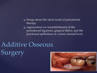
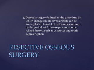
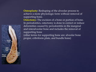
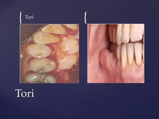
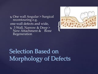
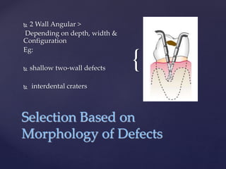
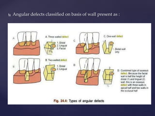
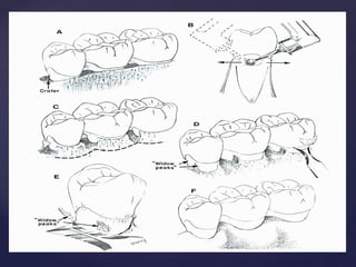
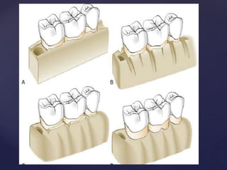
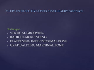
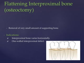
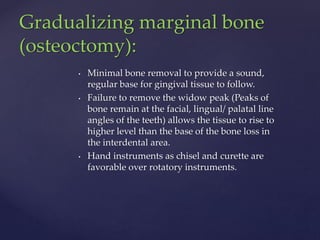
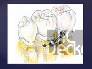
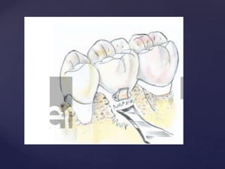
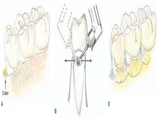
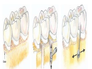
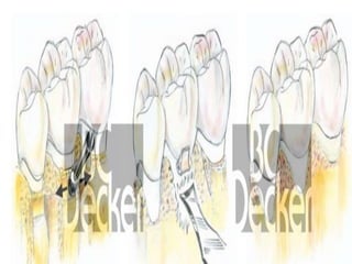
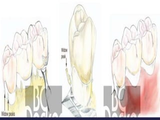
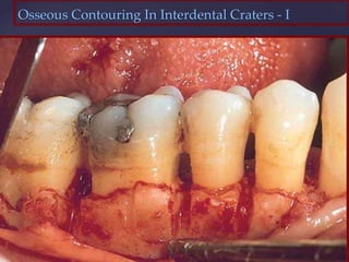
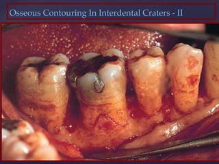
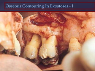
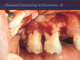
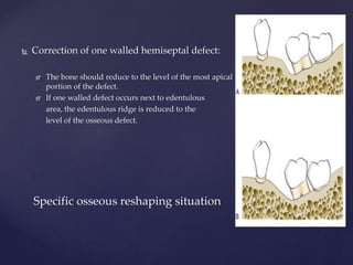
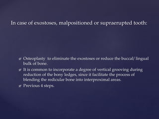
{
Resective osseous
surgery
chapter 60
CARANNZZA 12th
Dr Enas Musbah.
The principles of osseous surgery in
periodontal therapy were outlined by
Schluger (1949) and Goldman (1950).
 They pointed out that alveolar bone loss
caused by inflammatory periodontal
disease often results in an uneven outline
of the bone crest.
 the elimination of soft tissue pockets
often has to be combined with osseous
reshaping and the elimination of osseous
craters and angular bony
Osseous Surgery
 The discrepancies of bone and gingiva
to recurrences of pocket
 That is why we reshape the marginal bone to
resemble alveolar process undamaged by
periodontitis
 Usually we use apically displaced flap to
eliminate pocket and improves tissue contour.
 The end result is improve tissue contour to
provide easy maintainable environment
Rationale
 the more effective the periodontal maintenance
therapy,
the greater is the longitudinal stability of the surgical
result.
 The efficacy of osseous surgery therefore depends
on its ability to
affect pocket depth and to
promote periodontal maintenance for both patent and
the Periodontist.
Rationale ….cont…
{
1) The interproximal bone is
more coronal in position
than the labial or
lingual/palatal bone and
pyramidal in form
Normal Alveolar Bone
Morphology
{
 2. The form of the interdental
bone is depend on the tooth
form and the embrasure width.
 tapered tooth
more pyramidal the bony
 the wider the embrasure
 more flattened is the
interdental bone mesiodistally
and buccolingually.
Normal Alveolar Bone
Morphology…cont..
{
3.The position of the bony
margin mimics the contours of
the cementoenamel junction.
‘scalloping’ depend on root
form,
tooth and position within the
alveolus “dehiscense &
fenestration’
Normal Alveolar Bone
Morphology…cont..
 Osseous Surgery: defied as the
procedure by which changes in the
alveolar bone can be accomplished
to rid it of deformities induced by
the periodontal disease process or
other related factors such as
exostosis and tooth supraeruption.
 can be either additive or subtractive
in nature
Definitions……
 Additive Osseous Surgery eg bone graft
includes procedures directed at restoring the
alveolar bone to it’s original level.
 Subtractive Osseous Surgery
is designed to restore the form of pre-existing
alveolar bone to the level existing at the time of
surgery or slightly more apical to this level.
Additive Or Subtractive
Bone loss has been classified as either:
 Vertical bone loss.
 Horizontal bone loss results in a relative
thickening of marginal alveolar bone.
 Combination.
 brings about the ideal result of periodontal
therapy
 regeneration we reestablishment of the
periodontal ligament, gingival fibers, and the
junctional epithelium at a more coronal level.
Additive Osseous
Surgery
 Osseous surgery defined as :the procedure by
which changes in the alveolar bone can be
accomplished to rid it of deformities induced
by the periodontal disease process or other
related factors, such as exostoses and tooth
supra eruption
RESECTIVE OSSEOUS
SURGERY
Osteoplasty: Reshaping of the alveolar process to
achieve a more physiologic form without removal of
supporting bone.
Ostectomy: The excision of a bone or portion of bone.
In periodontics, ostectomy is done to correct or reduce
deformities caused by periodontitis in the marginal
and interalveolar bone and includes the removal of
supporting bone
(other terms for supporting bone are: alveolar bone
proper, cribriform plate, and bundle bone)
{ {
Tori
Tori
{
 are done when the other
method is not feasible
Subtractive Osseous Surgeries
{
 One wall Angular > Surgical
recontouring e.g.
one-wall defects and wide,
 3 Wall, Narrow & Deep >
New Attachment & Bone
Regeneration
Selection Based on
Morphology of Defects
{
 2 Wall Angular >
Depending on depth, width &
Configuration
Eg:
 shallow two-wall defects
 interdental craters
Selection Based on
Morphology of Defects
 Angular defects classified on basis of wall present as :
{
Terms that describe bone after reshaping:
• Ideal osseous form:
– The bone consistently more coronal on the interproximal surfaces
than on the facial and lingual surfaces. Similar interdental height,
with gradual, curved slops between interdental peaks.
• Flat architecture:
• Positive architecture:
• Negative architecture:
{
The bone consistently
more coronal on the
interproximal surfaces
than on the facial and
lingual surfaces.
Similar interdental
height, with gradual,
curved slops between
interdental peaks.
Ideal osseous form
{
The interdental
bone at the same
level with radicular
bone
Flat architecture
{
The radical bone is
apical to the
Interdental bone.
Positive architecture
{
The interdental
bone is more apical
than the radicular
bone.
Negative architecture
Terms that
describe bone
after reshaping:
Ideal osseous
form
Flat architecture:
Positive
architecture:
Negative
architecture
Terms that relate to the thoroughness of the osseous
reshaping techniques include “definitive” and
“compromise.”
 Definitive osseous reshaping implies that further
osseous reshaping would not improve the overall result.
 Compromise osseous reshaping indicates a bone pattern
that cannot be improved without significant osseous
removal that would be detrimental to the overall result.
 References to compromise and definitive osseous
architecture can be useful to the clinician as terms that
express the expected therapeutic result.
thoroughness of the
osseous reshaping techniques
• Procedure use to correct osseous defect have been classified
into two group:
– Osteoplasty: it is reshaping of bone without removing the
supporting bone.
– Osteoectomy: it is reshaping of bone with removal of supporting
bone.
Terminology
• The technique ostectomy is best applied to patient
with early to moderate bone loss(2-3mm)with
moderate –length root trunks that have bony defect
with one or two wall defect.
• Patient with advanced attachment loss and deep
intrabony defect are not candidate for the
procedure
• Two wall defects, or craters, occur at the expense of
the interseptal bone. As a result they have lingual
and buccal wall that extend from one tooth to the
adjacent tooth, the bacco-lingual contour that result
is opposite to contour of the CEJ of the teeth.
Factor in selection of
resective surgery
{
A and B, Diagram of facial
and interproximal bony
contours after flap
reflection.
Note the loss of some
interproximal bone and
cratering. C and D, Line
angles; this is only
Osteoplasty and has
resulted in a reversed
architecture.
E and F, Ostectomy on the
facial and lingual bone
and the removal of the
residual widow's peaks to
produce a positive bony
architecture
Effect of correction of craters
{
In crater if the facial
and lingual plate of
this bone is resected ,
the resultant
interproximal
contour would
become more
flattened
 If confining resection to ledges and the
interproximal lesion result in facial and lingual
bone form in which the interproximal bone is
located more apically than the bone on the facial or
lingual aspects of the tooth. The result would be
reversed or negative architecture
• Although the reversed architecture minimizes
the amount of ostectomy that is preformed , it
is not without consequences ( widow’s peaks)
facial and lingual line angel (attachment loss)
resorb the peak = pocket
• For the positive architecture ostectomy is
required to remove the widow’s peak as well
as some of the facial , lingual and palatal and
interproximal bone. and this will gives the
topography that resembles normal bone form
before disease .
 Probing and exploration are key aspects of the
examination
 Probing reveals the presence of :
 Pocket depth
 Base of pocket relative to mucogingival
junction and attachment level on the adjecent
teeth.
 Number of bony wall defects.
 The presence of furcation defect.
Examination and treatment
planning
 Trans-gingival probing (sounding):
 Under local anesthesia confirms the extent and
configuration of the intrabony component of the pocket
or furcation defects.
 The probe walks along the tissue-tooth interface to feel
the bony topography.
 The probe may pass horizontally through the tissue to
provide three-dimensional information regarding
bony contours
 Radiograph ( two dimension) cannot accurately
document the number of bony walls and the
presence or extend of bony lesion on the facial
/buccal or lingual/palatal walls.
 Well made radiograph provide useful
information about the extend of interproximal
bone loss, angular bone loss, caries, root trunk
length, and the root morphology.
• After oral hygiene instruction and
debridement , the response of the patient to
these treatment procedures is evaluated by
reexamination and recording the changes in
the periodontium.
• Because the extend of periodontal involvement
may vary from tooth to tooth in the same
patient.
• After resolution of edema and swelling, will
result in return to normal pocket depth and
configuration.
 In patient with moderate to advanced
periodontitis and bony defects, although the
overt sign of periodontitis may be reduced,
may display a persistence of pocket depth
bleeding on probing and suppuration. These
sign may indicate the presence of residual
plaque and calculus inability to instrument
deep pocket or patient unwillingness to
preform adequate oral hygiene
 Osseous resective surgery is also used to
facilitate certain restorative and prosthetic
dental procedure.
 Caries
 Fracture root of abutment teeth can be exposed
for removal.
 Bony exostoses
 Short anatomic crown can be lengthened
{
piezoelectric
surgical
instrument
STEPS IN RESECTIVE
OSSEOUS SURGERY
{ {
Rongeurs Low Speed Burs
{ {
Schluger and Sugarman Back-action chisels
{Ochsenbein chisels
Technique:
• VERTICAL GROOVING
• RADICULAR BLENDING
• FLATTENING INTERPROXIMAL BONE
• GRADUALIZING MARGINAL BONE
STEPS IN RESECTIVE OSSEOUS SURGERY continued
• It is the first step because it can define the general thickness
and subsequent form of alveolar housing.
• It is usually done by rotatory instruments as carbide or
diamond burs.
• it is designed to:
– Reduce the thickness of the alveolar housing.
– Provide relative prominence to the radicular aspect of the teeth.
– Provide continuity from interproximal surface onto the radicular
surface.
• Indications:
– Thick, bony margins, shallow crater formations.
– Areas require maximal osteoplasty and minimal osteoctomy.
• Contraindication:
– Areas with close root proximity or thin alveolar housing.
Vertical grooving (osteoplasty):
- It is an attempt to gradualize the bone over the entire radicular
surface to provide the best results from vertical grooving.
- It provides smooth, blended surface for good flap adaptation.
• Indications:
– Thick ledges of bone on the radicular surface.
• Contraindication:
– Minor vertical grooving or thin, fenestrated radicular bone.
• Both vertical grooving and radicular blending may be used for
treatment of:
– Shallow crater formation.
– Thick osseous ledges of bone in radicular surface.
– Class I and early class II furcation involvement.
Radicular blending (osteoplasty):
– Removal of very small amount of supporting bone.
• Indications:
 Interproximal bone varies horizontally.
 One-walled interproximal defect.
Flattening Interproximal bone
(osteoctomy)
• Minimal bone removal to provide a sound,
regular base for gingival tissue to follow.
• Failure to remove the widow peak (Peaks of
bone remain at the facial, lingual/ palatal line
angles of the teeth) allows the tissue to rise to
higher level than the base of the bone loss in
the interdental area.
• Hand instruments as chisel and curette are
favorable over rotatory instruments.
Gradualizing marginal bone
(osteoctomy):
Osseous Contouring In Interdental Craters - I
Osseous Contouring In Interdental Craters - II
Osseous Contouring In Exostoses - I
Osseous Contouring In Exostoses - II
Osseous Contouring In One Wall Vertical Defect - I
Osseous Contouring In One Wall Vertical Defect – I I
 Correction of one walled hemiseptal defect:
 The bone should reduce to the level of the most apical
portion of the defect.
 If one walled defect occurs next to edentulous
area, the edentulous ridge is reduced to the
level of the osseous defect.
Specific osseous reshaping situation
 Osteoplasty to eliminate the exostoses or reduce the buccal/ lingual
bulk of bone.
 It is common to incorporate a degree of vertical grooving during
reduction of the bony ledges, since it facilitate the process of
blending the redicular bone into interproximal areas.
 Previous 4 steps.
In case of exostoses, malpositioned or supraerupted tooth:
Exostoses – Surgical Planning
 Reduction of interdental walls of craters and
the one-walled component of angular defects
and walls, and grooving into sites of early
involvement.
 The walls of the crater may reduced at the
expense of the buccal, lingual or both walls.
 The reduction should be made to remove the
least amount of alveolar bone required to
produce a satisfactory form, prevent
furcation and blend the contour with
adjacent tooth.
 The selective reduction of bony defects by
ramping the bone to the palatal or lingual to
avoid involvement of the furcations.
In the absence of ledges or exostoses:
Interproximal osseous ramping. A. Presurgical view with 6 mm probing depth on
mesial of first molar. B. Deep two-wall intrabony defect between the second premolar
And first molar, hemiseptal defect between the two premolars and lingual exostosis.
C. Osseous resective surgery eliminated the interproximal osseous defects by ramping
to the lingual, corrected the reversed osseous topography and removed the osseous
ledges. D. Normal scalloped gingival morphology and good health 6 months after
osseous resective surgery
 Replacing the flap in areas that previously had
deep pockets may result initially in greater
postoperative pocket depth, although a
selective recession may diminish the depth
over time. minimizes postoperative
complications
 Positioning the flap apically to expose marginal
bone is results in more postsurgical resorption
of bone and patient discomfort
Flap Placement and Closure
 Suturing may be accomplished using a variety
of different suture materials and suture knots
Thanks
Pre-Surgical Photograph Post Surgical – 1Year
7. Remodeling Resective Osseous Surgery.pptx

7. Remodeling Resective Osseous Surgery.pptx

  • 1.
  • 2.
    The principles ofosseous surgery in periodontal therapy were outlined by Schluger (1949) and Goldman (1950).  They pointed out that alveolar bone loss caused by inflammatory periodontal disease often results in an uneven outline of the bone crest.  the elimination of soft tissue pockets often has to be combined with osseous reshaping and the elimination of osseous craters and angular bony Osseous Surgery
  • 3.
     The discrepanciesof bone and gingiva to recurrences of pocket  That is why we reshape the marginal bone to resemble alveolar process undamaged by periodontitis  Usually we use apically displaced flap to eliminate pocket and improves tissue contour.  The end result is improve tissue contour to provide easy maintainable environment Rationale
  • 4.
     the moreeffective the periodontal maintenance therapy, the greater is the longitudinal stability of the surgical result.  The efficacy of osseous surgery therefore depends on its ability to affect pocket depth and to promote periodontal maintenance for both patent and the Periodontist. Rationale ….cont…
  • 5.
    { 1) The interproximalbone is more coronal in position than the labial or lingual/palatal bone and pyramidal in form Normal Alveolar Bone Morphology
  • 6.
    {  2. Theform of the interdental bone is depend on the tooth form and the embrasure width.  tapered tooth more pyramidal the bony  the wider the embrasure  more flattened is the interdental bone mesiodistally and buccolingually. Normal Alveolar Bone Morphology…cont..
  • 7.
    { 3.The position ofthe bony margin mimics the contours of the cementoenamel junction. ‘scalloping’ depend on root form, tooth and position within the alveolus “dehiscense & fenestration’ Normal Alveolar Bone Morphology…cont..
  • 9.
     Osseous Surgery:defied as the procedure by which changes in the alveolar bone can be accomplished to rid it of deformities induced by the periodontal disease process or other related factors such as exostosis and tooth supraeruption.  can be either additive or subtractive in nature Definitions……
  • 10.
     Additive OsseousSurgery eg bone graft includes procedures directed at restoring the alveolar bone to it’s original level.  Subtractive Osseous Surgery is designed to restore the form of pre-existing alveolar bone to the level existing at the time of surgery or slightly more apical to this level. Additive Or Subtractive
  • 11.
    Bone loss hasbeen classified as either:  Vertical bone loss.  Horizontal bone loss results in a relative thickening of marginal alveolar bone.  Combination.
  • 12.
     brings aboutthe ideal result of periodontal therapy  regeneration we reestablishment of the periodontal ligament, gingival fibers, and the junctional epithelium at a more coronal level. Additive Osseous Surgery
  • 14.
     Osseous surgerydefined as :the procedure by which changes in the alveolar bone can be accomplished to rid it of deformities induced by the periodontal disease process or other related factors, such as exostoses and tooth supra eruption RESECTIVE OSSEOUS SURGERY
  • 15.
    Osteoplasty: Reshaping ofthe alveolar process to achieve a more physiologic form without removal of supporting bone. Ostectomy: The excision of a bone or portion of bone. In periodontics, ostectomy is done to correct or reduce deformities caused by periodontitis in the marginal and interalveolar bone and includes the removal of supporting bone (other terms for supporting bone are: alveolar bone proper, cribriform plate, and bundle bone)
  • 16.
  • 17.
    {  are donewhen the other method is not feasible Subtractive Osseous Surgeries
  • 21.
    {  One wallAngular > Surgical recontouring e.g. one-wall defects and wide,  3 Wall, Narrow & Deep > New Attachment & Bone Regeneration Selection Based on Morphology of Defects
  • 22.
    {  2 WallAngular > Depending on depth, width & Configuration Eg:  shallow two-wall defects  interdental craters Selection Based on Morphology of Defects
  • 23.
     Angular defectsclassified on basis of wall present as :
  • 24.
  • 26.
    Terms that describebone after reshaping: • Ideal osseous form: – The bone consistently more coronal on the interproximal surfaces than on the facial and lingual surfaces. Similar interdental height, with gradual, curved slops between interdental peaks. • Flat architecture: • Positive architecture: • Negative architecture:
  • 27.
    { The bone consistently morecoronal on the interproximal surfaces than on the facial and lingual surfaces. Similar interdental height, with gradual, curved slops between interdental peaks. Ideal osseous form
  • 28.
    { The interdental bone atthe same level with radicular bone Flat architecture
  • 29.
    { The radical boneis apical to the Interdental bone. Positive architecture
  • 30.
    { The interdental bone ismore apical than the radicular bone. Negative architecture
  • 31.
    Terms that describe bone afterreshaping: Ideal osseous form Flat architecture: Positive architecture: Negative architecture
  • 33.
    Terms that relateto the thoroughness of the osseous reshaping techniques include “definitive” and “compromise.”  Definitive osseous reshaping implies that further osseous reshaping would not improve the overall result.  Compromise osseous reshaping indicates a bone pattern that cannot be improved without significant osseous removal that would be detrimental to the overall result.  References to compromise and definitive osseous architecture can be useful to the clinician as terms that express the expected therapeutic result. thoroughness of the osseous reshaping techniques
  • 34.
    • Procedure useto correct osseous defect have been classified into two group: – Osteoplasty: it is reshaping of bone without removing the supporting bone. – Osteoectomy: it is reshaping of bone with removal of supporting bone. Terminology
  • 35.
    • The techniqueostectomy is best applied to patient with early to moderate bone loss(2-3mm)with moderate –length root trunks that have bony defect with one or two wall defect. • Patient with advanced attachment loss and deep intrabony defect are not candidate for the procedure • Two wall defects, or craters, occur at the expense of the interseptal bone. As a result they have lingual and buccal wall that extend from one tooth to the adjacent tooth, the bacco-lingual contour that result is opposite to contour of the CEJ of the teeth. Factor in selection of resective surgery
  • 42.
    { A and B,Diagram of facial and interproximal bony contours after flap reflection. Note the loss of some interproximal bone and cratering. C and D, Line angles; this is only Osteoplasty and has resulted in a reversed architecture. E and F, Ostectomy on the facial and lingual bone and the removal of the residual widow's peaks to produce a positive bony architecture Effect of correction of craters
  • 43.
    { In crater ifthe facial and lingual plate of this bone is resected , the resultant interproximal contour would become more flattened
  • 44.
     If confiningresection to ledges and the interproximal lesion result in facial and lingual bone form in which the interproximal bone is located more apically than the bone on the facial or lingual aspects of the tooth. The result would be reversed or negative architecture
  • 47.
    • Although thereversed architecture minimizes the amount of ostectomy that is preformed , it is not without consequences ( widow’s peaks) facial and lingual line angel (attachment loss) resorb the peak = pocket • For the positive architecture ostectomy is required to remove the widow’s peak as well as some of the facial , lingual and palatal and interproximal bone. and this will gives the topography that resembles normal bone form before disease .
  • 49.
     Probing andexploration are key aspects of the examination  Probing reveals the presence of :  Pocket depth  Base of pocket relative to mucogingival junction and attachment level on the adjecent teeth.  Number of bony wall defects.  The presence of furcation defect. Examination and treatment planning
  • 50.
     Trans-gingival probing(sounding):  Under local anesthesia confirms the extent and configuration of the intrabony component of the pocket or furcation defects.  The probe walks along the tissue-tooth interface to feel the bony topography.  The probe may pass horizontally through the tissue to provide three-dimensional information regarding bony contours
  • 51.
     Radiograph (two dimension) cannot accurately document the number of bony walls and the presence or extend of bony lesion on the facial /buccal or lingual/palatal walls.  Well made radiograph provide useful information about the extend of interproximal bone loss, angular bone loss, caries, root trunk length, and the root morphology.
  • 52.
    • After oralhygiene instruction and debridement , the response of the patient to these treatment procedures is evaluated by reexamination and recording the changes in the periodontium. • Because the extend of periodontal involvement may vary from tooth to tooth in the same patient. • After resolution of edema and swelling, will result in return to normal pocket depth and configuration.
  • 53.
     In patientwith moderate to advanced periodontitis and bony defects, although the overt sign of periodontitis may be reduced, may display a persistence of pocket depth bleeding on probing and suppuration. These sign may indicate the presence of residual plaque and calculus inability to instrument deep pocket or patient unwillingness to preform adequate oral hygiene
  • 55.
     Osseous resectivesurgery is also used to facilitate certain restorative and prosthetic dental procedure.  Caries  Fracture root of abutment teeth can be exposed for removal.  Bony exostoses  Short anatomic crown can be lengthened
  • 56.
  • 57.
    { { Rongeurs LowSpeed Burs
  • 58.
    { { Schluger andSugarman Back-action chisels
  • 59.
  • 60.
    Technique: • VERTICAL GROOVING •RADICULAR BLENDING • FLATTENING INTERPROXIMAL BONE • GRADUALIZING MARGINAL BONE STEPS IN RESECTIVE OSSEOUS SURGERY continued
  • 61.
    • It isthe first step because it can define the general thickness and subsequent form of alveolar housing. • It is usually done by rotatory instruments as carbide or diamond burs. • it is designed to: – Reduce the thickness of the alveolar housing. – Provide relative prominence to the radicular aspect of the teeth. – Provide continuity from interproximal surface onto the radicular surface. • Indications: – Thick, bony margins, shallow crater formations. – Areas require maximal osteoplasty and minimal osteoctomy. • Contraindication: – Areas with close root proximity or thin alveolar housing. Vertical grooving (osteoplasty):
  • 63.
    - It isan attempt to gradualize the bone over the entire radicular surface to provide the best results from vertical grooving. - It provides smooth, blended surface for good flap adaptation. • Indications: – Thick ledges of bone on the radicular surface. • Contraindication: – Minor vertical grooving or thin, fenestrated radicular bone. • Both vertical grooving and radicular blending may be used for treatment of: – Shallow crater formation. – Thick osseous ledges of bone in radicular surface. – Class I and early class II furcation involvement. Radicular blending (osteoplasty):
  • 65.
    – Removal ofvery small amount of supporting bone. • Indications:  Interproximal bone varies horizontally.  One-walled interproximal defect. Flattening Interproximal bone (osteoctomy)
  • 66.
    • Minimal boneremoval to provide a sound, regular base for gingival tissue to follow. • Failure to remove the widow peak (Peaks of bone remain at the facial, lingual/ palatal line angles of the teeth) allows the tissue to rise to higher level than the base of the bone loss in the interdental area. • Hand instruments as chisel and curette are favorable over rotatory instruments. Gradualizing marginal bone (osteoctomy):
  • 74.
    Osseous Contouring InInterdental Craters - I
  • 75.
    Osseous Contouring InInterdental Craters - II
  • 76.
    Osseous Contouring InExostoses - I
  • 77.
    Osseous Contouring InExostoses - II
  • 78.
    Osseous Contouring InOne Wall Vertical Defect - I
  • 79.
    Osseous Contouring InOne Wall Vertical Defect – I I
  • 80.
     Correction ofone walled hemiseptal defect:  The bone should reduce to the level of the most apical portion of the defect.  If one walled defect occurs next to edentulous area, the edentulous ridge is reduced to the level of the osseous defect. Specific osseous reshaping situation
  • 81.
     Osteoplasty toeliminate the exostoses or reduce the buccal/ lingual bulk of bone.  It is common to incorporate a degree of vertical grooving during reduction of the bony ledges, since it facilitate the process of blending the redicular bone into interproximal areas.  Previous 4 steps. In case of exostoses, malpositioned or supraerupted tooth:
  • 83.
  • 84.
     Reduction ofinterdental walls of craters and the one-walled component of angular defects and walls, and grooving into sites of early involvement.  The walls of the crater may reduced at the expense of the buccal, lingual or both walls.  The reduction should be made to remove the least amount of alveolar bone required to produce a satisfactory form, prevent furcation and blend the contour with adjacent tooth.  The selective reduction of bony defects by ramping the bone to the palatal or lingual to avoid involvement of the furcations. In the absence of ledges or exostoses:
  • 85.
    Interproximal osseous ramping.A. Presurgical view with 6 mm probing depth on mesial of first molar. B. Deep two-wall intrabony defect between the second premolar And first molar, hemiseptal defect between the two premolars and lingual exostosis. C. Osseous resective surgery eliminated the interproximal osseous defects by ramping to the lingual, corrected the reversed osseous topography and removed the osseous ledges. D. Normal scalloped gingival morphology and good health 6 months after osseous resective surgery
  • 86.
     Replacing theflap in areas that previously had deep pockets may result initially in greater postoperative pocket depth, although a selective recession may diminish the depth over time. minimizes postoperative complications  Positioning the flap apically to expose marginal bone is results in more postsurgical resorption of bone and patient discomfort Flap Placement and Closure
  • 87.
     Suturing maybe accomplished using a variety of different suture materials and suture knots
  • 88.

Editor's Notes

  • #81 Healing remodling in 6 month,,,attachment of flap to gingiva in 21 days