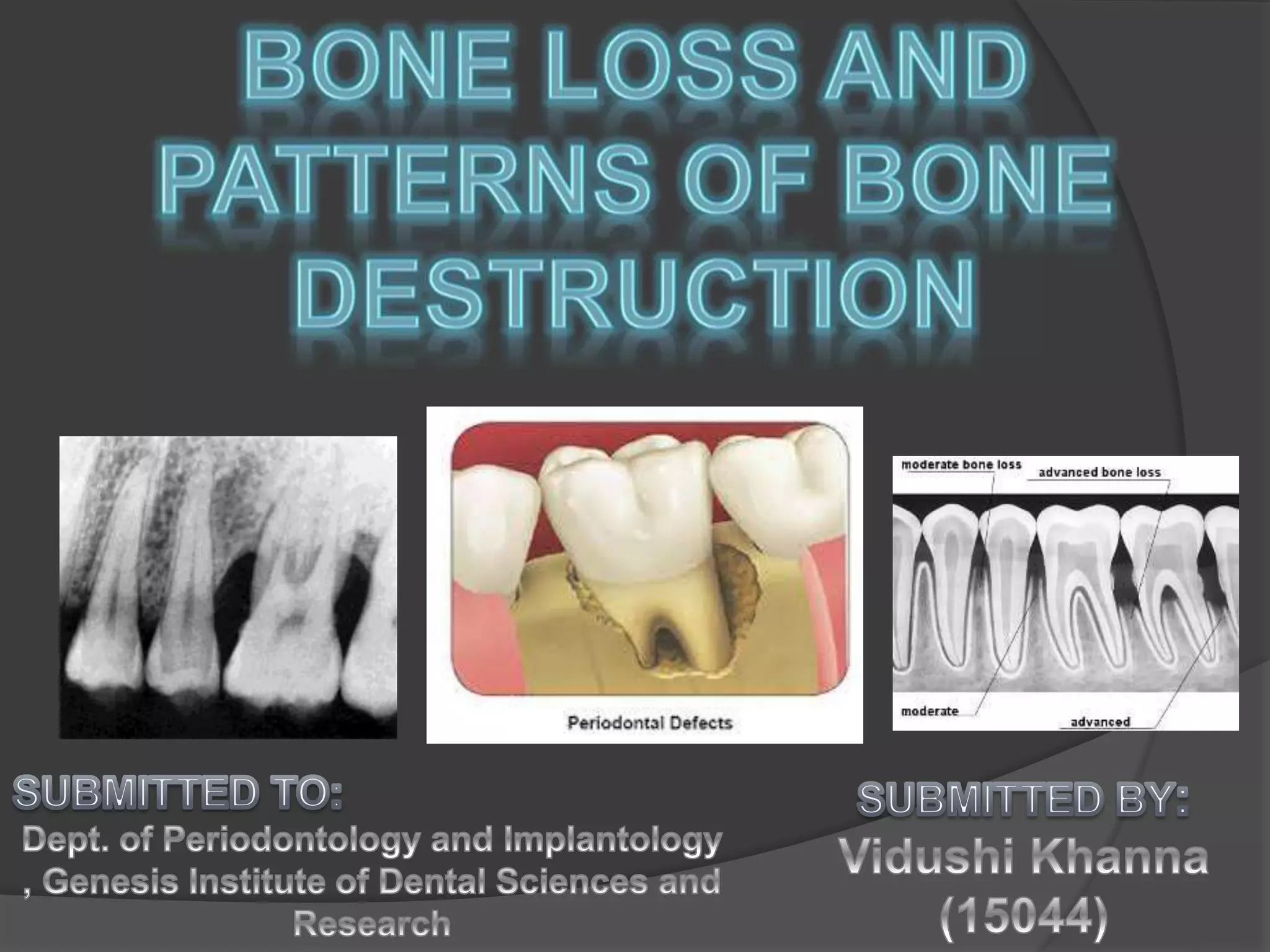
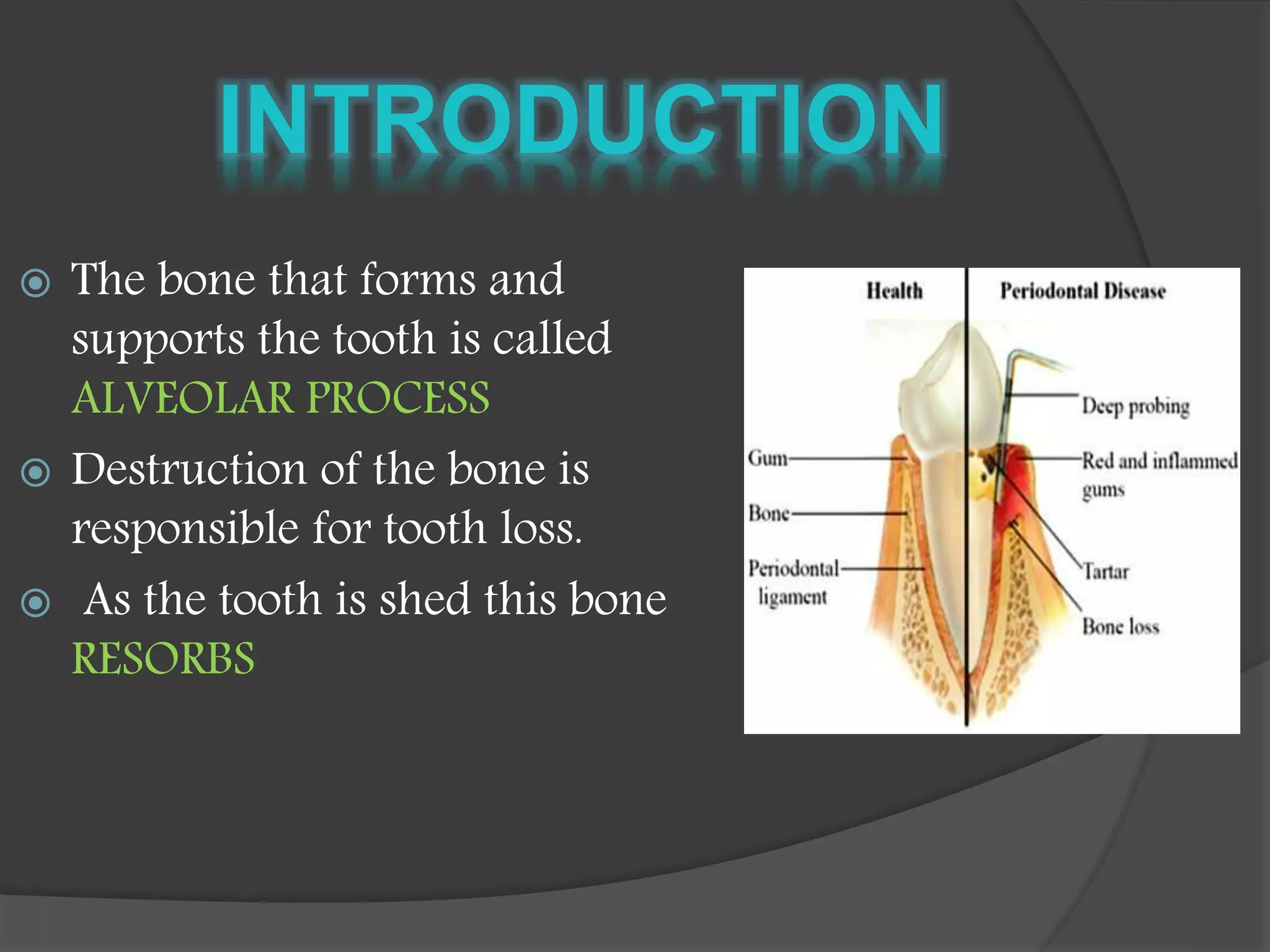
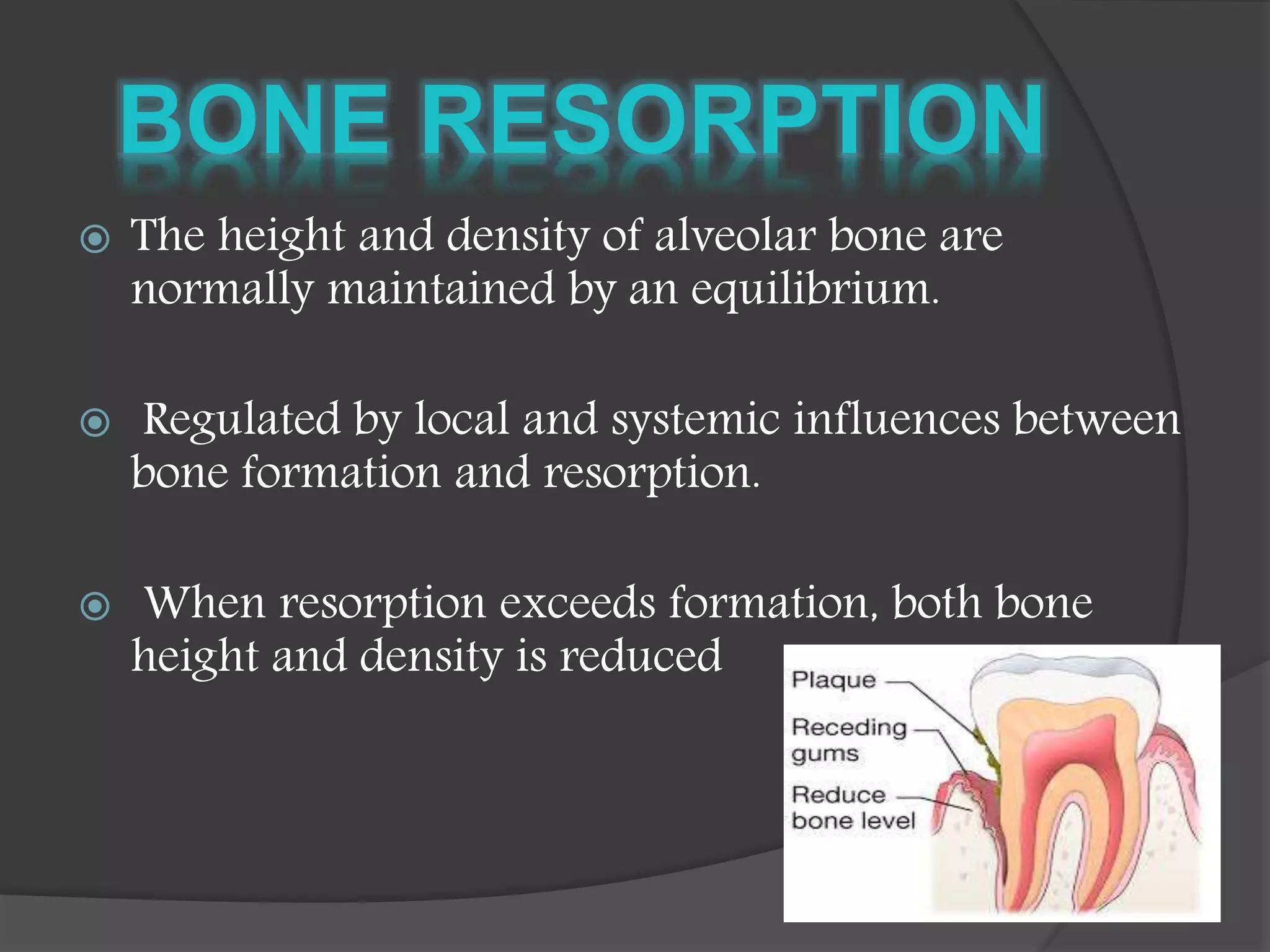
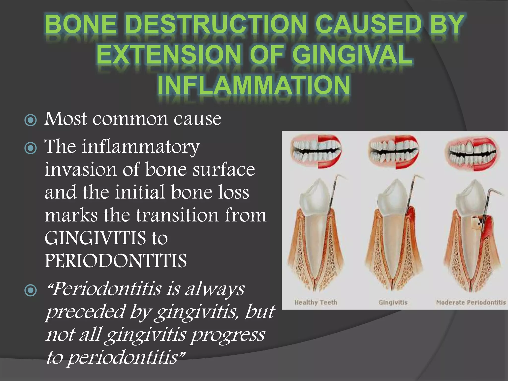
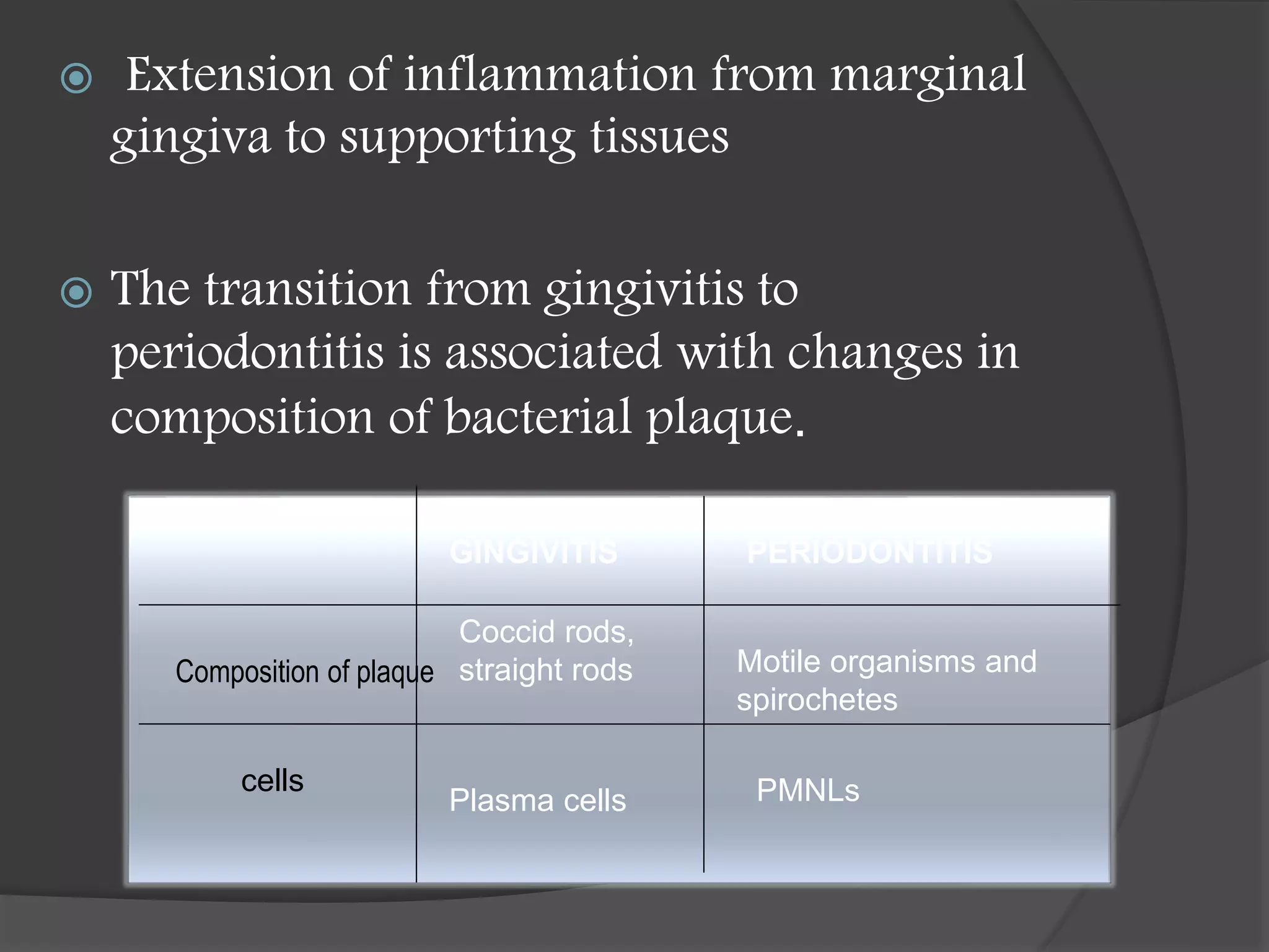
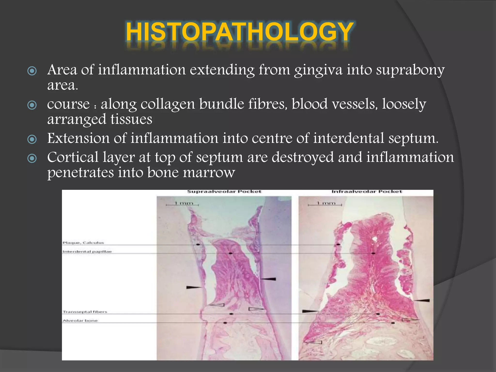
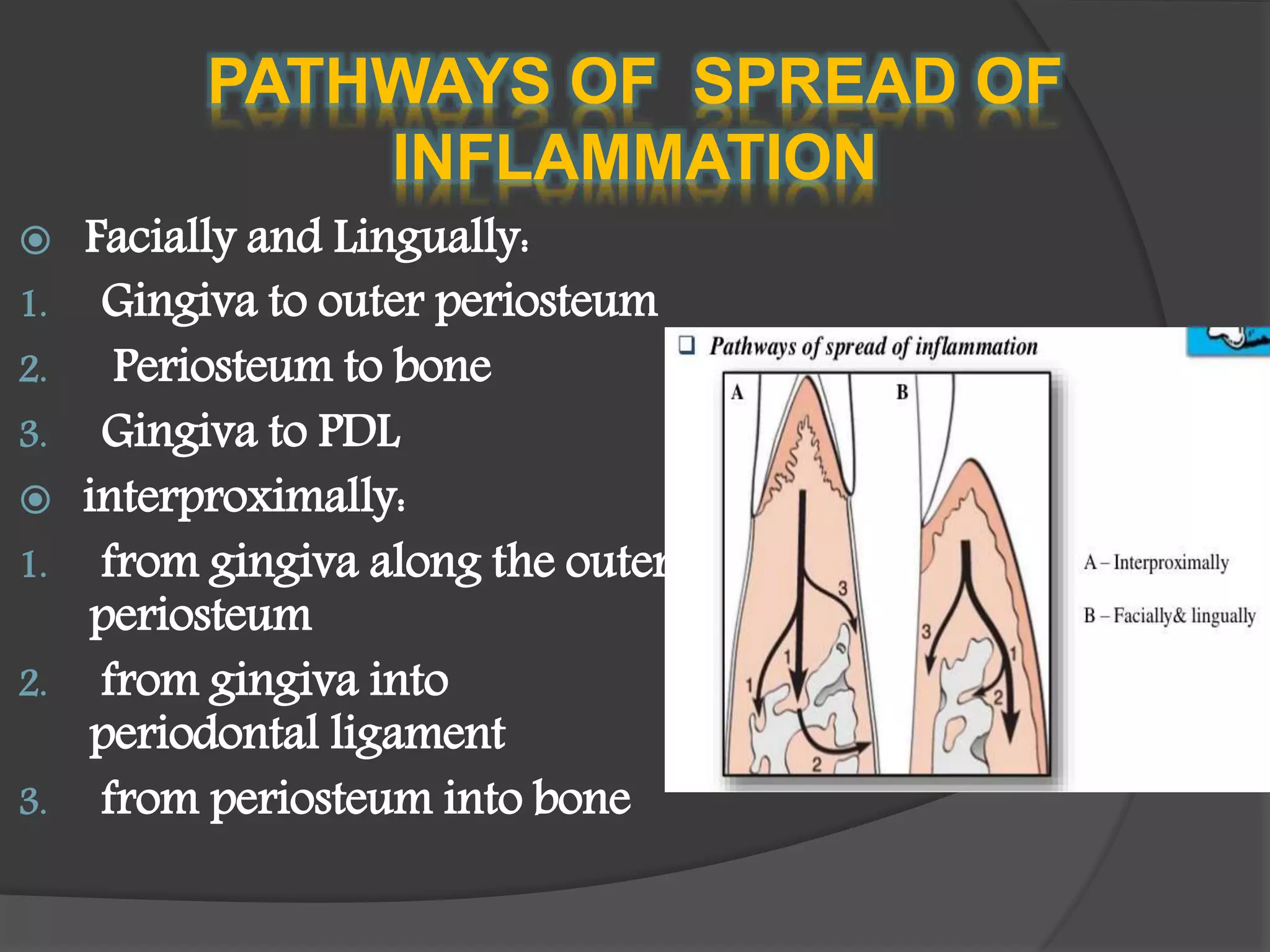
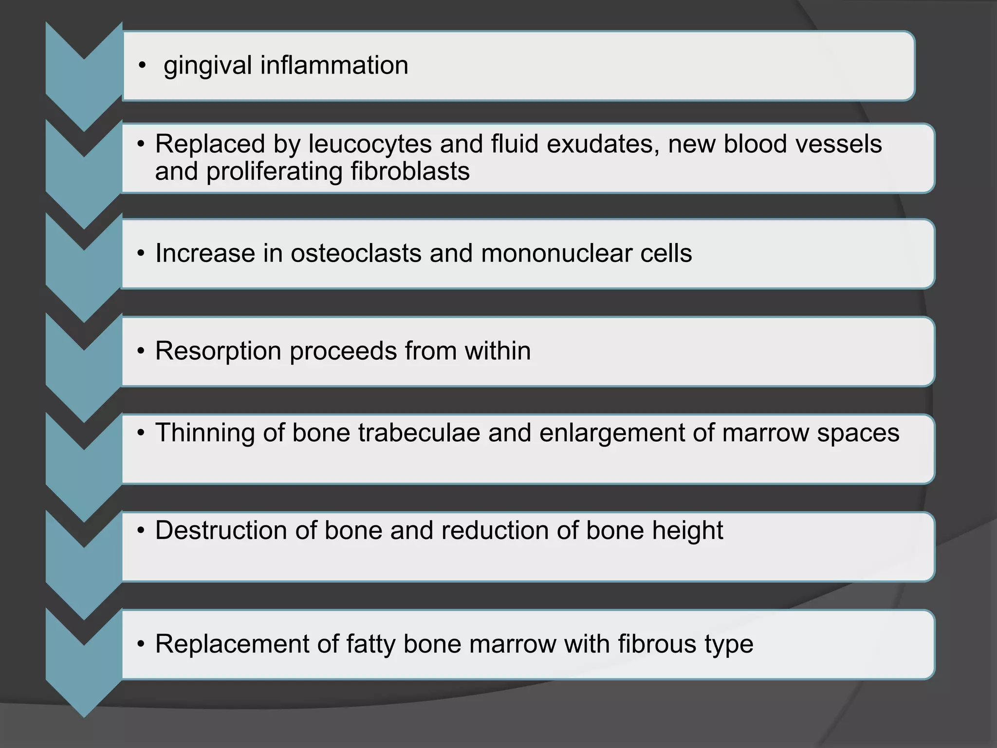
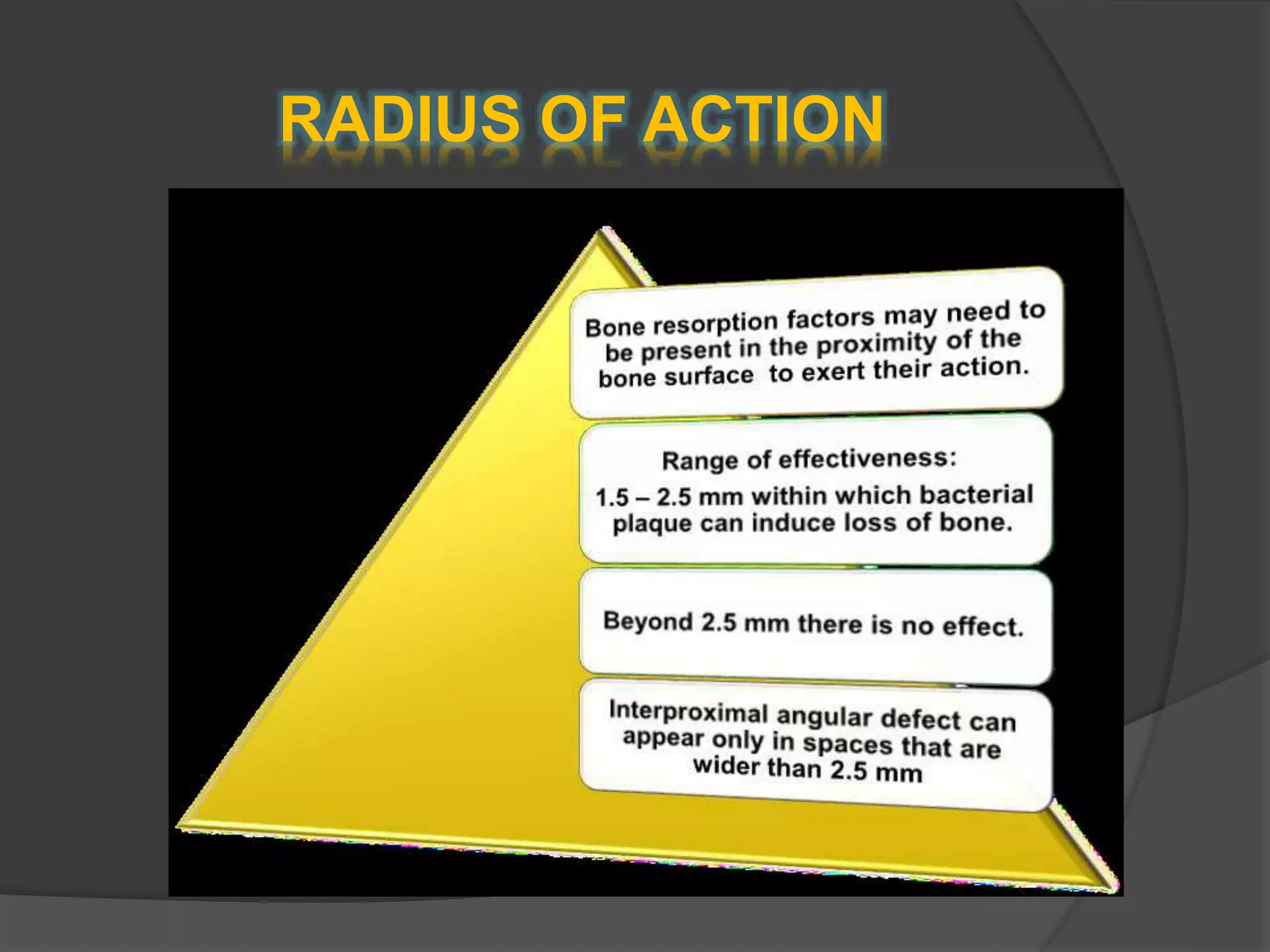
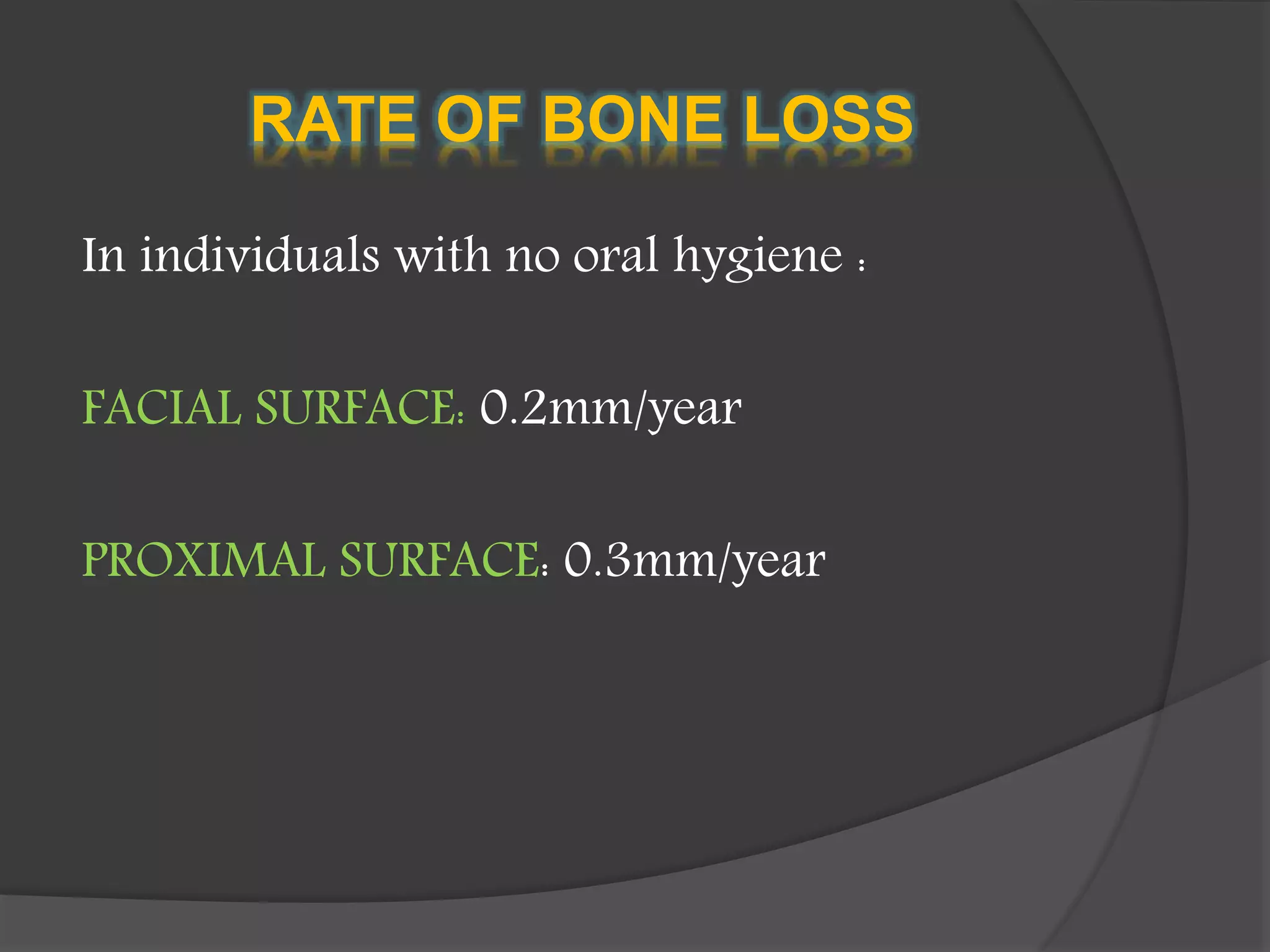
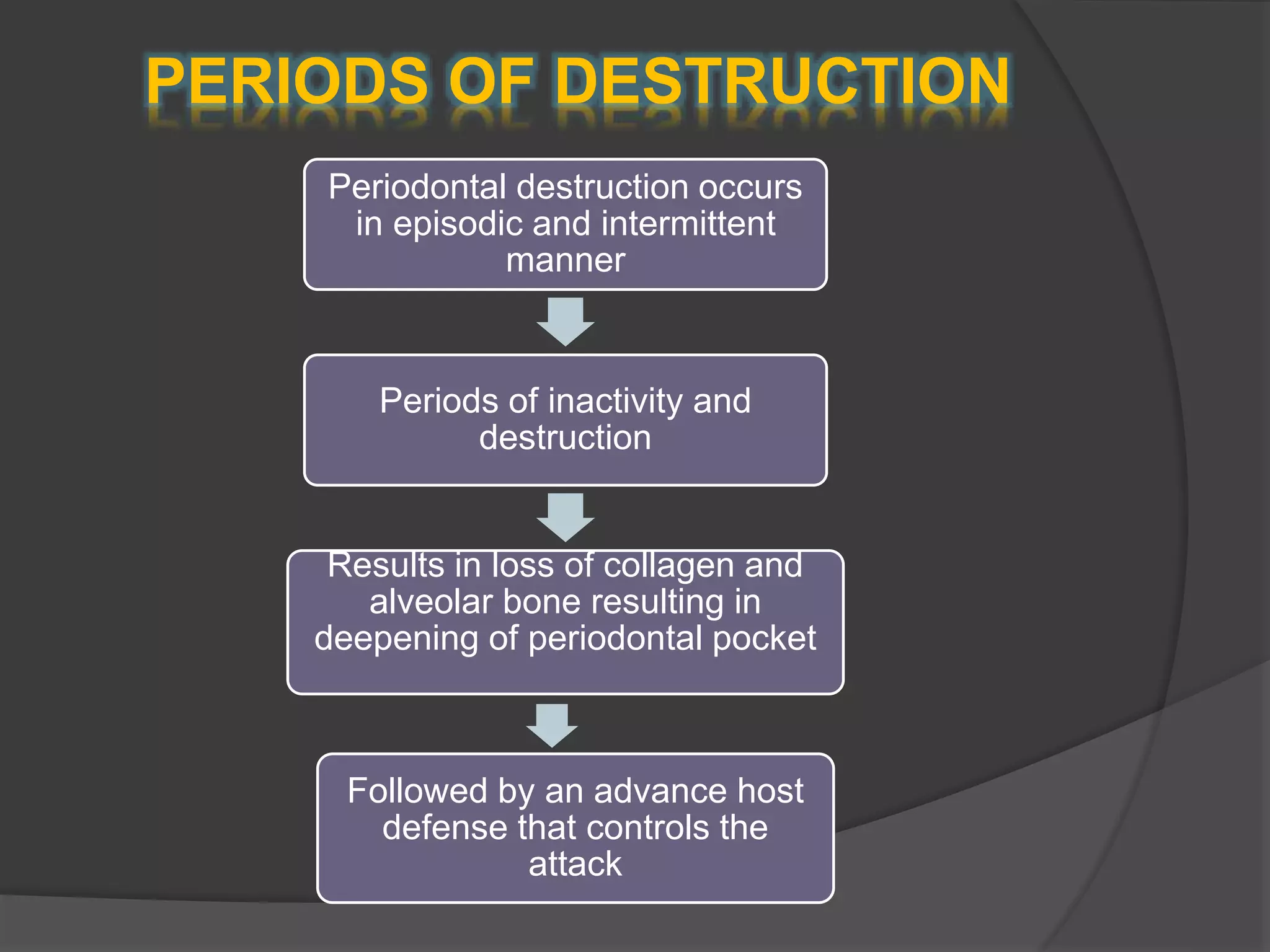
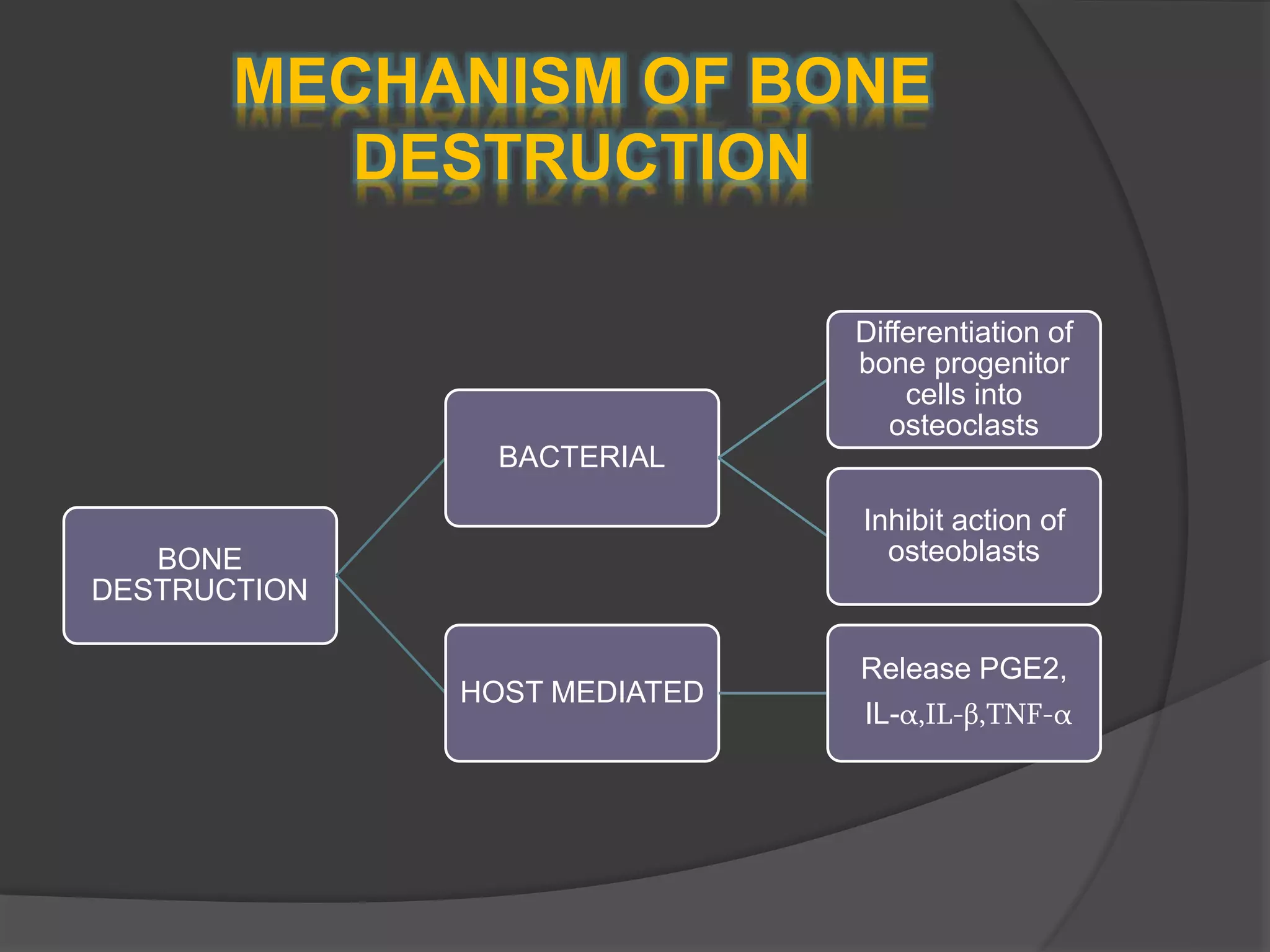
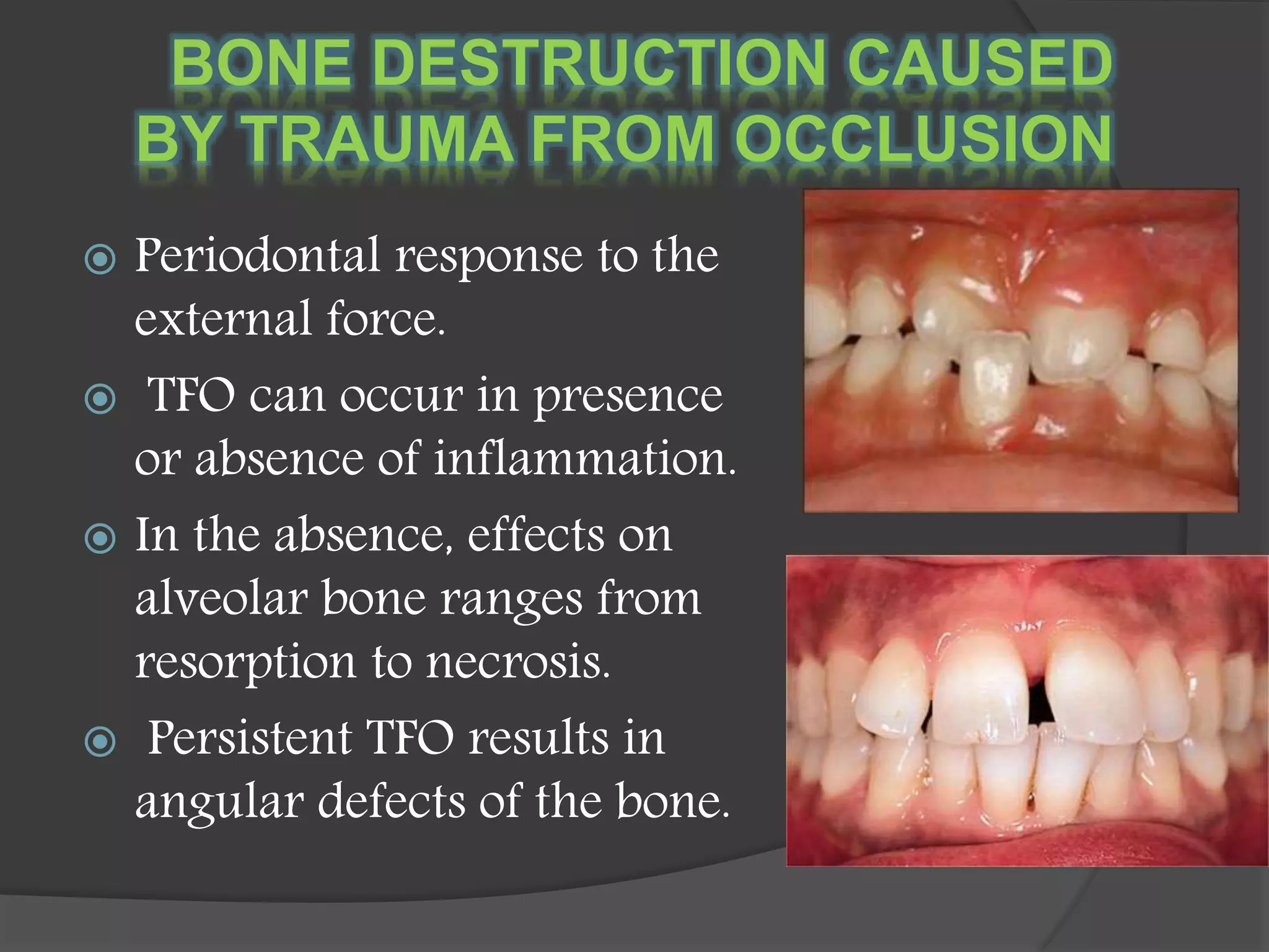
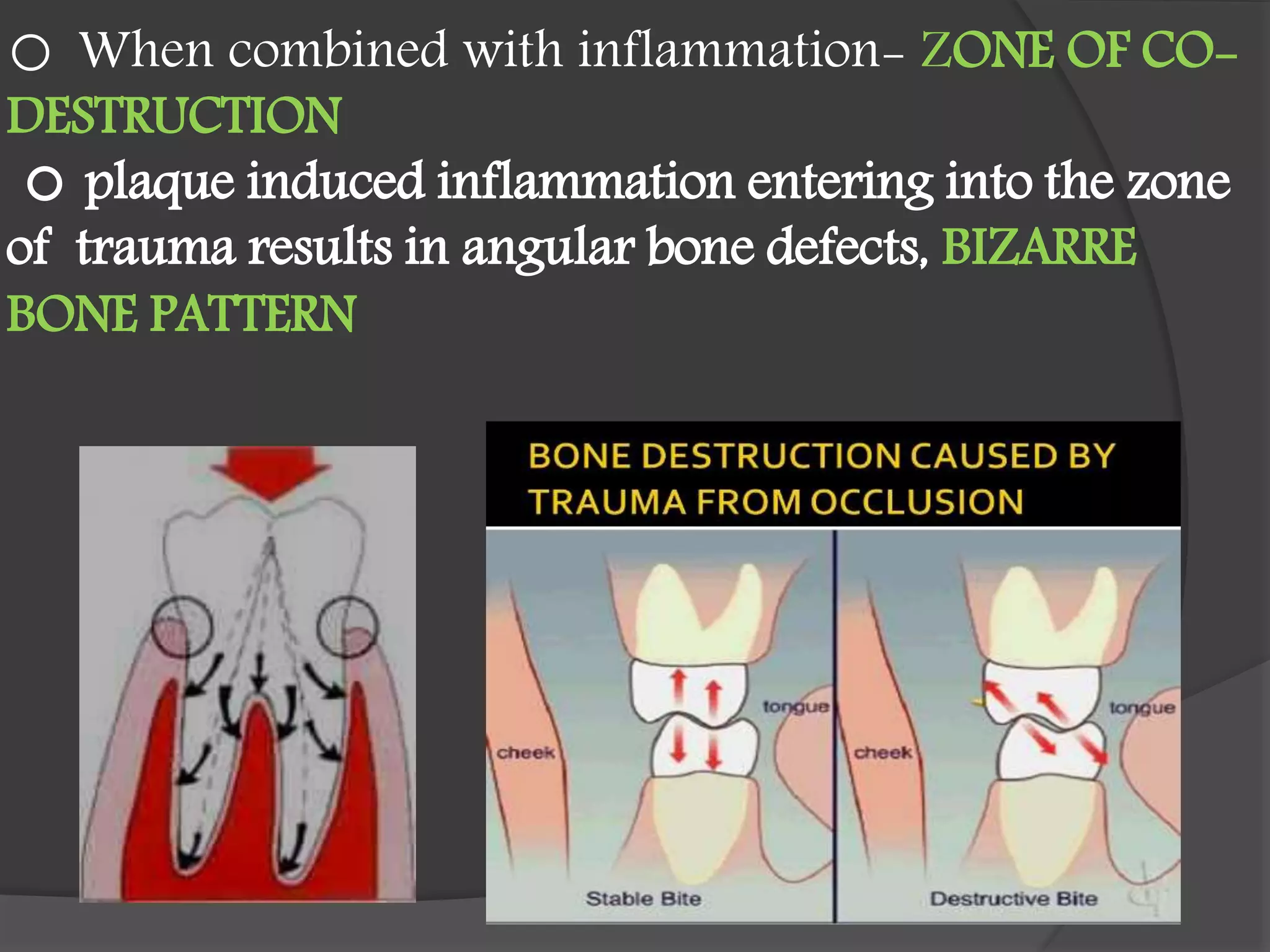
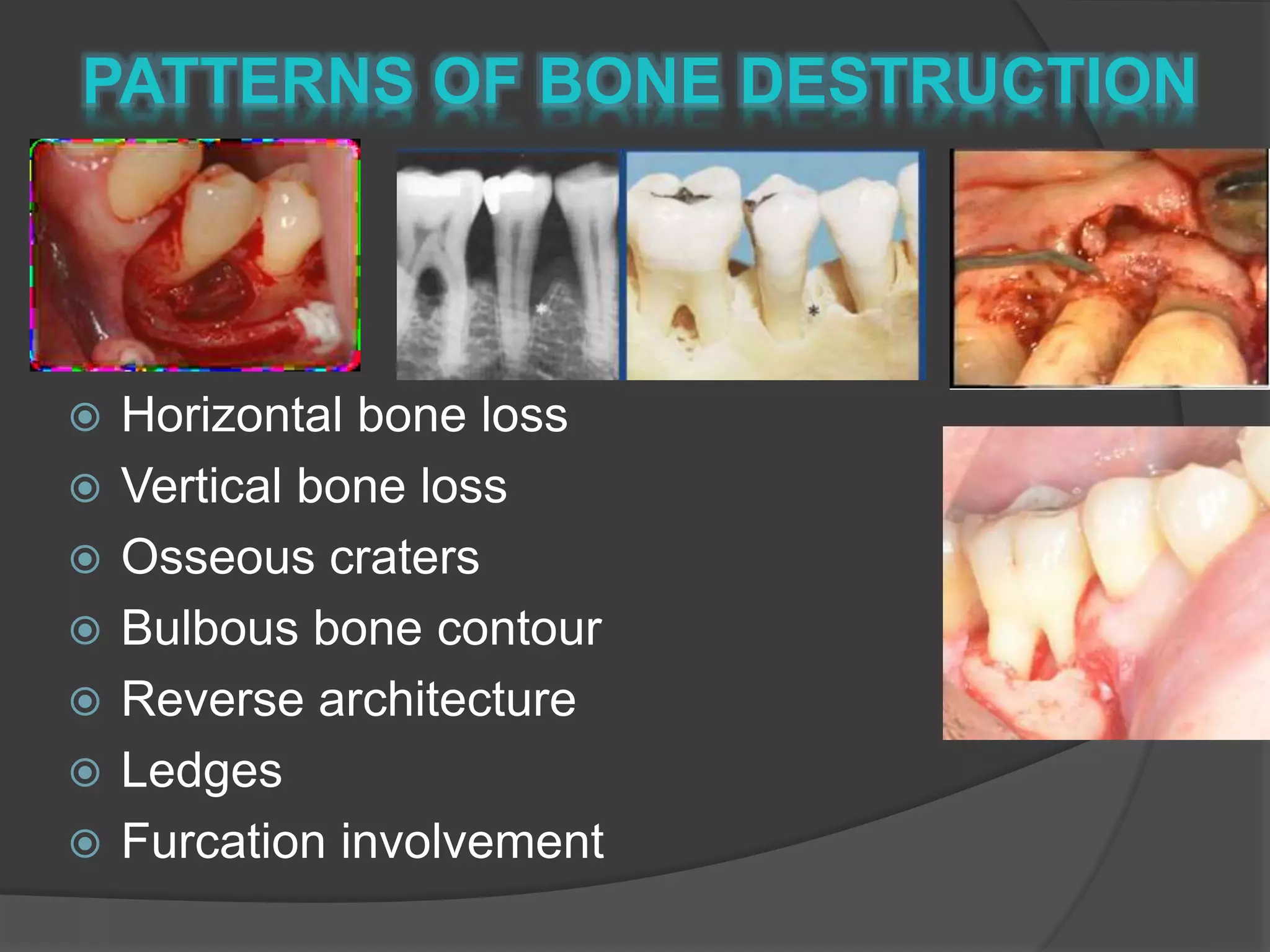
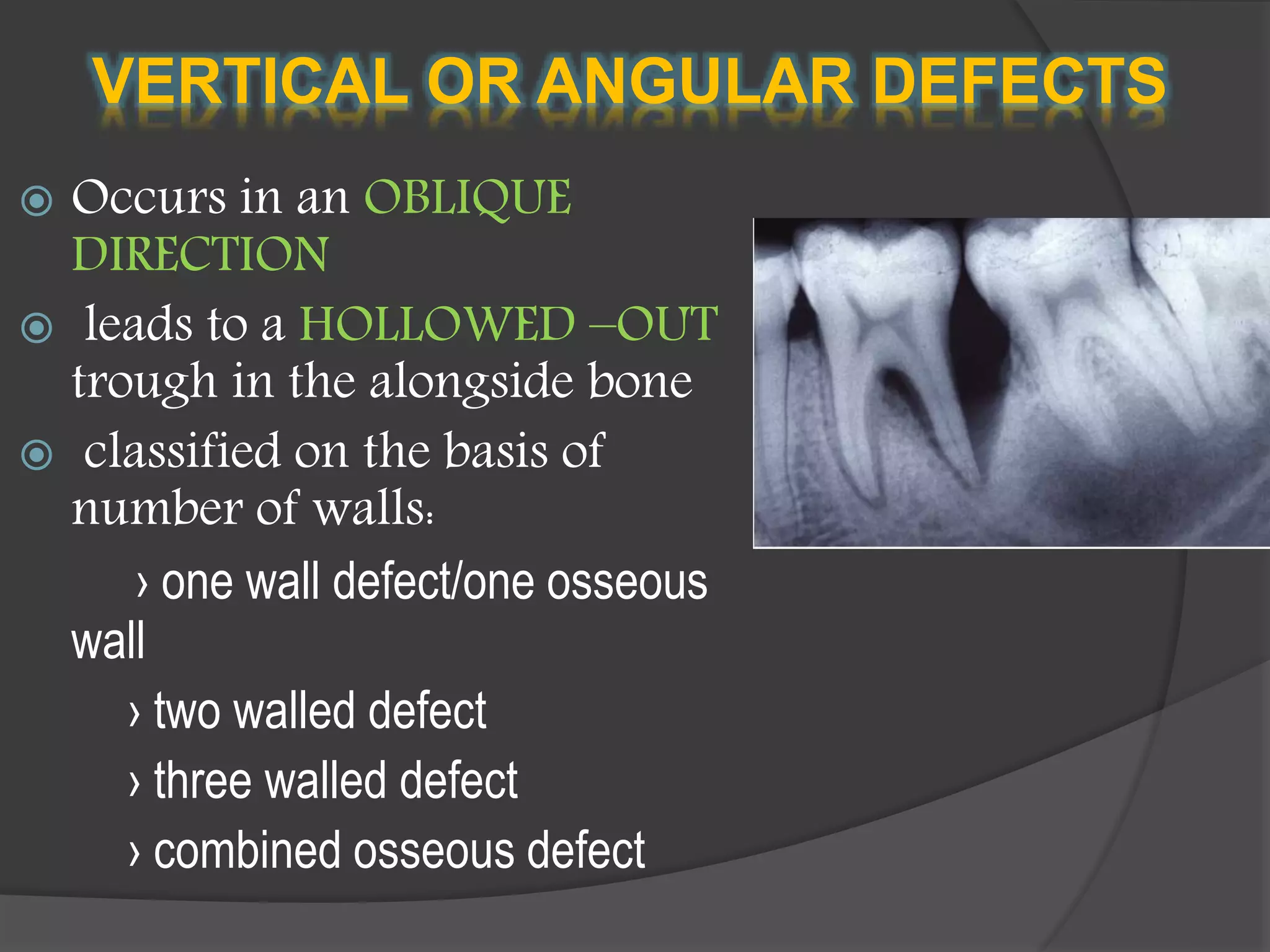
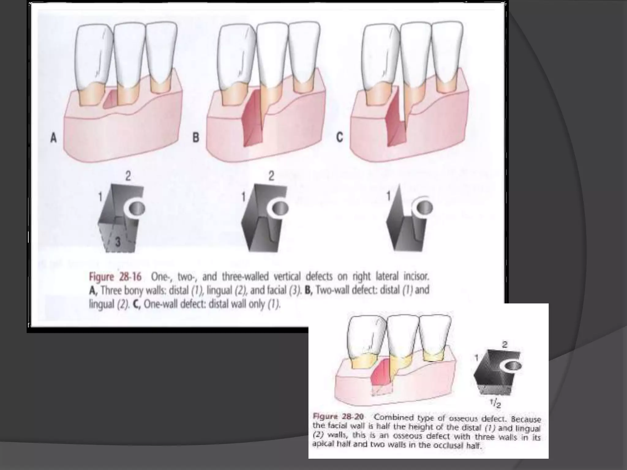
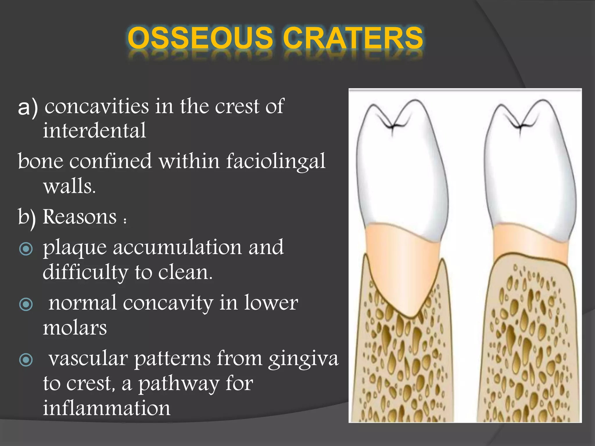
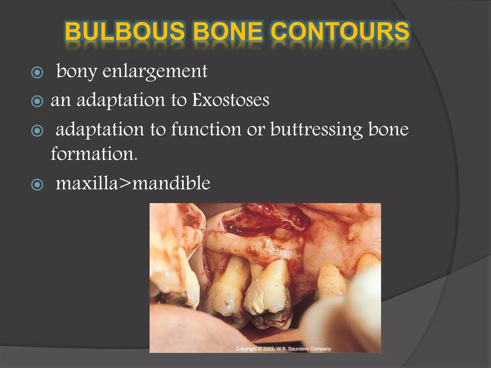
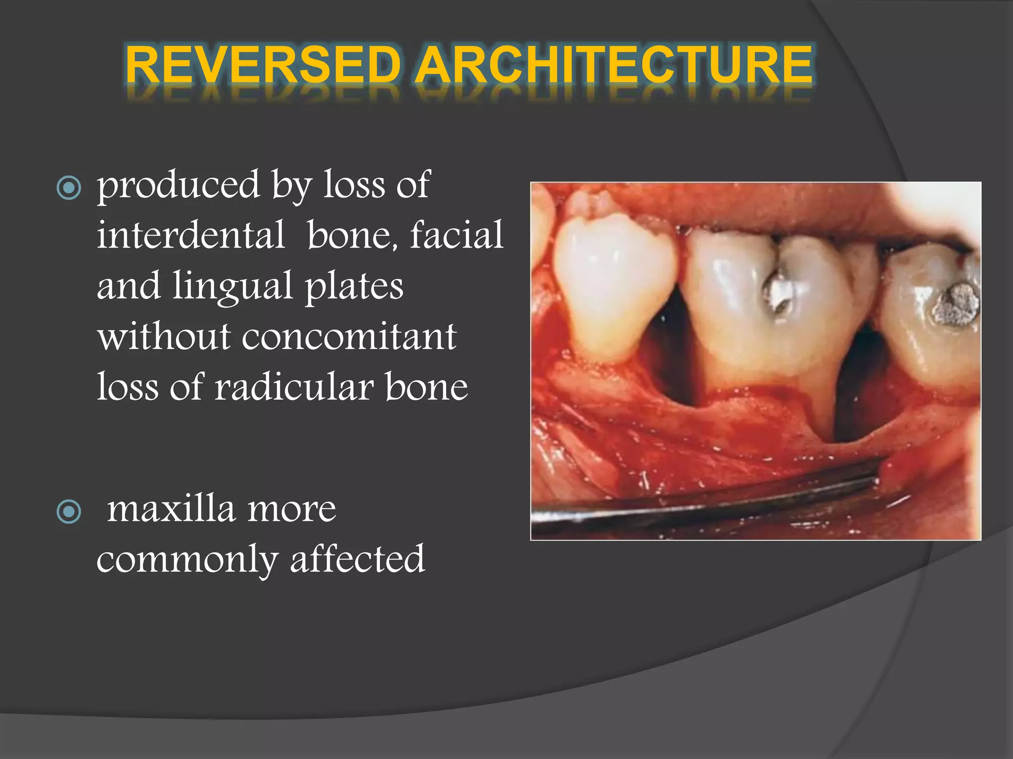
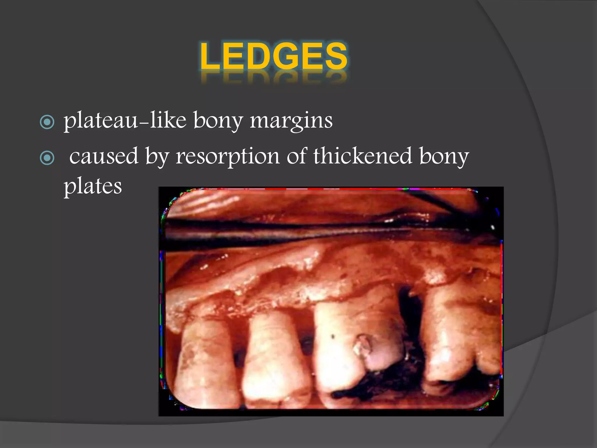
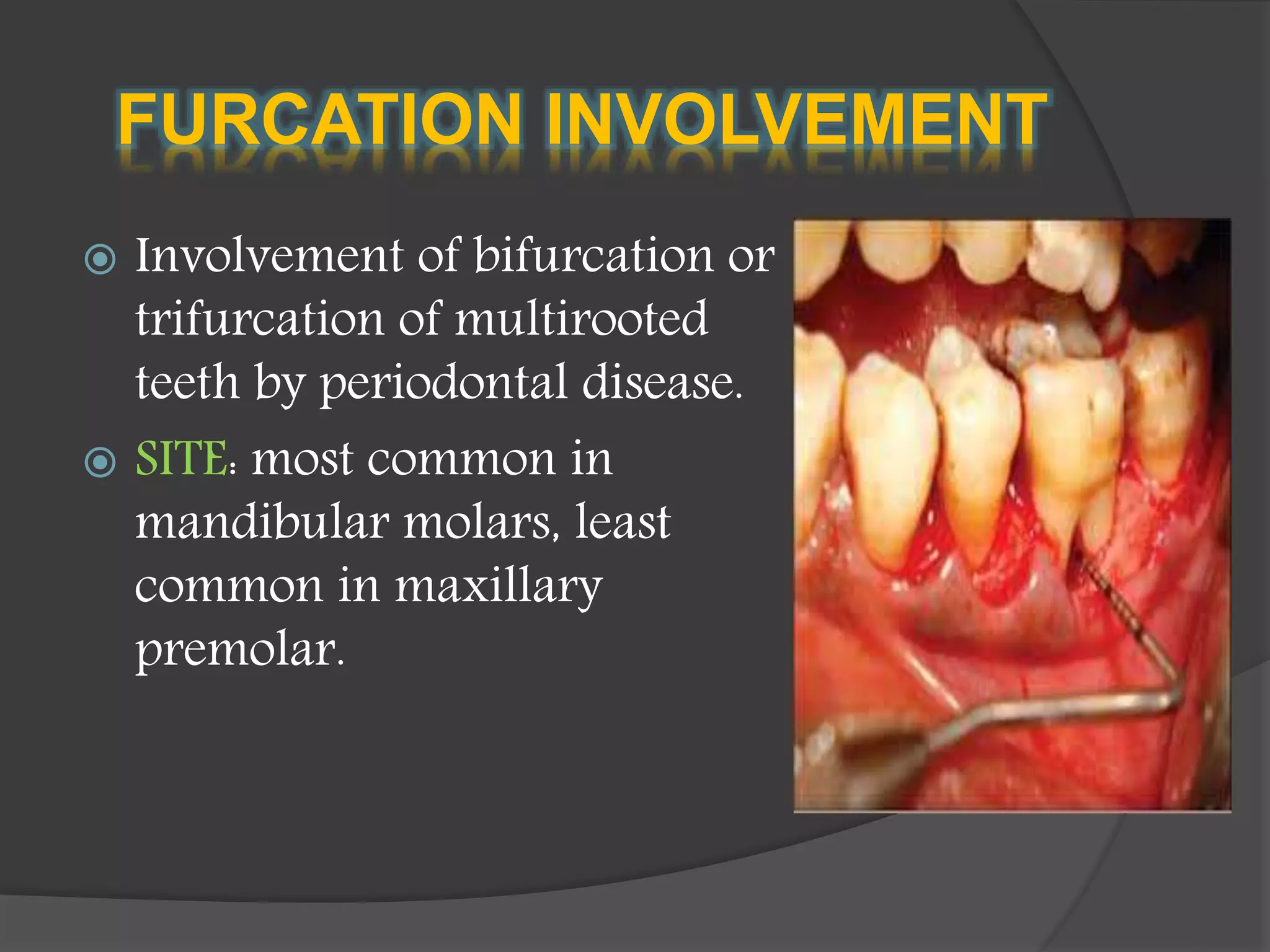
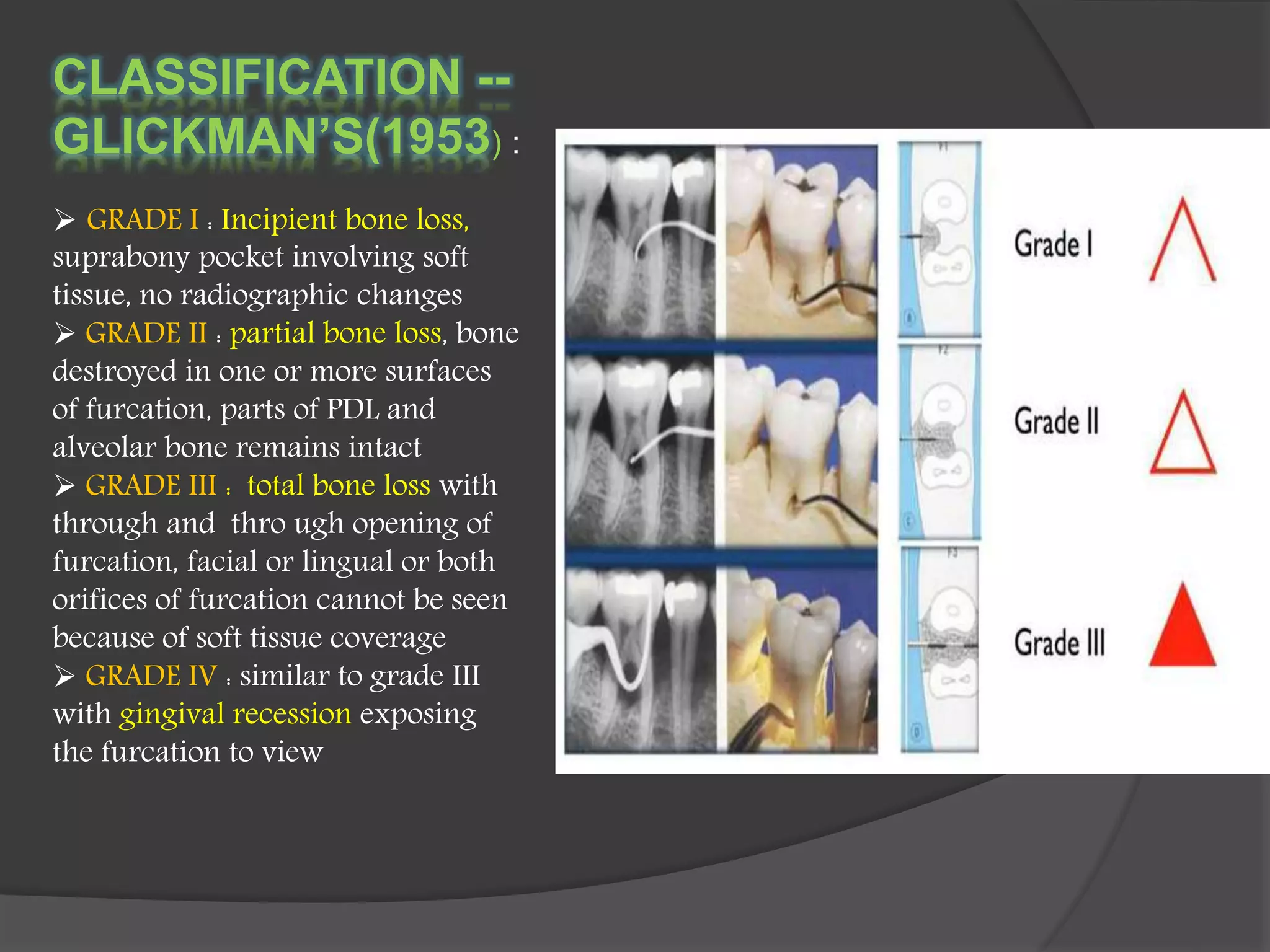
 The bone that forms and
supports the tooth is called
ALVEOLAR PROCESS
 Destruction of the bone is
responsible for tooth loss.
 As the tooth is shed this bone
RESORBS
 The height and density of alveolar bone are
normally maintained by an equilibrium.
 Regulated by local and systemic influences between
bone formation and resorption.
 When resorption exceeds formation, both bone
height and density is reduced
1) Extension Of Gingival Inflammation
2) Trauma from occlusion(TFO)
3) Systemic disorders
BONE DESTRUCTION CAUSED BY
EXTENSION OF GINGIVAL
INFLAMMATION
 Most common cause
 The inflammatory
invasion of bone surface
and the initial bone loss
marks the transition from
GINGIVITIS to
PERIODONTITIS
 “Periodontitis is always
preceded by gingivitis, but
not all gingivitis progress
to periodontitis”
 Extension of inflammation from marginal
gingiva to supporting tissues
 The transition from gingivitis to
periodontitis is associated with changes in
composition of bacterial plaque.
Composition of plaque
GINGIVITIS PERIODONTITIS
Coccid rods,
straight rods Motile organisms and
spirochetes
cells
Plasma cells PMNLs
HISTOPATHOLOGY
 Area of inflammation extending from gingiva into suprabony
area.
 course : along collagen bundle fibres, blood vessels, loosely
arranged tissues
 Extension of inflammation into centre of interdental septum.
 Cortical layer at top of septum are destroyed and inflammation
penetrates into bone marrow
PATHWAYS OF SPREAD OF
INFLAMMATION
 Facially and Lingually:
1. Gingiva to outer periosteum
2. Periosteum to bone
3. Gingiva to PDL
 interproximally:
1. from gingiva along the outer
periosteum
2. from gingiva into
periodontal ligament
3. from periosteum into bone
• gingival inflammation
• Replaced by leucocytes and fluid exudates, new blood vessels
and proliferating fibroblasts
• Increase in osteoclasts and mononuclear cells
• Resorption proceeds from within
• Thinning of bone trabeculae and enlargement of marrow spaces
• Destruction of bone and reduction of bone height
• Replacement of fatty bone marrow with fibrous type
RADIUS OF ACTION
RATE OF BONE LOSS
In individuals with no oral hygiene :
FACIAL SURFACE: 0.2mm/year
PROXIMAL SURFACE: 0.3mm/year
PERIODS OF DESTRUCTION
Periodontal destruction occurs
in episodic and intermittent
manner
Periods of inactivity and
destruction
Results in loss of collagen and
alveolar bone resulting in
deepening of periodontal pocket
Followed by an advance host
defense that controls the
attack
MECHANISM OF BONE
DESTRUCTION
BONE
DESTRUCTION
BACTERIAL
Differentiation of
bone progenitor
cells into
osteoclasts
Inhibit action of
osteoblasts
HOST MEDIATED
Release PGE2,
IL-α,IL-β,TNF-α
BONE DESTRUCTION CAUSED
BY TRAUMA FROM OCCLUSION
 Periodontal response to the
external force.
 TFO can occur in presence
or absence of inflammation.
 In the absence, effects on
alveolar bone ranges from
resorption to necrosis.
 Persistent TFO results in
angular defects of the bone.
o When combined with inflammation- ZONE OF CO-
DESTRUCTION
plaque induced inflammation entering into the zone
of trauma results in angular bone defects, BIZARRE
BONE PATTERN
BONE DESTRUCTION CAUSED
BY SYSTEMIC DISORDERS
 Possible relationship between periodontal bone
loss and systemic disorders.
 OSTEOPOROSIS : loss of bone mineral content and
structural bone changes. Risk factors ageing,
smoking, etc
 OSTEOPENIA : tooth mobility and tooth loss
 Hyperparathyroidism, leucopenia
1) Normal variation of alveolar bone:
 thickness width, crestal angulations of
interdental septa
 thickness of facial and lingual septa
 presence of fenestrations and dehiscence
 root and trunk anatomy
2) Exostoses:
 Exostoses are outgrowths of bone in varied
shapes and sizes
 They can occur as small nodules, sharp ridges,
spike like projections, or a combination of
these
3) Buttressing bone formation:
 bone formation sometimes occurs in an
attempt to buttress bony trabeculae weakened
by resorption.
 When this occurs within the jaw it is termed as
central buttressing bone
 when it occurs on external surface it is is
termed as peripheral buttressing formation
4) Food impaction:
 interdental bone defects occur where proximal
contact is abnormal or absent. In such areas
food impaction results in inverted bone
architecture
5) aggressive periodontitis:
 vertical or angular bone defects
 Horizontal bone loss
 Vertical bone loss
 Osseous craters
 Bulbous bone contour
 Reverse architecture
 Ledges
 Furcation involvement
HORIZONTAL BONE LOSS
 the most common pattern
 bone height is reduced, but bone margins
remain perpendicular to tooth surface.
 interdental septa, facial and lingual cortical
plates are affected
VERTICAL OR ANGULAR DEFECTS
 Occurs in an OBLIQUE
DIRECTION
 leads to a HOLLOWED –OUT
trough in the alongside bone
 classified on the basis of
number of walls:
› one wall defect/one osseous
wall
› two walled defect
› three walled defect
› combined osseous defect
OSSEOUS CRATERS
a) concavities in the crest of
interdental
bone confined within faciolingal
walls.
b) Reasons :
 plaque accumulation and
difficulty to clean.
 normal concavity in lower
molars
 vascular patterns from gingiva
to crest, a pathway for
inflammation
BULBOUS BONE CONTOURS
 bony enlargement
 an adaptation to Exostoses
 adaptation to function or buttressing bone
formation.
 maxilla>mandible
REVERSED ARCHITECTURE
 produced by loss of
interdental bone, facial
and lingual plates
without concomitant
loss of radicular bone
 maxilla more
commonly affected
LEDGES
 plateau-like bony margins
 caused by resorption of thickened bony
plates
FURCATION INVOLVEMENT
 Involvement of bifurcation or
trifurcation of multirooted
teeth by periodontal disease.
 SITE: most common in
mandibular molars, least
common in maxillary
premolar.
CLASSIFICATION --
GLICKMAN’S(1953) :
 GRADE I : Incipient bone loss,
suprabony pocket involving soft
tissue, no radiographic changes
 GRADE II : partial bone loss, bone
destroyed in one or more surfaces
of furcation, parts of PDL and
alveolar bone remains intact
 GRADE III : total bone loss with
through and thro ugh opening of
furcation, facial or lingual or both
orifices of furcation cannot be seen
because of soft tissue coverage
 GRADE IV : similar to grade III
with gingival recession exposing
the furcation to view
 Although periodontitis is an infectious
disease of the gingival tissue , changes that
occur in bone are crucial because
destruction of bone is responsible for tooth
loss.
 Bone loss patterns associated with
periodontal disease is varied and the type of
management depends upon the type of loss.
Bone loss and patterns of bone destruction

Bone loss and patterns of bone destruction

  • 2.
     The bonethat forms and supports the tooth is called ALVEOLAR PROCESS  Destruction of the bone is responsible for tooth loss.  As the tooth is shed this bone RESORBS
  • 3.
     The heightand density of alveolar bone are normally maintained by an equilibrium.  Regulated by local and systemic influences between bone formation and resorption.  When resorption exceeds formation, both bone height and density is reduced
  • 4.
    1) Extension OfGingival Inflammation 2) Trauma from occlusion(TFO) 3) Systemic disorders
  • 5.
    BONE DESTRUCTION CAUSEDBY EXTENSION OF GINGIVAL INFLAMMATION  Most common cause  The inflammatory invasion of bone surface and the initial bone loss marks the transition from GINGIVITIS to PERIODONTITIS  “Periodontitis is always preceded by gingivitis, but not all gingivitis progress to periodontitis”
  • 6.
     Extension ofinflammation from marginal gingiva to supporting tissues  The transition from gingivitis to periodontitis is associated with changes in composition of bacterial plaque. Composition of plaque GINGIVITIS PERIODONTITIS Coccid rods, straight rods Motile organisms and spirochetes cells Plasma cells PMNLs
  • 7.
    HISTOPATHOLOGY  Area ofinflammation extending from gingiva into suprabony area.  course : along collagen bundle fibres, blood vessels, loosely arranged tissues  Extension of inflammation into centre of interdental septum.  Cortical layer at top of septum are destroyed and inflammation penetrates into bone marrow
  • 8.
    PATHWAYS OF SPREADOF INFLAMMATION  Facially and Lingually: 1. Gingiva to outer periosteum 2. Periosteum to bone 3. Gingiva to PDL  interproximally: 1. from gingiva along the outer periosteum 2. from gingiva into periodontal ligament 3. from periosteum into bone
  • 9.
    • gingival inflammation •Replaced by leucocytes and fluid exudates, new blood vessels and proliferating fibroblasts • Increase in osteoclasts and mononuclear cells • Resorption proceeds from within • Thinning of bone trabeculae and enlargement of marrow spaces • Destruction of bone and reduction of bone height • Replacement of fatty bone marrow with fibrous type
  • 10.
  • 11.
    RATE OF BONELOSS In individuals with no oral hygiene : FACIAL SURFACE: 0.2mm/year PROXIMAL SURFACE: 0.3mm/year
  • 12.
    PERIODS OF DESTRUCTION Periodontaldestruction occurs in episodic and intermittent manner Periods of inactivity and destruction Results in loss of collagen and alveolar bone resulting in deepening of periodontal pocket Followed by an advance host defense that controls the attack
  • 13.
    MECHANISM OF BONE DESTRUCTION BONE DESTRUCTION BACTERIAL Differentiationof bone progenitor cells into osteoclasts Inhibit action of osteoblasts HOST MEDIATED Release PGE2, IL-α,IL-β,TNF-α
  • 14.
    BONE DESTRUCTION CAUSED BYTRAUMA FROM OCCLUSION  Periodontal response to the external force.  TFO can occur in presence or absence of inflammation.  In the absence, effects on alveolar bone ranges from resorption to necrosis.  Persistent TFO results in angular defects of the bone.
  • 15.
    o When combinedwith inflammation- ZONE OF CO- DESTRUCTION plaque induced inflammation entering into the zone of trauma results in angular bone defects, BIZARRE BONE PATTERN
  • 16.
    BONE DESTRUCTION CAUSED BYSYSTEMIC DISORDERS  Possible relationship between periodontal bone loss and systemic disorders.  OSTEOPOROSIS : loss of bone mineral content and structural bone changes. Risk factors ageing, smoking, etc  OSTEOPENIA : tooth mobility and tooth loss  Hyperparathyroidism, leucopenia
  • 17.
    1) Normal variationof alveolar bone:  thickness width, crestal angulations of interdental septa  thickness of facial and lingual septa  presence of fenestrations and dehiscence  root and trunk anatomy
  • 18.
    2) Exostoses:  Exostosesare outgrowths of bone in varied shapes and sizes  They can occur as small nodules, sharp ridges, spike like projections, or a combination of these
  • 19.
    3) Buttressing boneformation:  bone formation sometimes occurs in an attempt to buttress bony trabeculae weakened by resorption.  When this occurs within the jaw it is termed as central buttressing bone  when it occurs on external surface it is is termed as peripheral buttressing formation
  • 20.
    4) Food impaction: interdental bone defects occur where proximal contact is abnormal or absent. In such areas food impaction results in inverted bone architecture 5) aggressive periodontitis:  vertical or angular bone defects
  • 21.
     Horizontal boneloss  Vertical bone loss  Osseous craters  Bulbous bone contour  Reverse architecture  Ledges  Furcation involvement
  • 22.
    HORIZONTAL BONE LOSS the most common pattern  bone height is reduced, but bone margins remain perpendicular to tooth surface.  interdental septa, facial and lingual cortical plates are affected
  • 23.
    VERTICAL OR ANGULARDEFECTS  Occurs in an OBLIQUE DIRECTION  leads to a HOLLOWED –OUT trough in the alongside bone  classified on the basis of number of walls: › one wall defect/one osseous wall › two walled defect › three walled defect › combined osseous defect
  • 25.
    OSSEOUS CRATERS a) concavitiesin the crest of interdental bone confined within faciolingal walls. b) Reasons :  plaque accumulation and difficulty to clean.  normal concavity in lower molars  vascular patterns from gingiva to crest, a pathway for inflammation
  • 26.
    BULBOUS BONE CONTOURS bony enlargement  an adaptation to Exostoses  adaptation to function or buttressing bone formation.  maxilla>mandible
  • 27.
    REVERSED ARCHITECTURE  producedby loss of interdental bone, facial and lingual plates without concomitant loss of radicular bone  maxilla more commonly affected
  • 28.
    LEDGES  plateau-like bonymargins  caused by resorption of thickened bony plates
  • 29.
    FURCATION INVOLVEMENT  Involvementof bifurcation or trifurcation of multirooted teeth by periodontal disease.  SITE: most common in mandibular molars, least common in maxillary premolar.
  • 30.
    CLASSIFICATION -- GLICKMAN’S(1953) : GRADE I : Incipient bone loss, suprabony pocket involving soft tissue, no radiographic changes  GRADE II : partial bone loss, bone destroyed in one or more surfaces of furcation, parts of PDL and alveolar bone remains intact  GRADE III : total bone loss with through and thro ugh opening of furcation, facial or lingual or both orifices of furcation cannot be seen because of soft tissue coverage  GRADE IV : similar to grade III with gingival recession exposing the furcation to view
  • 31.
     Although periodontitisis an infectious disease of the gingival tissue , changes that occur in bone are crucial because destruction of bone is responsible for tooth loss.  Bone loss patterns associated with periodontal disease is varied and the type of management depends upon the type of loss.