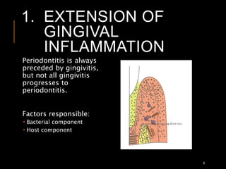
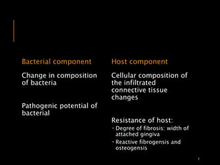
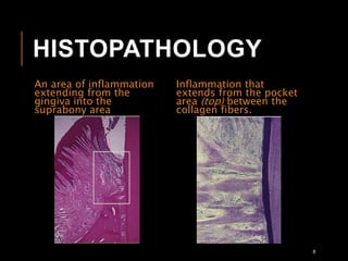
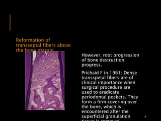
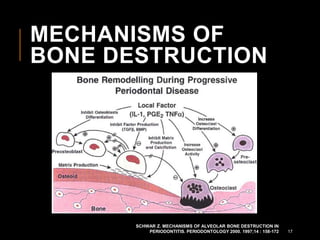
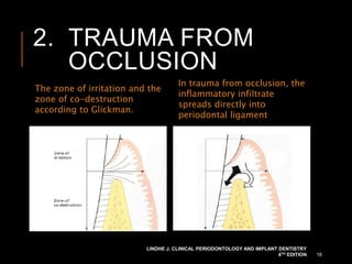
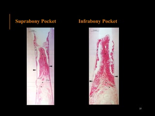
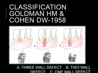
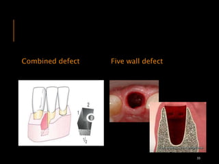
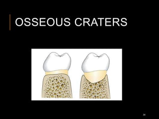
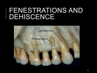
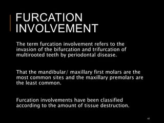
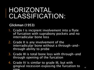
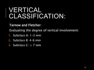
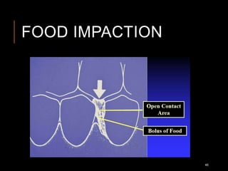
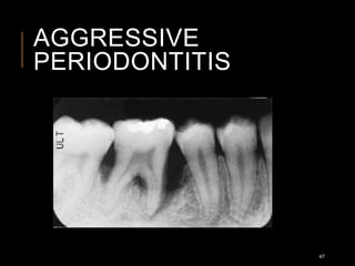
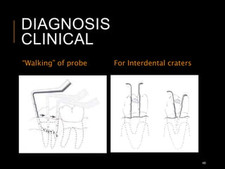
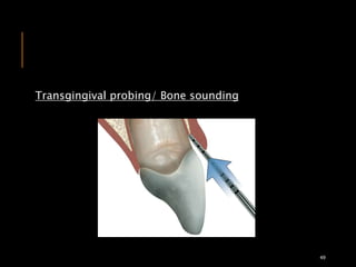
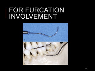
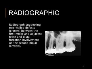
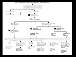
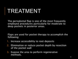
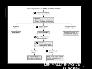
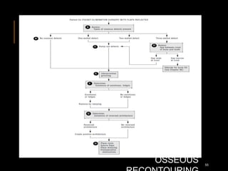
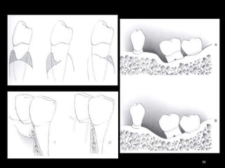
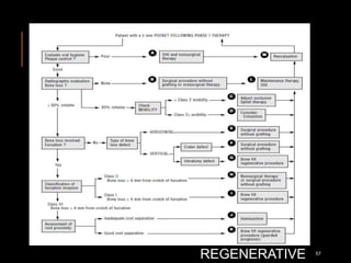
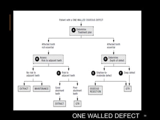
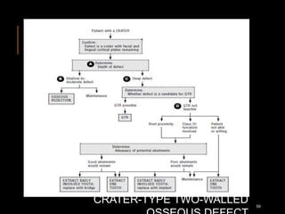
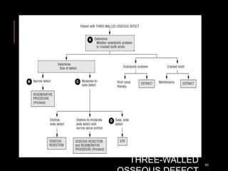
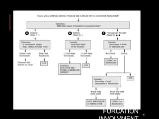
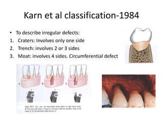
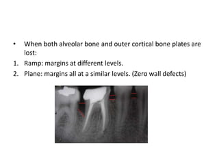
PERIODONTAL
BONE DEFECTS
Dr. Heenal Adhyaru
CONTENT
Causes of bone destruction:
 Bone destruction caused by extension of gingival inflammation
 Bone destruction caused by trauma from occlusion
Factors determining bone morphology in
periodontal disease
Bone destruction patterns in periodontal disease
Bone defects with particular disease
Diagnosis
Treatment
References
2
BONE DESTRUCTION
Bone
formation
Bone
resorption
3
The level of bone is the consequence of past
pathologic experiences, whereas changes in the
soft tissue of the pocket wall reflect the present
inflammatory condition.
When resorption exceeds formation, both bone
height and bone density may be reduced.
The destruction of bone is responsible for tooth
loss.
4
PATHWAYS OF
INFLAMMATION FROM
THE GINGIVA INTO THE
SUPPORTING
PERIODONTAL TISSUES
5
The pathway of the
spread of inflammation
of critical because it
affects the pattern of
bone destruction in
periodontal disease.
1. EXTENSION OF
GINGIVAL
INFLAMMATION
6
Periodontitis is always
preceded by gingivitis,
but not all gingivitis
progresses to
periodontitis.
Factors responsible:
 Bacterial component
 Host component
Bacterial component
Change in composition
of bacteria
Pathogenic potential of
bacterial
Host component
Cellular composition of
the infiltrated
connective tissue
changes
Resistance of host:
 Degree of fibrosis: width of
attached gingiva
 Reactive fibrogensis and
osteogensis
7
HISTOPATHOLOGY
An area of inflammation
extending from the
gingiva into the
suprabony area
Inflammation that
extends from the pocket
area (top) between the
collagen fibers.
8
Reformation of
transseptal fibers above
the bone margin
However, root progression
of bone destruction
progress.
Prichaid F in 1961: Dense
transspetal fibers are of
clinical importance when
surgical procedure are
used to eradicate
periodontal pockets. They
form a firm covering over
the bone, which is
encountered after the
superficial granulation 9
The cortical layer at the top of
the septum has been destroyed,
and the inflammation penetrates
into the marrow spaces
Osteoclasts and Howship
lacunae in the resorption of
crestal bone.
10
RADIUS OF ACTION
Garant and Cho suggested that locally produced
bone resorption factors may need to be present in
the proximity of the bone surface to exert their
action.
Page and Schroeder: 1.5 mm to 2.5 mm
Beyond 2.5 mm, there is no effect; interproximal
angular defects can appear only in spaces that are
wider than 2.5 mm, because narrower spaces would
be destroyed entirely.
Aggressive types of periodontitis: presence of
bacteria in the tissues. 11
12
A. Horizontal resorption
B. Horizontal resorption, incipient vertical resorption
C. Vertical resorption, bony pocket
RATE OF BONE LOSS
Loe and colleagues in 1986 in Sri Lankan tea
laborers with no oral hygiene and no dental care
found the rate of bone loss to average about
0.2 mm per year for facial surfaces and about
0.3 mm per year for proximal surfaces when
periodontal disease was allowed to progress
untreated
Gutman in 1978 stated that loss of attachment can
be equated with loss of bone, although attachment
loss precedes bone loss by about 6 to 8 months.
13
Loe and colleagues also identified the following
three subgroups of patients with periodontal
disease on the basis of the interproximal loss of
attachment and tooth mortality:
1. Approximately 8% of persons had a rapid
progression of periodontal disease that was
characterized by a yearly loss of attachment of
0.1 mm to 1.0 mm.
2. Approximately 81% of individuals had moderately
progressive periodontal disease with a yearly loss
of attachment of 0.05 mm to 0.5 mm.
3. The remaining 11% of persons had minimal or no
progression of destructive disease with a yearly
loss of attachment of 0.05 mm to 0.09 mm. 14
PERIODS OF
DESTRUCTION
Periodontal destruction occurs in an episodic,
intermittent manner, with periods of inactivity or
quiescence.
Theories:
1. Schoder-1980; Page RC-1982: Bursts of destructive
activity are associated with subgingival ulceration and
an acute inflammatory reaction, resulting in rapid loss
of alveolar bone.
2. Saymore GJ-1979: Bursts of destructive activity
coincide with the conversion of a predominantly T-
lymphocyte lesion to one with a predominantly B-
lymphocyte–plasma cell infiltrate 15
3. Newman MG-1979: Periods of exacerbation are
associated with an increase of the loose,
unattached, motile, gram-negative, anaerobic
pocket flora, whereas periods of remission
coincide with the formation of a dense,
unattached, nonmotile, gram-positive flora with
a tendency to mineralize.
4. Saglie-1987: The onset of periods of destruction
coincides with tissue invasion by one or several
bacterial species and that this is followed by an
advanced local host defense that controls the
attack.
16
MECHANISMS OF
BONE DESTRUCTION
SCHWAR Z. MECHANISMS OF ALVEOLAR BONE DESTRUCTION IN
PERIODONTITIS. PERIODONTOLOGY 2000. 1997;14 : 158-172 17
2. TRAUMA FROM
OCCLUSION
The zone of irritation and the
zone of co-destruction
according to Glickman.
In trauma from occlusion, the
inflammatory infiltrate
spreads directly into
periodontal ligament
LINDHE J. CLINICAL PERIODONTOLOGY AND IMPLANT DENTISTRY
4TH EDITION 18
The changes caused by trauma from occlusion vary
from increased compression and tension of the
periodontal ligament.
Persistent trauma from occlusion results in funnel-
shaped widening of the crestal portion of the
periodontal changes which may cause the bony
crest to have an angular shape, represent
adaptation of the periodontal tissues aimed at
“cushioning” increased occlusal forces, but the
modified bone shape may weaken tooth support
and cause tooth mobility.
19
Widening of PDL space
(Arrow)- “cushioning”
effect
These changes are
reversible in that they
can be repaired if the
offending forces are
removed.
20
FACTORS DETERMINING
BONE MORPHOLOGY IN
PERIODONTAL DISEASE
Normal variation in alveolar bone
 The thickness, width, and crestal angulation of the
interdental septa
 The thickness of the facial and lingual alveolar plates
 The presence of fenestrations and dehiscences
 The alignment of the teeth
 Root and root trunk anatomy
 Root position within the alveolar process
 Proximity with another tooth surface
21
NORMAL VARIATION
IN ALVEOLAR BONE
22
EXOSTOSES
23
BUTTRESSING BONE
FORMATION
Bone formation sometimes occurs in an attempt to
buttress bony trabeculae weakened by resorption.
When it occurs within the jaw, it is termed central
buttressing bone formation.
When it occurs on the external surface, it is referred
to as peripheral buttressing bone formation.
(Lipping)
24
25
BONE DESTRUTION
PATTERNS IN
PERIODONTAL
DISEASEPeriodontal disease alters the morphologic features
of the bone in addition to reducing bone height.
An understanding of the nature and pathogenesis
of these alterations is essential for effective
diagnosis and treatment.
26
CLASSIFICATION
Pritchard (1965)
Interproximal craters.
Inconstent margins.
Hemisepta.
Furcation involvement.
Intra bony defect (with
three osseous walls).
Combination of above.
Fenestration.
Dehiscence.
Glickman (1964)
Osseous craters.
Intra bony defect.
Bulbous bony contours.
Hemisepta.
In consistent margins.
Ledges.
Horizontal bone loss
27
HORIZONTAL BONE
LOSS
28
VERTICAL OR
ANGULAR DEFECTS
29
Suprabony Pocket Infrabony Pocket
Base of pocket coronal to level of
alveolar bone.
Base of pocket is apical to crest of
alveolar bone.
Pattern of bone destruction is
horizontal.
Pattern of bone destruction is
vertical.
Interproximally transseptal fibers
are arranged horizontally in the
space between the base of the
pocket and the alveolar bone.
Interproximally transseptal fibers
are oblique rather than horizontal.
On the facial and lingual surface,
the PDL fibers beneath the pocket
follow their normal horizontal
oblique course between the
healthy cementum of the tooth
and the alveolar bone.
On the facial and lingual surface,
the PDL fibers follow the angular
bone pattern of the adjacent bone.
They extend from the healthy
cementum between the base of the
pocket along the base and over the
crest to join with the outer
periosteum (over the bone).
30
Suprabony Pocket Infrabony Pocket
31
CLASSIFICATION
GOLDMAN HM &
COHEN DW-1958
A. THREE WALL DEFECT. B. TWO WALL
32
Combined defect Five wall defect
33
OSSEOUS CRATERS
34
The following reasons for the higher
frequency of interdental craters have
been suggested:
1. The interdental area collects plaque
and is difficult to clean.
2. The normal flat or even concave
faciolingual shape of the interdental
septum in lower molars may favor
crater formation.
3. Vascular patterns from the gingiva to
the center of the crest may provide a
pathway for inflammation.
35
BULBOUS BONE
CONTOUR
36
REVERSED
ARCHITECTURE
37
Peaks of bone typically remain at the facial and
lingual/palatal line angles of the teeth are known as
widow’s peaks.
38
LEDGES
Ledges are plateau
like bone margins
caused by resorption
of thickened bony
plates.
39
FENESTRATIONS AND
DEHISCENCE
40
FURCATION
INVOLVEMENT
The term furcation involvement refers to the
invasion of the bifurcation and trifurcation of
multirooted teeth by periodontal disease.
That the mandibular/ maxillary first molars are the
most common sites and the maxillary premolars are
the least common.
Furcation involvements have been classified
according to the amount of tissue destruction.
41
HORIZONTAL
CLASSIFICATION:
Glickman (1953)
1. Grade I is incipient involvement into a flute
of furcation with suprabony pockets and no
interradicular bone loss
2. Grade II is any involvement of the
interradicular bone without a through-and-
through ability to probe
3. Grade III is total bone loss with through-and
through opening of the furcation
4. Grade IV is similar to grade III, but with
gingival recession exposing the furcation to 42
43
VERTICAL
CLASSIFICATION:
Tarnow and Fletcher:
Evaluating the degree of vertical involvement:
1. Subclass A: 1–3 mm
2. Subclass B: 4–6 mm
3. Subclass C: ≥ 7 mm
44
BONE DEFECTS WITH
PARTICULAR
DISEASE
1. Trauma from occlusion
2. Food impaction
3. Aggressive periodontitis
45
FOOD IMPACTION
46
AGGRESSIVE
PERIODONTITIS
47
DIAGNOSIS
CLINICAL
“Walking” of probe For Interdental craters
48
Transgingival probing/ Bone sounding
49
FOR FURCATION
INVOLVEMENT
50
RADIOGRAPHIC
Radiograph suggesting
two-walled defects
(craters) between the
first molar and adjacent
teeth and distal
furcation involvement
on the second molar
(arrows).
51
52
TREATMENT
The periodontal flap is one of the most frequently
employed procedures particularly for moderate to
deep pockets in posterior areas.
Flaps are used for pocket therapy to accomplish the
following:
1. Increase accessibility to root deposits
2. Elimination or reduce pocket depth by resection
of the pocket wall
3. Expose the area to perform regenerative
methods.
53
MINIMALLY INVASIVE 54
OSSEOUS 55
56
BONE FILL
REGENERATIVE 57
ONE WALLED DEFECT 58
CRATER-TYPE TWO-WALLED 59
THREE-WALLED 60
FURCATION 61
REEVALUATION OF THE
PATIENT WITH BONE
FILLS REGENERATION
PROCEDURE
62
Karn et al classification-1984
• To describe irregular defects:
1. Craters: Involves only one side
2. Trench: involves 2 or 3 sides
3. Moat: involves 4 sides. Circumferential defect
• When both alveolar bone and outer cortical bone plates are
lost:
1. Ramp: margins at different levels.
2. Plane: margins all at a similar levels. (Zero wall defects)
REFERENCES
1. Carranza’s Clinical Periodontology 10th edition
2. Edward S. Cohen, Atlas of Cosmetic and
Reconstructive Periodontal Surgery, Third Edition
3. Lindhe J. Clinical Periodontology and Implant
Dentistry 4th edition
4. Schwar Z. Mechanisms of alveolar bone
destruction in periodontitis. Periodontology
2000. 1997;14 : 158-172
5. Khairnar M. Classification of Food Impaction -
Revisited and its Management. Indian J Dent Adv
2013; 5(1): 1113-1119
6. Hall WB. Critical Decisions in Periodontogy. 4th
edition 65

Periodontal bone defects

  • 1.
  • 2.
    CONTENT Causes of bonedestruction:  Bone destruction caused by extension of gingival inflammation  Bone destruction caused by trauma from occlusion Factors determining bone morphology in periodontal disease Bone destruction patterns in periodontal disease Bone defects with particular disease Diagnosis Treatment References 2
  • 3.
  • 4.
    The level ofbone is the consequence of past pathologic experiences, whereas changes in the soft tissue of the pocket wall reflect the present inflammatory condition. When resorption exceeds formation, both bone height and bone density may be reduced. The destruction of bone is responsible for tooth loss. 4
  • 5.
    PATHWAYS OF INFLAMMATION FROM THEGINGIVA INTO THE SUPPORTING PERIODONTAL TISSUES 5 The pathway of the spread of inflammation of critical because it affects the pattern of bone destruction in periodontal disease.
  • 6.
    1. EXTENSION OF GINGIVAL INFLAMMATION 6 Periodontitisis always preceded by gingivitis, but not all gingivitis progresses to periodontitis. Factors responsible:  Bacterial component  Host component
  • 7.
    Bacterial component Change incomposition of bacteria Pathogenic potential of bacterial Host component Cellular composition of the infiltrated connective tissue changes Resistance of host:  Degree of fibrosis: width of attached gingiva  Reactive fibrogensis and osteogensis 7
  • 8.
    HISTOPATHOLOGY An area ofinflammation extending from the gingiva into the suprabony area Inflammation that extends from the pocket area (top) between the collagen fibers. 8
  • 9.
    Reformation of transseptal fibersabove the bone margin However, root progression of bone destruction progress. Prichaid F in 1961: Dense transspetal fibers are of clinical importance when surgical procedure are used to eradicate periodontal pockets. They form a firm covering over the bone, which is encountered after the superficial granulation 9
  • 10.
    The cortical layerat the top of the septum has been destroyed, and the inflammation penetrates into the marrow spaces Osteoclasts and Howship lacunae in the resorption of crestal bone. 10
  • 11.
    RADIUS OF ACTION Garantand Cho suggested that locally produced bone resorption factors may need to be present in the proximity of the bone surface to exert their action. Page and Schroeder: 1.5 mm to 2.5 mm Beyond 2.5 mm, there is no effect; interproximal angular defects can appear only in spaces that are wider than 2.5 mm, because narrower spaces would be destroyed entirely. Aggressive types of periodontitis: presence of bacteria in the tissues. 11
  • 12.
    12 A. Horizontal resorption B.Horizontal resorption, incipient vertical resorption C. Vertical resorption, bony pocket
  • 13.
    RATE OF BONELOSS Loe and colleagues in 1986 in Sri Lankan tea laborers with no oral hygiene and no dental care found the rate of bone loss to average about 0.2 mm per year for facial surfaces and about 0.3 mm per year for proximal surfaces when periodontal disease was allowed to progress untreated Gutman in 1978 stated that loss of attachment can be equated with loss of bone, although attachment loss precedes bone loss by about 6 to 8 months. 13
  • 14.
    Loe and colleaguesalso identified the following three subgroups of patients with periodontal disease on the basis of the interproximal loss of attachment and tooth mortality: 1. Approximately 8% of persons had a rapid progression of periodontal disease that was characterized by a yearly loss of attachment of 0.1 mm to 1.0 mm. 2. Approximately 81% of individuals had moderately progressive periodontal disease with a yearly loss of attachment of 0.05 mm to 0.5 mm. 3. The remaining 11% of persons had minimal or no progression of destructive disease with a yearly loss of attachment of 0.05 mm to 0.09 mm. 14
  • 15.
    PERIODS OF DESTRUCTION Periodontal destructionoccurs in an episodic, intermittent manner, with periods of inactivity or quiescence. Theories: 1. Schoder-1980; Page RC-1982: Bursts of destructive activity are associated with subgingival ulceration and an acute inflammatory reaction, resulting in rapid loss of alveolar bone. 2. Saymore GJ-1979: Bursts of destructive activity coincide with the conversion of a predominantly T- lymphocyte lesion to one with a predominantly B- lymphocyte–plasma cell infiltrate 15
  • 16.
    3. Newman MG-1979:Periods of exacerbation are associated with an increase of the loose, unattached, motile, gram-negative, anaerobic pocket flora, whereas periods of remission coincide with the formation of a dense, unattached, nonmotile, gram-positive flora with a tendency to mineralize. 4. Saglie-1987: The onset of periods of destruction coincides with tissue invasion by one or several bacterial species and that this is followed by an advanced local host defense that controls the attack. 16
  • 17.
    MECHANISMS OF BONE DESTRUCTION SCHWARZ. MECHANISMS OF ALVEOLAR BONE DESTRUCTION IN PERIODONTITIS. PERIODONTOLOGY 2000. 1997;14 : 158-172 17
  • 18.
    2. TRAUMA FROM OCCLUSION Thezone of irritation and the zone of co-destruction according to Glickman. In trauma from occlusion, the inflammatory infiltrate spreads directly into periodontal ligament LINDHE J. CLINICAL PERIODONTOLOGY AND IMPLANT DENTISTRY 4TH EDITION 18
  • 19.
    The changes causedby trauma from occlusion vary from increased compression and tension of the periodontal ligament. Persistent trauma from occlusion results in funnel- shaped widening of the crestal portion of the periodontal changes which may cause the bony crest to have an angular shape, represent adaptation of the periodontal tissues aimed at “cushioning” increased occlusal forces, but the modified bone shape may weaken tooth support and cause tooth mobility. 19
  • 20.
    Widening of PDLspace (Arrow)- “cushioning” effect These changes are reversible in that they can be repaired if the offending forces are removed. 20
  • 21.
    FACTORS DETERMINING BONE MORPHOLOGYIN PERIODONTAL DISEASE Normal variation in alveolar bone  The thickness, width, and crestal angulation of the interdental septa  The thickness of the facial and lingual alveolar plates  The presence of fenestrations and dehiscences  The alignment of the teeth  Root and root trunk anatomy  Root position within the alveolar process  Proximity with another tooth surface 21
  • 22.
  • 23.
  • 24.
    BUTTRESSING BONE FORMATION Bone formationsometimes occurs in an attempt to buttress bony trabeculae weakened by resorption. When it occurs within the jaw, it is termed central buttressing bone formation. When it occurs on the external surface, it is referred to as peripheral buttressing bone formation. (Lipping) 24
  • 25.
  • 26.
    BONE DESTRUTION PATTERNS IN PERIODONTAL DISEASEPeriodontaldisease alters the morphologic features of the bone in addition to reducing bone height. An understanding of the nature and pathogenesis of these alterations is essential for effective diagnosis and treatment. 26
  • 27.
    CLASSIFICATION Pritchard (1965) Interproximal craters. Inconstentmargins. Hemisepta. Furcation involvement. Intra bony defect (with three osseous walls). Combination of above. Fenestration. Dehiscence. Glickman (1964) Osseous craters. Intra bony defect. Bulbous bony contours. Hemisepta. In consistent margins. Ledges. Horizontal bone loss 27
  • 28.
  • 29.
  • 30.
    Suprabony Pocket InfrabonyPocket Base of pocket coronal to level of alveolar bone. Base of pocket is apical to crest of alveolar bone. Pattern of bone destruction is horizontal. Pattern of bone destruction is vertical. Interproximally transseptal fibers are arranged horizontally in the space between the base of the pocket and the alveolar bone. Interproximally transseptal fibers are oblique rather than horizontal. On the facial and lingual surface, the PDL fibers beneath the pocket follow their normal horizontal oblique course between the healthy cementum of the tooth and the alveolar bone. On the facial and lingual surface, the PDL fibers follow the angular bone pattern of the adjacent bone. They extend from the healthy cementum between the base of the pocket along the base and over the crest to join with the outer periosteum (over the bone). 30
  • 31.
  • 32.
    CLASSIFICATION GOLDMAN HM & COHENDW-1958 A. THREE WALL DEFECT. B. TWO WALL 32
  • 33.
    Combined defect Fivewall defect 33
  • 34.
  • 35.
    The following reasonsfor the higher frequency of interdental craters have been suggested: 1. The interdental area collects plaque and is difficult to clean. 2. The normal flat or even concave faciolingual shape of the interdental septum in lower molars may favor crater formation. 3. Vascular patterns from the gingiva to the center of the crest may provide a pathway for inflammation. 35
  • 36.
  • 37.
  • 38.
    Peaks of bonetypically remain at the facial and lingual/palatal line angles of the teeth are known as widow’s peaks. 38
  • 39.
    LEDGES Ledges are plateau likebone margins caused by resorption of thickened bony plates. 39
  • 40.
  • 41.
    FURCATION INVOLVEMENT The term furcationinvolvement refers to the invasion of the bifurcation and trifurcation of multirooted teeth by periodontal disease. That the mandibular/ maxillary first molars are the most common sites and the maxillary premolars are the least common. Furcation involvements have been classified according to the amount of tissue destruction. 41
  • 42.
    HORIZONTAL CLASSIFICATION: Glickman (1953) 1. GradeI is incipient involvement into a flute of furcation with suprabony pockets and no interradicular bone loss 2. Grade II is any involvement of the interradicular bone without a through-and- through ability to probe 3. Grade III is total bone loss with through-and through opening of the furcation 4. Grade IV is similar to grade III, but with gingival recession exposing the furcation to 42
  • 43.
  • 44.
    VERTICAL CLASSIFICATION: Tarnow and Fletcher: Evaluatingthe degree of vertical involvement: 1. Subclass A: 1–3 mm 2. Subclass B: 4–6 mm 3. Subclass C: ≥ 7 mm 44
  • 45.
    BONE DEFECTS WITH PARTICULAR DISEASE 1.Trauma from occlusion 2. Food impaction 3. Aggressive periodontitis 45
  • 46.
  • 47.
  • 48.
  • 49.
  • 50.
  • 51.
    RADIOGRAPHIC Radiograph suggesting two-walled defects (craters)between the first molar and adjacent teeth and distal furcation involvement on the second molar (arrows). 51
  • 52.
  • 53.
    TREATMENT The periodontal flapis one of the most frequently employed procedures particularly for moderate to deep pockets in posterior areas. Flaps are used for pocket therapy to accomplish the following: 1. Increase accessibility to root deposits 2. Elimination or reduce pocket depth by resection of the pocket wall 3. Expose the area to perform regenerative methods. 53
  • 54.
  • 55.
  • 56.
  • 57.
  • 58.
  • 59.
  • 60.
  • 61.
  • 62.
    REEVALUATION OF THE PATIENTWITH BONE FILLS REGENERATION PROCEDURE 62
  • 63.
    Karn et alclassification-1984 • To describe irregular defects: 1. Craters: Involves only one side 2. Trench: involves 2 or 3 sides 3. Moat: involves 4 sides. Circumferential defect
  • 64.
    • When bothalveolar bone and outer cortical bone plates are lost: 1. Ramp: margins at different levels. 2. Plane: margins all at a similar levels. (Zero wall defects)
  • 65.
    REFERENCES 1. Carranza’s ClinicalPeriodontology 10th edition 2. Edward S. Cohen, Atlas of Cosmetic and Reconstructive Periodontal Surgery, Third Edition 3. Lindhe J. Clinical Periodontology and Implant Dentistry 4th edition 4. Schwar Z. Mechanisms of alveolar bone destruction in periodontitis. Periodontology 2000. 1997;14 : 158-172 5. Khairnar M. Classification of Food Impaction - Revisited and its Management. Indian J Dent Adv 2013; 5(1): 1113-1119 6. Hall WB. Critical Decisions in Periodontogy. 4th edition 65

Editor's Notes

  • #4 There is equilibrium the amount of bone formation and resorption Formation: osteoblasts Resorption: osteoclasts Regulation: local and systemic factors Bone remodeling: bone is constantly resorbed on a particular bony surface, followed by a phase of bone formation Difference with modeling: is the process of bone to shape itself to its maximal strength
  • #5 The degree of bone loss is not necessarily correlated with the depth of periodontal pocket, severity of ulceration of the pocket wall or presence or absence of pus Responsible to mobility and tooth loss
  • #6 Interproximally: Gingival to bone Bone to PDL Gingiva to PDL Facially/lingually Gingiva to outer periosteum Periosteum to bone Gingiva to bone
  • #7 Most common cause of bone destruction Extension of gingival inflammation from marginal gingiva to supporting periodontal tissue
  • #8 Increased spirochetes and motile Decreased straight rods and cocci LPS, tissue penetration Stages of gingivitis: initial: PMNs Early: Lymphocytes 3. Established: Plasma cells Ruben M 1981: a fibrin-fibrinolytic system has been mentioned as walling off the advancing lesion
  • #9 Extension of inflammation: collagen fibers and associated blood vessels Partially destroyed collagen fibers- disorganized granular fragments interpersed among the inflammatory cells and edema
  • #11 Thinning of the surrounding bone Enlargement of marrow spaces Fatty marrow replaced with fibrotic tissue Destruction required viable bone Amount of inflammation is correlated with degree of bone loss
  • #24 Exotoses are outgrowths of bone of varied size and shape Nery EB-1977: palatal exotoses: 40% of human skulls Types: small or large nodules. Sharp ridges, spike like projection or combination Pack1991: developed after placement of free gingival grafts
  • #25 Modeling
  • #26 Lipping of facial bone A. Peripheral buttressing bone formation along the external surface of the facial bony plate and at the crest. Note the deformity in the bone produced by the buttressing bone formation and the bulging of the mucosa B. detailed view showing lipping and deformity produced by buttressing bone formation.
  • #29 Most common pattern of bone loss in periodontal disease Bone reduce in height, margins are perpendicular to the tooth surface Facial and lingual plates and interdental septa involved but not at equal around the tooth
  • #30 Bone loss in oblique direction Base of the defect is located apical to the surrounding bone Intra bony defects-all vertical defects: three wall defect
  • #33  Vertical defects: distal and mesial surface of molars Three wall: three wall present: intrabony defect…now all vertical defects are know was intrabony defects Mesial surface of second and third molar One wall defect: hemiseptum.
  • #34 Combined: the no of walls in the apical portion of the defect may be greater than that in its occlusal portion
  • #35 Osseous craters are concavities in the crest of the interdental bone confined within the facial and lingual walls Occurance: 1/3rd of all defects, 2/3rd of mandibular defects, Most common in post. 85% similar facial and lingual height, 15% facial>lingual
  • #37 Adaptation to function Maxilla>mandible
  • #38 Reverse architecture defects are produced by loss of interdental bone, including the facial and lingual plates, without concomitant loss of radicular bone, thereby reversing the normal architecture Maxilla> mandibular
  • #39 After the surgical resective procedure for osseous craters, reversed architecture will result. Soft tissue follow the contour of the bony architecture
  • #41 Dehiscences and fenestrations reflect anatomical variations concerning the shape and the morphology of the alveolar bone Isolated areas in which the root is denuded of bone and the root surface is convered only by the periosteum and the overlying gingiva are termed fenestrations The marginal bone is intact, when the denuded areas extend through the marginal bone the defect is called a dehiscence Complicate outcome during healing process For implant –through flapless approach
  • #49 Clinical diagnosis Radiographic Final with opening of the flap Probe insertion parallel to the vertical axis of the tooth…walked circumferentially around each surface of teeth to detect the areas of deepest penetration. Interdental craters- oblique positioning of the probe reaches the depth of the crater
  • #50 Under anesthesia
  • #51 Naber’s probe
  • #52 Radiographs are an indispensable aid in identifying the presence of pathosis and the conditions that affect the prognosis and treatment of periodontosis. As with pocket probing, there are restrictions in interpretation. Exposure guidelines and processing procedures must be followed to achieve adequate contrast. Proper angulation of the film and of the head of the x-ray machine minimizes distortion. A properly placed radiograph has few overlapping contact areas, and the entire tooth is visible on periapical views. See for…crown tooth ratio Lamina dura…widening atropy..periapical pathology Orientation of roots..approximation to the sinus
  • #53 Additional digital radiography
  • #55 The MIS flap is closed using a vertical mattress suture. regenerative procedure is performed. This can be an osseous graft stabilized with Vicryl or the use of a growth enhancer (Emdogain), or a combination of both
  • #56 Osseous recontouring facilitate the closure of flaps and permit the most ideal access for plaque removal by the patient after healing. B. No osseous defect..correct the reverse architecture
  • #57 Osseous craters One wall defect adjacent to missing tooth Correction of exotoses
  • #58 Periodontal pockets greater than 5 mm following phase I therapy are difficult for patients to maintain and may be susceptible to further breakdown. These areas should be evaluated and considered for either pocket reduction or bone fill regenerative surgery. Increased success can be achieved with a decision-making process aimed at controlling those clinical factors that contribute to less favorable outcomes with bone fill regenerative procedures. The decision pathway includes evaluation of important criteria such as plaque control, amount and type of bone loss, tooth mobility, degree of furcation involvement, and root proximity.