Periodontal microsurgery
•
20 likes•705 views

This document provides an overview of periodontal microsurgery. It begins with an introduction to microsurgery, discussing the rationale and historical background. It then covers principles of microsurgery including magnification systems, microsurgical instruments, and indications for periodontal microsurgery. The document discusses loupes, the surgical operating microscope, and three-dimensional on-screen microsurgery systems. It also covers hand control, microsurgical instruments including blades, scissors, and needles, and techniques for microsurgery.

Recommended
More Related Content
What's hot
What's hot (20)
Similar to Periodontal microsurgery
Similar to Periodontal microsurgery (20)
More from Dr.R.Dhivya.,MDS
Recently uploaded
Recently uploaded (20)
Periodontal microsurgery
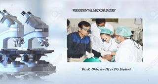
- 1. PERIODONTAL MICROSURGERY Dr. R. Dhivya – III yr PG Student 1
- 2. Introduction Definition and terminologies Rationale for microsurgery Historical Background Principles of microsurgery Elements - Microsurgical Triad Clinical Philosophy Magnification Systems Hand control during microsurgery Microsurgical instruments Indications Of Periodontal Microsurgery Video Limitations of microsurgery Recent Advances Conclusion References Contents 2
- 3. INTRODUCTION LASERS MICROSURGERY LOCAL DRUG DELIVERY IMPLANTOLOGY MUCOGINGIVAL SURGERY TISSUE ENGINEERING 3
- 4. PERIODONTAL MICROSURGERY There has been a tremendous advancement in the medical and dental fields to meet the patient’s expectations and to achieve much desired therapeutic goals. Microsurgery is an advanced surgical technique in which the normal vision is enhanced through magnification. The use of microsurgical instruments, improved view of root surfaces permit more definitive removal of calculus and better smoothness of root. Studies have demonstrated improved vascularization, enhanced mobility of flaps, and hence, possibility of obtaining primary wound closure, less post-operative discomfort, thus providing better esthetic results. 4 “Events that spawn a paradigm begin with a vision.”
- 5. Definition and terminologies In 1979, Daniel RK defined microsurgery in broad terms as “Surgery performed under magnification by the Microscope”. In 1980, Serafin described microsurgery as a methodology – “A modification and refinement of existing surgical techniques using magnification to improve visualization, with applications to all specialities.” Periodontal microsurgery is the refinement of basic surgical techniques made possible by the improvement in visual acuity gained with the use of the surgical microscope - INT JOURNAL OF MICRO DENT 2009 5
- 6. Rationale for microsurgery Reduce the amount of trauma to the tissue Minimal manipulation of the tissue Better blood perfusion and healing is faster with minimal scar formation Least possible trauma to the graft Less bleeding with clear vision Healing by primary intention Patient acceptance 6
- 7. Historical Background • In 1694, Amsterdam merchant Anton van Leeuwenhook constructed the first compound lens microscope. • Saemisch, a German ophthalmologist, introduced simple binocular loupes to ophthalmic surgery in 1876. • In 1921, Carl Nylen, who is considered the father of microsurgery, first used a binocular microscope for ear surgery. • During 1950s, Barraquer began using the microscope for corneal surgery. • Apotheker and Jako first introduced the microscope to dentistry in 1978. • During 1992, Carr published an article outlining the use of the surgical microscope during endodontic procedures. • In 1993, Shanelec and Tibbetts presented a continuing education course on periodontal microsurgery at the annual meeting of the American Academy of Periodontology. 7
- 8. Principles of microsurgery Microsurgery incorporates three important principles : Improvement of motor skills, thereby enhancing surgical ability. An emphasis on passive wound closure with exact primary apposition of the wound edge. The application of microsurgical instrumentation and suturing to reduce tissue trauma. 8
- 9. Elements of microsurgery Microsurgical Triad– Kim et al (2001) Magnification – Perceive two closely lying objects seperately Illumination – Visualization of fine details Instruments – To perform micro-surgeries 9
- 10. Clinical Philosophy Consistent application of the philosophy and techniques learned in basic microsurgery education is necessary for the operator to attain a level of experience and competence needed for various periodontal surgical procedures. Effective periodontal microsurgery allows the operator to consistently achieve clinical results that were once thought to be unlikely. Becoming a clinically proficient periodontal microsurgeon requires a willingness to adopt new values and ideas. The development of new thought patterns regarding surgical esthetics is necessary and attention must be paid to microanatomy, tissue manipulation, and surgical craftsmanship 10
- 11. Magnification Systems Loupes Surgical operating Microscope (SOM) Three dimensional on – screen microsurgery system (TOMS) Magnification system 11
- 12. Loupes Loupes are the most common form of magnification used in dentistry. Fundamentally, loupes are two monocular microscopes with side by- side lenses that are angled to focus on an object. Surgical loupes for magnifi cation enable the clinician to experience the ergonomic benefi ts of an increased working distance from viewing object as well as improved visual acuity. The pattern of convergent lens system is called a Keplerian optical system. Three types of Keplerian loupes commonly used in periodontics are: i) Simple loupes, ii) Compound loupes, and iii) Prism loupes. 12
- 13. Simple loupes Simple loupes are primitive magnifiers with limited capabilities, consisting of a pair of single, positive, side-by-side meniscus lenses. The disadvantage of simple loupe is that they are highly subjected to spherical and chromatic aberration, which distorts the image of the object that is being viewed. In spite of its cost advantages, the size and weight limitations make simple loupes impractical for magnifi cation beyond 1.5 diameters. Advantage - Low cost. Disadvantage - Subjected to spherical and chromatic aberration, that ultimately distorts the image and color of the object that is being viewed. - Their size and weight limits the practical application in dentistry, which is beyond magnifi cation range of 1.5 × diameters, hence distorting the image 13
- 14. compound loupes • The compound loupes are commonly mounted in or on the eyeglasses and can be adjusted to clinical needs without excessive increase in size or weight. • Compound lenses can be achromatic (limits the effects of chromatic and spherical aberration and brings two wavelengths into focus in the same plane), which is an important feature for any magnifying loupe used in periodontics periodontics. Advantages • Better magnifi cation • Wider depths of field • Longer working distances, and Larger fields of view Disadvantages • There is lack of variable magnifi cation. • Individual light source may be required. • Protective coating of anti-refl ective material to prevent loss of light transmitted 14
- 15. PRISM LOUPES These loupes produce superior magnification since they contain Schmidt or roof-top prisms. Other technical advantages include: Better magnification, larger surgical view with wider depths of field, and longer working distances. Furthermore, because of the shorter barrels of the prism loupes, these loupes can be easily mounted on either eyeglass frames or head bands. The incorporation of coaxial fiber optic lights in prism telescopic loupes has improved the operative site illumination to a greater extent. 15
- 16. Loupe magnification range • The surgical loupes provide a wide range of magnification ×1.5 to ×10. • In most of the periodontal procedures, prism telescopic loupes of 4x magnification, even though lower than the operating surgical microscope, provide an effective combination of magnification, field of view, and depth of focus. • The major disadvantage of loupes is that the clinician’s eyes must converge to view on the operate field, which can result in eye strain, fatigue, and even vision changes when poorly designed loupes are used. • But, loupes are less expensive and initially easier to use. 16
- 17. Principal Optical features of Loupes Working distance Working Range Convergence angle Field of view Interpupillary distance Viewing angle Choice of loupes Text book of Lindhe – 5th Edition 17
- 18. Surgical operating microscope – som “Pushing the boundaries of the possible in periodontics” • Surgical microscope utilizes the ‘Galilean optical principles.’ • Optimal magnification factor for the periodontal surgery ranges from ×5 to ×12. • The microscope mountings are available for ceiling, wall mount, or on the floor. • Clinicians are not affected by the weight of the instrument or the challenges of maintaining a stabilized field of vision since they are external to the body. • Surgical microscope has both maneuverability and stability. • The fiber optic technology has improved the methods of focusing light on specifi c areas. • Documentation of periodontal pathology and procedures of all types and video-documentation are also possible 18
- 19. System Components of Surgical Microscope Magnification Changer Eyepiece Objective Lens / Fine focus Binocular Suspension system Tiltable Viewing Tube Tiltable Viewing tube 19
- 20. ERGONOMIC POSTURE POR OPTIMAL PERFORMANCE 20
- 21. Advantages of surgical microscope - Greater operator eye comfort because of the parallel viewing optics of the Galilean system as well as the range of variable magnification. - Excellent coaxial fiber optic illumination - Countless accessories such as still and video cameras for case documentaries Disadvantages - It is an expensive equipment. 21
- 22. Loupes Vs. Operating Microscope Loupes Operating Microscopes 1.5x to 10x magnification 2.5x to 20x magnification Need additional illumination for magnifications of 4x or greater Use excellent coaxial Fiber-optic illumination, hence does not need additional light source Operator eye comfort is less as the eyes must converge to view the image High comfort as it has parallel binoculars Initially easy to use Basic training required to use surgical microscope Less expensive Main disadvantage is that these are expensive Cannot provide variable magnification Has the advantage of providing variable magnifications 22
- 23. TOMS -Three Dimensional On-Screen Microsurgery System • TOMS is a three dimentional system used for better visualization of the surgical area through the monitor, so that direct viewing through microscope can be avoided and thereby reduces eye strain. • The system consists of two single chip video cameras mounted on to the custom fit eyepiece adapters, a dual camera-controller, a record image processor, a VCR for optional recording, digital monitor, synchronizing signal emitter and 120 MHz shutter glasses. • The greatest advantage is that they helps in providing a clear and accurate sense of depth perception. • Drawbacks 1. Technique sensitive 2. High cost 3. Restricted areas of vision 4. Time consuming 5. Loss of visual reference points. 23
- 24. Hand control during microsurgery HAND CONTROL HAND GRIP PHYSIOLOGIC TREMOR 24
- 25. Hand control during microsurgery Physiologic tremor • Finger movements controlled by the long flexor and extensor muscles that move our fingers are relatively crude. • Thus, active finger extensions, or flexions, are likely to be relatively crude. • However, when the wrist is stabilized by resting on a flat surface, angled in a dorsi flection position at approximately 20 degrees, more accurate, finely controlled finger movement can be accomplished because of the reduction in muscle tremor provided by this “platform.” • In microsurgery, the hand should either directly or indirectly rest on an immovable surface or unwanted movements will occur. • Only the fingertips move. All movements should be efficient and economical and should be made with a unity of effort toward purposeful, deliberate motions. • There are several factors that can influence a surgeon’s physiologic tremor, including anxiety, recent exercise, alcohol, smoking, caffeine, heavy meals, hypoglycemia, and medication usage. • To avoid these tremors, microsurgeons should have a relaxed mind,comfortable posture, well – supported hand and stable hold on the instrument. 25
- 26. Hand grips • Basic hand skills in the United States have been associated with and thought of as an extension of penmanship. • With the increased use of keyboards for computers and text messaging on mobile devices, educational curricula no longer stress penmanship. • This may play a role in the lack of basic hand skills in the “writing” or penmanship position. • The acquisition of poor ergonomic habits prior to and during dental education may increase the time it takes for postgraduate residents to become proficient in microsurgery. • The most commonly advocated precision grip for microsurgical procedures is the pen grip or internal precision grip , which provides a greater stability in comparison to any other hand grip due to the tripod formed by the fingers, while the middle finger holds the instrument. • It is best to start with the pen grip until basic manipulations are mastered and more freehand positions can be initiated. 26
- 28. Microsurgical instruments Microsurgical Blade / knives Microsurgical scissors Microsurgical needle holders Microsurgical needles Microsurgical sutures Microsurgical Knots Circular in cross section 15 cm in Length The working tips are much smaller Manufactured under magnification to high tolerances. Needle holders and tissue forceps are made of titanium. Resistant to distortion, non magnetized and are lighter. Shorter instruments with a rectangular cross-sectional design are not ideal for microsurgery. 28
- 29. • These knives have their characteristic ability to create clean incisions to prepare the sharp flap margins for healing by primary intention. • Using Castroviejo microsurgical scalpel, incisions are made at 90 degrees angles to the surface. • Magnification permits easy identification of ragged wound edges for trimming and freshening. • Various types of knives such as crescent, lamellar, blade breaker, sclera, and spoon knife can be used. • They offer the dual advantage of extreme sharpness and minimal size. Microsurgical blade / knives 1- blade breaker; 2-crescent; 3-minicrescent; 4- 260° spoon; 5- lamella and 6- sclera 29
- 30. • Common characteristics of these knives are their extreme sharpness and small size. This enables precise incisions and maneuvers in small areas . • The blade-breaker knife has a handle onto which a piece of an ophthalmic razor blade is affixed. • This allows for infinite angulations of the blade. • This knife is often used in place of a no. 15 blade. • The crescent knife can be used for intrasulcular procedures. It is available with one-piece handles or as a removable blade. • It can be used in connective tissue graft procedures to obtain the donor graft, to tunnel under tissue, and to prepare the recipient site. • The spoon knife is beveled on one side, allowing the knife to track through the tissue adjacent to bone. • It is frequently used in microsurgical procedures to undermine tissue, enhancing the placement of a connective tissue graft. Spoon knife shown in sulcular undermining incision. 30
- 31. Microsurgical scissors i. Micro scissors ii. Extra fine micro scissors (straight) iii. Extra fine micro scissors (curved) - Scissors such as the micro–vannas tissue scissors are used for removal of small fragments of tissue. Microsurgical NEEDLE HOLDERS They are designed to hold the fine needles. They differ in the way they grasp the needle - e.g. a grasp with flat surface if a flat needle is used. The working tips of needle holder are much smaller and are made up of Titanium 31
- 32. Microsurgical needles Every surgical needle has three distinct elements : 1. The Point 2. The Body 3. The Attachment The point : It extends from the tip of the needle to the maximum cross section of the body of the needle. It is designed to penetrate specific types of tissue. There are several types : Reverse cut – Minimal trauma and early regeneration of tissue Taper point / Round needle – Minimal tissue cutting and Smallest puncture hole Taper cut – Dense fibrous tissue and periosteum Blunt Point needle- Dissect through the friable tissue rather than cut through them. 32
- 33. The body : • It comprises slightly more than the middle third of the needle. This is the portion of the needle that is grasped by the needle holder during suturing • The size of the body as close as possible to the diameter of the suture material. The Attachment : • (Swaged end) is a method if attaching the needle and sutured together in a continuous unit that is convenient to use and minimize tissue trauma. • Significant size - ( 16 to 19 mm). • Periodontists frequently use a reverse cutting needle 33
- 34. Factors to consider in selection of suture needles: • Chord length : The straight – line-distance from the point of a curved needle to the swage. • Needle length : The distance measured along the radius of the needle from the point to end. • Radius : The distance from the center of the circle to the body of the needle if the curvature of the needle were to make a full circle. • Diameter : The thickness or guage of the needle wire. 34
- 35. Microsurgical sutures One of the three basic premises of microsurgery is 1. Attention to passive wound closure. 2. The desired result is exact primary apposition of the wound edge. 3. Ideally, the incisions should be almost invisible and closed with precisely placed, small sutures with minimal tissue damage and no bleeding An ideal suture material is sterile, easy to handle, minimally reactive in tissue, resistant to shrinkage in tissues, and capable of holding securely when knotted without fraying or cutting. Ideally, the needle and the suture material should be the same size. 35
- 36. • Suture size is stated numerically, as in 3-0 or 7-0. • The larger the no. of zeros , the smaller the diameter of sutures. • The smaller the size of the suture, the less tensile strength the suture will have depending on the procedure being performed. • Most Periodontal microsurgical suturing is done with sutures ranging in size from 6-0 to 9-0. • Suture bite size should be approximately 1.5 times the tissue thickness to achieve proper wound approximation. (top) 4-0 Vicryl on a FS-2 cutting needle; (bottom) 6-0 polypropylene on a KV-11 taper cutting needle. (top to bottom) 4-0 Vicryl, 6-0 polypropylene, 7-0 PDS-II, 8-0 nylon, 10-0 nylon. 36
- 37. • The suture of choice in microsurgery is a monofilament suture material such as polypropylene or polydioxanone. • These materials are bacteriostatic and noninflammatory, hold a knot extremely well, and are easily removed. • Monofilament materials are preferred as polyfilament threads are characterized by a high capillarity and wicking effect. • Suturing techniques are completely different in macrosurgery and microsurgery. The geometry of microsurgical suturing consists of the following points: • 1. Needle angle of entry and exit of slightly less than 90 degrees 2. Suture bite size of approximately 1.5 times the tissue thickness 3. Equal bite sizes (symmetry) on both sides of the wound 4. Needle passage perpendicular to the wound 37
- 38. Microsurgical knots OLD PHRASE – “Watch one , Do one , Teach one” Two basic knots employed in microsurgery are the square knot or reef knot and surgeon’s knot. The reef knot is composed of two single loops thrown in opposite directions. It lies flat when tied well and is ideal for passive wound closure. As postsurgical edema occurs, the reef knot opens sightly then becomes self-locking . The surgeon’s knot is composed of two double loops thrown in opposite directions. The first double throw is less likely to loosen when performing the second throw, making it is easier to control tissue apposition 38
- 39. INDICATIONS OF PERIODONTAL MICROSURGERY Scaling and Root planing Periodontal Flap surgery Mucogingival Surgery Ridge Augumentation Sinus Lift Procedures Crown Lengthening Root Surface Conditioning Interdental Papillary reconstruction Entire Papilla Preservation Technique (EPP) Minimal Invasive Surgical technique (MIST) Implant Surgery 39
- 40. Scaling and root planing • The critical determinant of the success of periodontal therapy is the thoroughness of debridement of the root surface (Lindhe et al. 1984). • Accessibility and visibility in deep subgingival pockets, furcation areas, and interdental areas can remarkably be improved using magnifications • It is clear that magnification around ×4–10 greatly improves the surgeon’s ability to create a clean, smooth root surface. • It can help to detect islands of biofilm, calculus, or material alba clinging to the root surface and facilitate removal from areas which were normally not visible to the naked eyes Lang et al - 2015 40
- 41. Periodontal Flap surgery • Several authors have proposed the use of microsurgical approach for the treatment of isolated or multiple intrabony defects. • The advantages of microsurgical approach in regenerative therapy relate to improved illumination and magnification of the surgical field that permits proper access to and debridement of the intrabony defect with an increased accuracy and minimal trauma. • Furthermore, the competency to achieve and maintain a primary wound closure minimizes bacterial contamination, and thereby provides more favorable conditions for periodontal regeneration. ADVANTAGE OF MICROSURGERY • Minimal marginal tissue recession and thus Improved esthetics • very limited intra and postoperative morbidity, high patient acceptance and satisfaction Cortellini and Tonetti 1999 41
- 42. • Harrel et al - 1999 in his study showed that probing depth reduction and clinical attachment level gain for the regenerative procedures performed with microsurgical approach has been found similar to those achieved with conventional surgical approach. • Bunckle et al – 1995 in his study with enamel matrix proteins have shown that enamel matrix derivative could exert better biologic activity in microsurgically treated sites because of reduced tissue trauma and vessel injury to improve vascularization and achieve primary wound closure, which allows optimal retention of enamel matrix derivatives. • Liu et al – 2016 in his recent meta-analysis found no significant differences in treatment of intrabony defects treated with minimally invasive periodontal surgery (MIPS) plus biomaterials and MIPS alone for the observed parameters (probing depth, clinical attachment level, marginal recession, and radiographic bone fill), pointing out that costs and benefits should be considered substantially while deciding a regenerative therapeutic modality. • Isolated interproximal defects that are usually limited to interproximal site are considered ideal for bone grafting with MIPS. • Generalized horizontal bone loss and multiple interconnected intrabony defects are a contraindication for MIPS and are best managed with more conventional surgical approaches 42
- 43. Mucogingival Surgery • To achieve an excellent result in terms of both esthetics and function, it is fundamental to perform extremely fine and accurate incisions, meticulous suturing to promote stabilization and immobilization of the graft and precise closure of wound margins. • Therefore, the use of surgical microscope in mucogingival therapy might be helpful for those sites where esthetics demand complete and perfect coverage. • Periodontal microsurgery performed by a trained and skilled surgeon offers an improved outcome of the root coverage procedures. advantage • increased vascularization of the grafts, • relatively better percentages of root coverage, • a significant increase in width and thickness of keratinized tissue, • an improved esthetic outcome, • and decreased patient morbidity. Free gingival graft Double papilla flaps Apical or coronal repositioned flaps Connective tissue grafts Pedicle or sliding flaps 43
- 44. FREE GINGIVAL GRAFT WITH PALATAL DONAR SITE CLOSURE Hedge et al - 2009 44
- 45. Laterally positioned flap - Zuchelli et al - 2004 LMCAF – with Loupe and microsurgical instruments as successful treatment option in class III Recession defect 45
- 46. Connective tissue graft and Double papilla Shanelac et al - 2015 46
- 47. Ridge augumentation Walter et al - 2008 47
- 48. Sinus Lift procedure • One of the novel applications of microsurgery is in the sinus lift procedure with a success rate of 97%. • The surgical microscope can aid indirect visualization of the sinus membrane and minimizes the risk of perforations. • Incorporation of microsurgical techniques for an improvement of altered sensation due to implants encroaching on the inferior alveolar nerve even without unscrewing them has also been reported Kumar et al - 2005 48
- 49. Crown Lengthening • Although the comparative studies of Crown lengthening with microsurgical methods are limited, it seems logical to substantiate the fact that magnification is beneficial in such procedures. Tibettes et al - 2015 49
- 50. Root Surface Conditioning • Furthermore, root preparation can be done with microultrasonic instruments. The smaller size (about ) 0.2–0.6 mm in diameter) and variable power settings (25,000)to more than 40,000 cycles per second) of these instruments allows subgingival treatment in deep pockets with less chances of overinstrumentation of the root surface. • Moreover, these instruments have active working sides on all surfaces; deliver ultrasonically activated lavage in the working area and can be used with minimal water spray • In conclusion, magnification improves the root surface debridement by enhancing clinician’s ability to differentiate the calculus from tooth surface and biofilm to the microscopic level, which reveals morphological contours of both supragingival and subgingival tooth surfaces and accurately procreates working end angles during instrumentation Lindhe et al - 1984 50
- 51. Interdental Papillary reconstruction Microsurgical techniques have been developed to replace the lost interdental papilla, which can create phonetic problems, saliva bubbles, and cosmetic deficiencies. A papillary deficiency can be created through iatrogenic surgical removal, as part of tissue collapse following extraction, with periodontal pocket elimination surgery,with periodontal bone loss and with orthodontic separation of overlapped teeth. Success in the treatment of black triangle with periodontal microsurgery is a significant leap in the field of perio-aesthetics, making it a realistic possibility. Andrade et al - 2010 51
- 52. Entire Papilla Preservation Technique (EPP) •Recently, a new microsurgical approach for periodontal regeneration named “Entire Papilla Preservation Technique” (EPP) has been described in literature. •In this technique, an interdental tunnel is made through the defect associated papilla by giving a beveled vertical releasing incision in the buccal gingiva of the adjacent interdental space. •After granulation tissue removal and root surface debridement, regenerative materials such as bone grafts and enamel matrix derivative are applied. •Primary advantage of EPP technique is enhanced wound stability and limited premature exposure of regenerative biomaterials Aslan et al - 2017 52
- 53. Minimal Invasive Surgical technique (MIST) • The minimally invasive surgical technique (MIST, Cortellini and Tonetti, 2007) is a concept that was designed, especially for isolated intrabony defects for periodontal regeneration. • It is based on minimal reflection of very short buccal and lingual flaps with minimal mesiodistal and coronoapical extensions, the aim being to expose the coronal edge of the residual bone crest that include the defect-associated interdental papilla. • Modified minimally invasive surgical technique (M-MIST) has been proposed by Cortellini and Tonetti, 2009, for use in combination with enamel matrix derivatives (amelogenins). • The overall idea of the M-MIST is to provide a very small interdental access to the defect only from the buccal side, following which the supracrestal interdental tissue is dissected from the granulation tissue by means of a mini-blade, and regenerative material of choice applied. • Passive closure by internal mattress sutures is preferred 53
- 54. Implant Surgery • Surgical microscope can be a valuable tool in implant dentistry. • Different stages of implant treatment ranging from implant placement to implant recovery and peri-implantitis management may be accomplished with more precision under magnification. • The microscope may be a valuable tool in visualizing the last threads of the implant for subcrestal placement, implant recovery with minimal trauma to adjacent tissues, management of peri-implantitis, visualization of the sinus membrane during sinus lift procedures, and minimizing the risk of perforations or tears Shanelec et al - 2005 54
- 55. video 55
- 56. Drawbacks of microsurgery • As we upgrade our surgical maneuvers with the aid of microsurgical concepts, there are a few shortcomings of this modus operandi, which need to be considered prior to its application. • It is much more demanding and technique-sensitive; the cost incurred to establish a microsurgical set up is also high. • Magnification systems used also pose some difficulties including restricted area of vision, loss of depth of field as magnification increases, and loss of visual reference points. • An experienced team approach mandates microsurgery and is time-consuming to develop. • Physiologic tremor control for finer movements intra-operatively and a steep learning curve are required for clinical proficiency. 56
- 57. RECENT ADVANCES IN MICROSCOPES • Zeiss OPMI PROErgo • Mechanical optical rotating assembly interface (MORA Interface) • Periodontal endoscope • Varioscope • Infrared 800, flow 800, and blue 400 fluorescence 57
- 58. Conclusion • Aesthetic preservation and improvement have become an integral part of today’s periodontal treatment. • Evaluation of periodontal aesthetic procedures has driven largely by the patient’s increased awareness of and desire for aesthetically pleasing smiles. • Patients are demanding a youthful attractive smile that includes healthy gingiva with ideal contours and texture. • Periodontal microsurgery is definitely a must for perio-aesthetics. • The improved visual acuity provided by magnification opens a whole new world for those who make effort and take time to become proficient in microsurgical principles and procedures. • The promising periodontal microsurgery will provide new possibilities to improve the therapeutic results for variety of periodontal surgeries. 58
- 59. Reference Textbook of periodontology – Carranza - 10th ed. Textbook of Periodontal Diseases: Basic Phenomena, Clinical Management, and Occlusal and Restorative Interrelationships - Page and Schluger - 2nd ed. Cohen – Atlas of Cosmetic & Reconstructive periodontal Surgery – 2nd ed. Hall WB. Critical Decisions in Periodontogy - 4th edition. Tetbook of Periobasics: A textbook of periodontics and implantology – 5st Edition Francetti L, Del Fabbro M, Calace S, Testori T, Weinstein RL. Microsurgical treatment of gingival recession: controlled clinical study. Int J Periodontics Restorative Dent 2005;25:181-8. 59
- 60. • Tibbetts LS, Shanelec D. Periodontal microsurgery. Dent Clin North Am 1988;42:339-59. • Michaelides PL. Use of the operating microscope in dentistry. J Calif Dent Assoc 1996;24:45-50. • Shanelec DA, Tibetts LS. A perspective on the future of periodontal microsurgery. Periodontol 2000 1996;11:58- 64. • Belcher JM. A perspective on periodontal microsurgery. Int J Periodontics Restorative Dent 2001;21:191-6. • Shanelec DA. Optical principles of loupes. J Calif Dent Assoc 1992;20:25-32. • RaiTioji MV. Periodontal microsurgery. Anna Essen Dent 2011;1:127-9. • Venugopal K. Periodontal microsurgery-A perspective. Periodontics 2012;1:68-72. 60
- 61. • Francetti L, Del Fabbro M, Calace S, Testori T, Weinstein RL. Microsurgical treatment of gingival recession: A controlled clinical study. Int J Periodontics Restorative Dent 2005;25:181-8. • Burkhardt R, Lang NP. Coverage of localized gingival recessions: Comparison of micro- and macro surgical techniques. J Clin Periodontol 2005;32:287-935 • Cortellini P, Tonetti MS. A minimally invasive surgical technique with an enamel matrix derivative in the regenerative treatment of intra-bony defects: A novel approach to limit morbidity. J Clin Periodontol 2007;34:87-93 • Cortellini P, Tonetti MS. Microsurgical approach to periodontal regeneration. Initial evaluation in a case cohort. J Periodontol 2001;72:559-69. 61
- 62. • Andrade PF, Grisi MF, Maracaccini AM, Fernandes PG, Reino DM, Souza SL, et al. Comparison between micro- and macrosurgical techniques for the treatment of localized gingival recessions using coronally repositioned fl aps and enamel matrix derivative. J Periodontol 2010;81:1572-9. • 20. Franken RJ, Gupta SC, Banis JC Jr, Th omas SV, Derr JW, Klein SA, et al. Microsurgery without a microscope: Laboratory evaluation of a three-dimensional on-screen microsurgery system. Microsurgery 1995;16:746-51. • Wachtel H, Schenk G, Böhm S, Weng D, Zuhr O, Hürzeler MB. Microsurgical access flap and enamel matrix derivative for the treatment of periodontal intrabony defects: A controlled clinical study. J Clin Periodontol 2003;30:496-504. • Kotschy P. Optimal root cleaning and microinvasive periodontal pocket surgery with microscope controlled glass bead blasting. Int J Microdentistry 2010;2:48-55 62
- 63. • Kumar A, Bains VK, Jhingran R, Srivastava R, Madan R, Rizvi I, et al. Patient-centered microsurgical management of gingival recession using coronally advanced flap with either platelet-rich fibrin or connective tissue graft: A comparative analysis. Contemp Clin Dent 2017;8:293-304. • Agarwal SK, Jhingran R, Bains VK, Srivastava R, Madan R, Rizvi I, et al Patient-centered evaluation of microsurgical management of gingival recession using coronally advanced flap with platelet-rich fibrin or amnion membrane: A comparative analysis. Eur J Dent 2016;10:121-33. • Nordland WP, Sandhu HS, Perio C. Microsurgical technique for augmentation of the interdental papilla: Three case reports. Int J Periodontics Restorative Dent 2008;28:543-9. • Bittencourt S, Del Peloso Ribeiro E, Sallum EA, Nociti FH Jr., Casati MZ. Surgical microscope may enhance root coverage with subepithelial connective tissue graft: A randomized-controlled clinical trial. J Periodontol 2012;83:721-3 63
- 64. 64
Editor's Notes
- Modern periodontology is closely linked to both plastic surgery and esthetic dentistry. Periodontal plastic microsurgery incorporates the use of a surgical microscope in an attempt to increase visibility, thereby minimizing soft tissue trauma and enhance surgical results. Periodontal plastic microsurgery incorporates the use of a surgical microscope in an attempt to increase visibility, thereby minimizing soft tissue trauma and enhance surgical results. The use of surgical operating microscope, microsurgical instruments has opened a new era in periodontal plastic surgery. Further, the successful use of the surgical microscope in periodontal surgery is less documented with only few studies addressing the advantages of the application of magnification to periodontal surgery. Here, we present an overview of magnifying tools available and their applications in the specialty of periodontics. The success criteria of treatments performed to improve esthetics may be quite different compared to those surgical procedures whose main goals are to improve periodontal health and restore compromised function.
- Magnification for microsurgical procedure was introduced to medicine during the late nineteenth century
- For a basic understanding of the fine finger movements necessary with the use of microscopic magnification, some important aspects of hand function must be reviewed.
- Instruments should be circular in cross section to allow for a smooth rotation movement. The working tips of microsurgical instruments are much smaller than those of regular instruments. To provide consistent manipulation of tissues, needles, and sutures, most microsurgical instruments are manufactured under magnification to high tolerances. Needle holders and tissue forceps are made of titanium. Properly cared for, such instruments are resistant to distortion from repeated use and sterilization, are non magnetized, and are lighter than surgical stainless steel instruments. Shorter instruments, as well as instruments with a rectangular cross-sectional design,do not allow as precise manipulation and therefore are not ideal for microsurgery
- In the last decades, a modification in the existing surgical procedures and their clinical effectiveness for periodontal regeneration of intrabony defects has been extensively studied.[
- It has been reported that root instrumentation is effective when done under illumination along with an improved early healing index and less postoperative pain