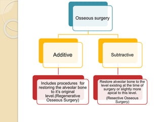
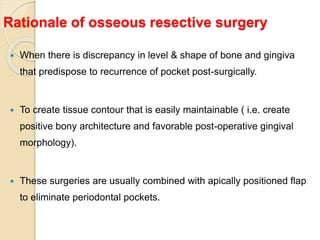
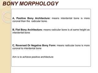
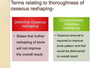
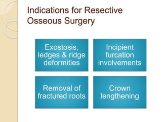
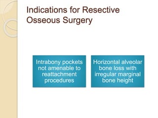
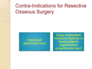
Resective
Osseous
Surgery
Contents
• Definition
• Historical prospective
• Osteoplasty, Ostectomy
• Rationale
• Indications
• Contra- Indications
• Factors affecting selection of cases
• Presurgical evaluation
• Steps of Resective Surgery
• Postsurgical maintainance
• Advantages
• Causes of Failure
The goal of
Osseous surgery
is:
To get rid of bony
deformities produced
by periodontal disease
Osseous surgery
Additive
Includes procedures for
restoring the alveolar bone
to it’s original
level.(Regenerative
Osseous Surgery)
Subtractive
Restore alveolar bone to the
level existing at the time of
surgery or slightly more
apical to this level.
(Resective Osseous
Surgery)
Resective
Osseous Surgery
Definitions
AAP’s glossary of terms (2001) -
Procedures to modify bone altered by
periodontal disease, either by reshaping the
alveolar process to achieve physiologic
form without the removal of alveolar
supporting bone, or by the removal of some
alveolar bone, thus changing the position of
the crestal bone relative to the tooth root.
Sims and Carranza (1996) - defined osseous surgery
as the procedure by which changes in the alveolar
bone can be accomplished to rid it of deformities
induced by the periodontal disease process or other
related factors, such as exostosis and tooth
supraeruption.
What both definitions
have
in common ?
The goal of osseous surgery is to
produce osseous contours that
are consistent with the shape and
form of the healthy gingival
tissues as gingival contours
always follow the bony
contours
Historical
prospecti
ve
Schluger (1949)
• First described Osseous Resective
therapy
• Identified the rationale and technique of
osseous resective surgery for pocket
elimination
Friedman (1955)
• Defined two procedures
as osteoplasty and
osteoectomy.
Osteoplasty & Ostectomy (Friedman 1955)
OSTEOPLASTY
Refers to reshaping the bone without
removing tooth supporting bone.
OSTECTOMY OR OSTEOECTOMY
Includes reshaping with removal of tooth supporting
bone.
Height of supporting bone decreases.
Rationale of osseous resective surgery
 When there is discrepancy in level & shape of bone and gingiva
that predispose to recurrence of pocket post-surgically.
 To create tissue contour that is easily maintainable ( i.e. create
positive bony architecture and favorable post-operative gingival
morphology).
 These surgeries are usually combined with apically positioned flap
to eliminate periodontal pockets.
BONY MORPHOLOGY
A, Positive Bony Architecture: means interdental bone is more
coronal than the radicular bone.
B, Flat Bony Architecture: means radicular bone is at same height as
interdental bone
C, Reversed Or Negative Bony Form: means radicular bone is more
coronal to interdental bone
Aim is to achieve positive architecture
Terms relating to thoroughness of
osseous reshaping-
Definitive Osseous
reshaping
• States that further
reshaping of bone
will not improve
the overall result.
Compromised
Osseous
reshaping
• Osseous removal is
required to improve
bone pattern and that
would be detrimental
to overall result.
Indications for Resective
Osseous Surgery
Patients with
moderate
bone loss
One walled
bone
defects.
Shallow
craters up to
3mm depth
Crown
lengthening
Indications for Resective
Osseous Surgery
Exostosis,
ledges & ridge
deformities
Incipient
furcation
involvements
Removal of
fractured roots
Crown
lengthening
Indications for Resective
Osseous Surgery
Intrabony pockets
not amenable to
reattachment
procedures
Horizontal alveolar
bone loss with
irregular marginal
bone height
Contra-Indications for Resective
Osseous Surgery
Advanced
attachment loss
Deep multiwalled
intrabony defects( in
these patients
regenerative
surgeries are best)
Contra-Indications for Resective
Osseous Surgery
When there is close
proximity of roots
When it can cause
recession, produce
unacceptable aesthetic
results so
contraindicated in
anterior aesthetic areas.
FACTORS IN SELECTION OF
RESECTIVE OSSEOUS SURGERY
◦ 1. Early to moderate bone loss (2-3mm) with moderate root
trunk length, and one or two walls bone defect. During
ostectomy , there is approximately 0.6mm of attachment loss
(Selipsky HS; 1976). So resective surgery is not indicated in
patients with advanced attachment loss. Regenerative therapy
is preferred in such patients or osteoplasty can be done along
with regenerative procedures to reduce bony ledges and to
facilitate flap closure and to achieve proper gingival contours
◦ 2. Two wall defects or craters should be treated with resection
of ledges and ostectomy in facial, lingual, palatal surface and
interproximal areas to obtain positive architecture. Otherwise,
presence of window’s peak causes recurrence of interproximal
pockets.
 WIDOW’S PEAK- These are residual peaks of
cortical bone that remain left over facial &
lingual /palatal line angle (as in craters).
Gingiva will heal over these peaks, but with
time these bony peaks resorbs leading to
pocket formation on interproximal areas. So
ostectomy should be done to remove these
widow peaks
◦ 3. In maxillary arch, the palatal
approach may be indicated as
opposed to a more accessible
buccal surface for esthetic reasons &
for being a more conservative
approach.
◦ 4. If one-walled defect is present
next to edentulous ridge, the ridge is
reduced to the level of bone defect
Selection Based on Morphology of Defects
 One wall Angular - Osseous resection
 3 Wall, Narrow & Deep - Bone Regeneration
 2 Wall Angular - Depending on depth, width &
configuration
(Osseous resection or regeneration)
PRE-SURGICAL EXAMINATION
 RADIOGRAHS – to see bone loss ( amount & pattern of bone loss).
 Periodontal probing- to check pocket depth & furcation involvement.
 TRANSGINGIVAL PROBING/ BONE SOUNDING- to check bone
topography and type of bony defect
But best evaluation of type of defect is done by raising the flap during
surgery
Instruments used for resective
osseous surgery
Hand, Rotary, Piezoelectric surgical techniques
 Hand instruments - bone rongeurs, bone files
and bone chisels
 Rotary instruments - burs & micromotor.
 Hand instruments are best for ostectomy
procedures.
 Rotary instruments are good for osteoplasy.
STEPS IN RESECTIVE OSSEOUS SURGERY
 VERTICAL GROOVING
 RADICULAR BLENDING
 FLATTENING INTERPROXIMAL BONE
 GRADUALIZING MARGINAL BONE
Not all steps are necessary in every case
First 2 steps involve osteoplasty
Last 2 steps involve ostectomy.
1. Vertical grooving (osteoplasty) : “first step”
 Done by instruments as carbide or diamond
burs.
It is designed to:
 Reduce thickness of alveolar housing.
 Provide relative prominence to radicular part
of the teeth.
 Provide smooth continuity from interproximal
area onto the radicular area
1. Vertical grooving (osteoplasty) : “first step”
 Indications:
Thick margins of bone, shallow craters
Areas that require minimal amount of
osteoctomy and maximal osteoplasty
 Contraindication:
Areas with close root proximity or thin
alveolar housing.
2. Radicular blending (osteoplasty) :
 Continuity of vertical grooving
 Gradualize the entire bone over the radicular
surface to give the best results from vertical
grooving.
 Provides smooth surface for good flap adaptation
and the best post-operative gingival contours
2. Radicular blending (osteoplasty) :
 Indications:
Presence of thick ledges of bone on radicular
surface.
 Contraindication:
Thin, fenestrated radicular bone
2. Radicular blending (osteoplasty) :
 Both vertical grooving and radicular blending may
be used for treatment of:
1) Shallow crater formation.
2) Thick bony ledges in radicular surface.
3) Class I and early class II furcation.
3. Flattening Interproximal bone (osteoctomy):
 Very small amount of supporting bone is removed
 Indications:
1) Coronally placed one wall edge of 3 wall angular
defect
2) One-walled interproximal defect (preferably
shallow as deep defect lead to compromised osseou
architecture)
4. Gradualizing marginal bone (osteoctomy):
 Minimal bone removal to provide a smooth,
regular base for gingival tissue to follow.
 Failure to remove the widow peak’s led to
recurrence of pockets
 Hand instruments as chisel and curette are
preferred over rotary instruments to prevent
excess bone loss.
Post Surgery maintainance
Suture removal after
1 week
Attachment of flap to
bone is completed in
14-21 days.
Scaling after every 2
weeks till healing is
complete
Waiting period of
minimum of 6 weeks
after surgery to
proceed for dental
restoration
Advantages
* Produces immediate reduction in probing
depth;
* Improves access for daily oral hygiene;
* Preserves gingival width via apically positioned
flaps;
* Permits recontouring of bone anomalies (e.g.,
tori, ledges);
*Allows access for root resection and hemisection;
* Permits access for correction of radicular
anomalies (e.g., cervical enamel projections,
enamel pearls etc.);
* Facilitates recontouring of restorative overhangs
CAUSES OF FAILURE OF RESECTIVE OSSEOUS SURGERY
 Poor post-operative plaque control.
 Failure to create ideal bone form.
Other causes can be :
»Improper Flap management
» Sequestration and resorption of bone following surgery.
» Improper suturing
» Exposure of thin bone during or after surgery.
» Flap necrosis.
» Post-surgical infection
» Root caries or pulpal problems.
Thank you

Resective osseous surgery

  • 1.
  • 3.
    Contents • Definition • Historicalprospective • Osteoplasty, Ostectomy • Rationale • Indications • Contra- Indications • Factors affecting selection of cases • Presurgical evaluation • Steps of Resective Surgery • Postsurgical maintainance • Advantages • Causes of Failure
  • 4.
    The goal of Osseoussurgery is: To get rid of bony deformities produced by periodontal disease
  • 5.
    Osseous surgery Additive Includes proceduresfor restoring the alveolar bone to it’s original level.(Regenerative Osseous Surgery) Subtractive Restore alveolar bone to the level existing at the time of surgery or slightly more apical to this level. (Resective Osseous Surgery)
  • 6.
  • 7.
    Definitions AAP’s glossary ofterms (2001) - Procedures to modify bone altered by periodontal disease, either by reshaping the alveolar process to achieve physiologic form without the removal of alveolar supporting bone, or by the removal of some alveolar bone, thus changing the position of the crestal bone relative to the tooth root.
  • 8.
    Sims and Carranza(1996) - defined osseous surgery as the procedure by which changes in the alveolar bone can be accomplished to rid it of deformities induced by the periodontal disease process or other related factors, such as exostosis and tooth supraeruption.
  • 9.
    What both definitions have incommon ? The goal of osseous surgery is to produce osseous contours that are consistent with the shape and form of the healthy gingival tissues as gingival contours always follow the bony contours
  • 10.
  • 11.
    Schluger (1949) • Firstdescribed Osseous Resective therapy • Identified the rationale and technique of osseous resective surgery for pocket elimination Friedman (1955) • Defined two procedures as osteoplasty and osteoectomy.
  • 12.
    Osteoplasty & Ostectomy(Friedman 1955) OSTEOPLASTY Refers to reshaping the bone without removing tooth supporting bone. OSTECTOMY OR OSTEOECTOMY Includes reshaping with removal of tooth supporting bone. Height of supporting bone decreases.
  • 13.
    Rationale of osseousresective surgery  When there is discrepancy in level & shape of bone and gingiva that predispose to recurrence of pocket post-surgically.  To create tissue contour that is easily maintainable ( i.e. create positive bony architecture and favorable post-operative gingival morphology).  These surgeries are usually combined with apically positioned flap to eliminate periodontal pockets.
  • 14.
    BONY MORPHOLOGY A, PositiveBony Architecture: means interdental bone is more coronal than the radicular bone. B, Flat Bony Architecture: means radicular bone is at same height as interdental bone C, Reversed Or Negative Bony Form: means radicular bone is more coronal to interdental bone Aim is to achieve positive architecture
  • 15.
    Terms relating tothoroughness of osseous reshaping- Definitive Osseous reshaping • States that further reshaping of bone will not improve the overall result. Compromised Osseous reshaping • Osseous removal is required to improve bone pattern and that would be detrimental to overall result.
  • 16.
    Indications for Resective OsseousSurgery Patients with moderate bone loss One walled bone defects. Shallow craters up to 3mm depth Crown lengthening
  • 17.
    Indications for Resective OsseousSurgery Exostosis, ledges & ridge deformities Incipient furcation involvements Removal of fractured roots Crown lengthening
  • 18.
    Indications for Resective OsseousSurgery Intrabony pockets not amenable to reattachment procedures Horizontal alveolar bone loss with irregular marginal bone height
  • 19.
    Contra-Indications for Resective OsseousSurgery Advanced attachment loss Deep multiwalled intrabony defects( in these patients regenerative surgeries are best)
  • 20.
    Contra-Indications for Resective OsseousSurgery When there is close proximity of roots When it can cause recession, produce unacceptable aesthetic results so contraindicated in anterior aesthetic areas.
  • 21.
    FACTORS IN SELECTIONOF RESECTIVE OSSEOUS SURGERY ◦ 1. Early to moderate bone loss (2-3mm) with moderate root trunk length, and one or two walls bone defect. During ostectomy , there is approximately 0.6mm of attachment loss (Selipsky HS; 1976). So resective surgery is not indicated in patients with advanced attachment loss. Regenerative therapy is preferred in such patients or osteoplasty can be done along with regenerative procedures to reduce bony ledges and to facilitate flap closure and to achieve proper gingival contours
  • 22.
    ◦ 2. Twowall defects or craters should be treated with resection of ledges and ostectomy in facial, lingual, palatal surface and interproximal areas to obtain positive architecture. Otherwise, presence of window’s peak causes recurrence of interproximal pockets.
  • 23.
     WIDOW’S PEAK-These are residual peaks of cortical bone that remain left over facial & lingual /palatal line angle (as in craters). Gingiva will heal over these peaks, but with time these bony peaks resorbs leading to pocket formation on interproximal areas. So ostectomy should be done to remove these widow peaks
  • 24.
    ◦ 3. Inmaxillary arch, the palatal approach may be indicated as opposed to a more accessible buccal surface for esthetic reasons & for being a more conservative approach.
  • 25.
    ◦ 4. Ifone-walled defect is present next to edentulous ridge, the ridge is reduced to the level of bone defect
  • 26.
    Selection Based onMorphology of Defects  One wall Angular - Osseous resection  3 Wall, Narrow & Deep - Bone Regeneration  2 Wall Angular - Depending on depth, width & configuration (Osseous resection or regeneration)
  • 27.
    PRE-SURGICAL EXAMINATION  RADIOGRAHS– to see bone loss ( amount & pattern of bone loss).  Periodontal probing- to check pocket depth & furcation involvement.  TRANSGINGIVAL PROBING/ BONE SOUNDING- to check bone topography and type of bony defect But best evaluation of type of defect is done by raising the flap during surgery
  • 28.
    Instruments used forresective osseous surgery Hand, Rotary, Piezoelectric surgical techniques  Hand instruments - bone rongeurs, bone files and bone chisels  Rotary instruments - burs & micromotor.  Hand instruments are best for ostectomy procedures.  Rotary instruments are good for osteoplasy.
  • 29.
    STEPS IN RESECTIVEOSSEOUS SURGERY  VERTICAL GROOVING  RADICULAR BLENDING  FLATTENING INTERPROXIMAL BONE  GRADUALIZING MARGINAL BONE Not all steps are necessary in every case First 2 steps involve osteoplasty Last 2 steps involve ostectomy.
  • 30.
    1. Vertical grooving(osteoplasty) : “first step”  Done by instruments as carbide or diamond burs. It is designed to:  Reduce thickness of alveolar housing.  Provide relative prominence to radicular part of the teeth.  Provide smooth continuity from interproximal area onto the radicular area
  • 31.
    1. Vertical grooving(osteoplasty) : “first step”  Indications: Thick margins of bone, shallow craters Areas that require minimal amount of osteoctomy and maximal osteoplasty  Contraindication: Areas with close root proximity or thin alveolar housing.
  • 32.
    2. Radicular blending(osteoplasty) :  Continuity of vertical grooving  Gradualize the entire bone over the radicular surface to give the best results from vertical grooving.  Provides smooth surface for good flap adaptation and the best post-operative gingival contours
  • 33.
    2. Radicular blending(osteoplasty) :  Indications: Presence of thick ledges of bone on radicular surface.  Contraindication: Thin, fenestrated radicular bone
  • 34.
    2. Radicular blending(osteoplasty) :  Both vertical grooving and radicular blending may be used for treatment of: 1) Shallow crater formation. 2) Thick bony ledges in radicular surface. 3) Class I and early class II furcation.
  • 35.
    3. Flattening Interproximalbone (osteoctomy):  Very small amount of supporting bone is removed  Indications: 1) Coronally placed one wall edge of 3 wall angular defect 2) One-walled interproximal defect (preferably shallow as deep defect lead to compromised osseou architecture)
  • 36.
    4. Gradualizing marginalbone (osteoctomy):  Minimal bone removal to provide a smooth, regular base for gingival tissue to follow.  Failure to remove the widow peak’s led to recurrence of pockets  Hand instruments as chisel and curette are preferred over rotary instruments to prevent excess bone loss.
  • 37.
    Post Surgery maintainance Sutureremoval after 1 week Attachment of flap to bone is completed in 14-21 days. Scaling after every 2 weeks till healing is complete Waiting period of minimum of 6 weeks after surgery to proceed for dental restoration
  • 38.
    Advantages * Produces immediatereduction in probing depth; * Improves access for daily oral hygiene; * Preserves gingival width via apically positioned flaps; * Permits recontouring of bone anomalies (e.g., tori, ledges);
  • 39.
    *Allows access forroot resection and hemisection; * Permits access for correction of radicular anomalies (e.g., cervical enamel projections, enamel pearls etc.); * Facilitates recontouring of restorative overhangs
  • 40.
    CAUSES OF FAILUREOF RESECTIVE OSSEOUS SURGERY  Poor post-operative plaque control.  Failure to create ideal bone form.
  • 41.
    Other causes canbe : »Improper Flap management » Sequestration and resorption of bone following surgery. » Improper suturing » Exposure of thin bone during or after surgery. » Flap necrosis. » Post-surgical infection » Root caries or pulpal problems.
  • 42.

Editor's Notes

  • #8 OSSEO US S.: Procedures to modify bone support altered by periodontal disease, either by reshaping the alveolar process to achieve physiologic form without the removal of alveolar supporting bone, or by the removal of some alveolar bone, thus changing the position of the crestal b,'nc relative to the tooth root.
  • #37 (Peaks of bone remain at the facial, lingual/ palatal line angles of the teeth)