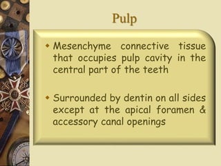
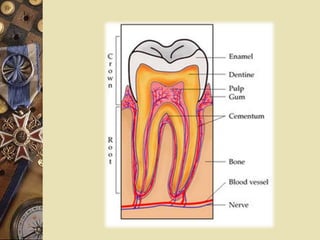
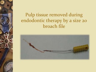
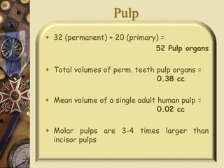
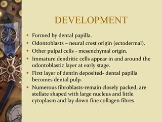
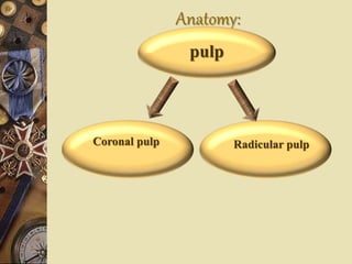
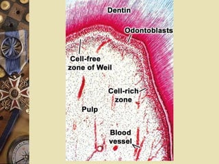
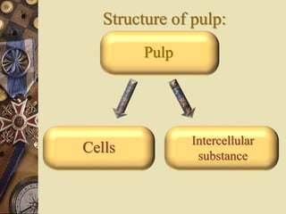
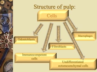
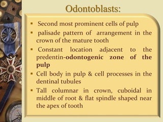
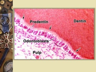
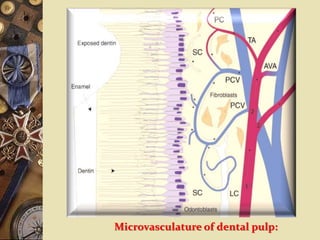
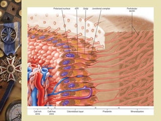
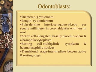
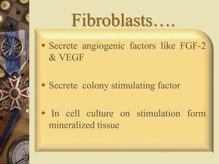
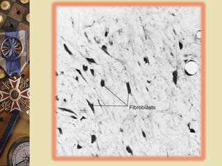
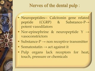
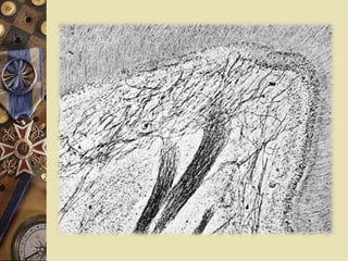
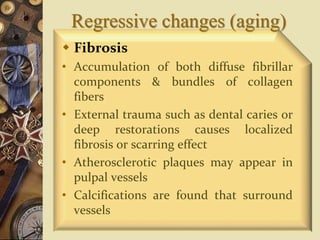
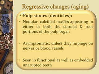
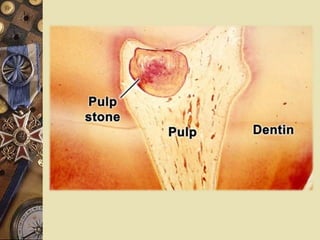
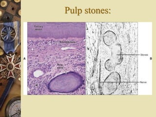
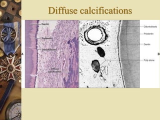
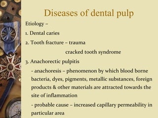
Pulp
Dr.Smitha Rao
Pulp
 Mesenchyme connective tissue
that occupies pulp cavity in the
central part of the teeth
 Surrounded by dentin on all sides
except at the apical foramen &
accessory canal openings
Pulp tissue removed during
endodontic therapy by a size 20
broach file
Pulp
 32 (permanent) + 20 (primary) =
52 Pulp organs
 Total volumes of perm. teeth pulp organs =
0.38 cc
 Mean volume of a single adult human pulp =
0.02 cc
 Molar pulps are 3-4 times larger than
incisor pulps
DEVELOPMENT
 Formed by dental papilla.
 Odontoblasts – neural crest origin (ectodermal).
 Other pulpal cells - mesenchymal origin.
 Immature dendritic cells appear in and around the
odontoblastic layer at early stage.
 First layer of dentin deposited- dental papilla
becomes dental pulp.
 Numerous fibroblasts-remain closely packed, are
stellate shaped with large nucleus and little
cytoplasm and lay down fine collagen fibres.
DEVELOPMENT
 Developing pulp has high GAG content.
 Chondroitin sulphate is higher with little hyaluronan,
while opposite is true for mature pulp.
 Enamel organ extends as HERS and determines final
morphology of the space (pulp chamber and root canals).
 Once full length of roots is established, the developmental
stage of dental pulp can be considered complete.
Anatomy:
pulp
Coronal pulp Radicular pulp
Coronal pulp
 Has 6 surfaces
 Has pulp horns which are protrusions
that extend into the cusps of each crown
 Number depends on the cuspal number
 Pulp becomes smaller with age
Radicular pulp
 Extends from the cervical region of the
crown to the root apex
 Anterior teeth-single
 Posterior teeth-multiple
 Continuous with the periapical
connective tissues through the
apical foramen
Histology
4 zones:
 Odontoblastic zone
 Cell free zone of weil
 Cell-rich zone
 Pulp Core
Structure of pulp:
Intercellular
substance
Pulp
Cells
Structure of pulp:
Cells
Odontoblasts
Fibroblasts
Immunocompetent
cells
Macrophages
Undifferentiated
ectomesenchymal cells
Odontoblasts:
 Second most prominent cells of pulp
 palisade pattern of arrangement in the
crown of the mature tooth
 Constant location adjacent to the
predentin-odontogenic zone of the
pulp
 Cell body in pulp & cell processes in the
dentinal tubules
 Tall columnar in crown, cuboidal in
middle of root & flat spindle shaped near
the apex of tooth
Microvasculature of dental pulp:
Odontoblasts:
Diameter -5-7micronm
Length-25-40micronm
Pulp-dentine interface-59,000-76,000 per
square millimeter in coronaldentin with less in
root
Active cell-elongated ,basally placed nucleus &
a basophilic cytoplasm
Resting cell-stubby,little cytoplasm &
haematoxyphilic nucleus
Transitional stage-intermediate beteen active
& resting stage
Odontoblasts:
 Undergo apoptotic cell death –shown by
marker bcl-2
 Release interleukin -8
 Nitric oxide synthetase have been
identified in odontoblasts
Fibroblasts:
 Cells occurring in greatest numbers
in the pulp
 More in number in the coronal
portion of pulp where they form the
cell-rich zone
 Form &maintain the pulp matrix,
which consists of collagen & ground
substance
Fibroblasts:
 They function in collagen fiber
formation
 In young pulp the cells divide & are
active in protein synthesis
 In older pulp they appear rounded
or spindle shaped with short
processes & exhibit fewer
intracellular organelles –fibrocytes
Fibroblasts:
Immature pulp - cellular elements
predominate
Mature pulp - fibrous components
predominate
Dual function; In addition to
forming the pulp matrix, capable of
ingesting &degrading the same
matrix
Fibroblasts….
 Secrete angiogenic factors like FGF-2
& VEGF
 Secrete colony stimulating factor
 In cell culture on stimulation form
mineralized tissue
Macrophages
 Oval or spindle shaped
 Macrophages are present in the normal
dental pulp & are more dense around the
blood vessels of the inner pulp & around
the odontoblast layer
Dendritic cells:
 Bone marrow- derived, antigen
presenting dendrite cells are found
in & around the odontoblast layer in
nonerupted teeth & in erupted teeth
beneath the odontoblast layer
 Function is similar to Langerhans
cells in that they capture & present
foreign antigen to the T cells
Dendritic cells:
 Participate in immunosurveillance
& increase in number in carious
teeth
 Dendritic cells & macrophages
constitute some 8% of the total
cell population, with dendritic
cells exceeding macrophages
Undifferentiated
ectomesenchymal cells
 Totipotent cell
 Depending upon the stimulus these
cells may give rise to odontoblasts ,
fibroblasts or macrophages.
 Represent the pool from which
connective tissue cells of the pulp are
derived
Matrix & ground substance
 Collagen-typeI & typeIII
 Ground substance composed of
glycosaminoglycans, glycoproteins & water
 Ground substance lend support to the cells of
the pulp& also serve as transport medium of
nutrients from the blood vessels to the cells &
transport of metabolites from cells to blood
vessels
Pulpal stem cells
 Migrate to the site of injured
odontoblasts & produce dentin
 TGF beta 1 & BMP-2 involved in the
migration of stem cells
 Pluripotent-adipocytes & neural cells
Pulpal stem cells
 Produce dentinogenesis &
osteogenesis
 Dentin sialoprotein present
 Dentonin stimulate dental pulp stem
cell
 Pulp tisue of 3rd molars - viable after
cryopreservation
Functions of pulp
1. Inductive: interact with oral
epithelial cells leading to
differentiation of dental lamina &
enamel organ formation
2. Formative: formation of dentin
3. Sensory: sensation of tooth is felt
through the nerves of the pulp
Functions of pulp
4. Nutritive: supplies nutrition to the
dentin through blood vessels &
odontoblastic processes & maintain the
vitality of tooth
5. Defensive or reparative: protects it
self & vitality of the tooth by producing
reparative dentin
Blood vessels of the dental pulp:
 Supply oxygen & nutrients where they are
most needed during dentinogenesis
 Pulpal blood flow-
1.in arterioles-0.3to1mm/sec
2.In venules-approx.0.15mm/sec
3.In capillaries-approx.0.08mm/sec
 Arise from inferior or superior alveolar
artery
Blood vessels of the dental pulp
Vessels possess 3 layers:
1. Tunica intima-squamous or cuboid
cells
2. Tunica media-5microm thick&consists
of one to three layers of smooth muscle
cells
3. Tunica adventitia-made up of few
collagen fibres
Blood vessels of the dental pulp:
 Pericyte lies on the surface of vessels
 Arterio-venous shunts regulate blood flow
 Blood capillaries – 8-10microm in
diameter
 Fenestrated capillaries-rapid transport of
metabolites during dentinogenesis
Nerves of the dental pulp
 Nerves enter pulp through apical
foramen, along with afferent blood
vessels & together form the “neuro-
vascular bundle”
 Nerve bundles that enter the pulp
consists principally of sensory afferents of
the trigeminal nerve & sympathetic
branches from the superior cervical
ganglion
Nerves of the dental pulp
 In the crown there is a pronounced
plexus of nerves beneath the
odontoblasts, known as the plexus of
Raschkow – occupy the cell-
free zone of weil & can be demonstrated
in silver stained sections under light
microscope or by immunocytochemical
techniques
 In the root-no corresponding plexus
Nerves of the dental pulp
 Each bundle contains myelinated &
unmyelinated axons
 Myelinated axons loose myelin-as the
nerve ascend coronally
 Substance-P, 5-hydroxy tryptamine,
vasoactive peptide, somatostatin &
prostaglandins as well as acetycholine
& nor epinephrine –found throughout
pulp
Nerves of the dental pulp :
 Neuropeptides-- Calcitonin gene related
peptide (CGRP) & Substance-P→
potent vasodilators
 Nor-epinephrine & neuropeptide Y →
vasoconstrictors
 Substance-P → non receptive transmitter
 Somatostatin → act against it
 Pulp organs lack receptors for heat,
touch, pressure or chemicals
Regressive changes (aging)
 Fibrosis
• Accumulation of both diffuse fibrillar
components & bundles of collagen
fibers
• External trauma such as dental caries or
deep restorations causes localized
fibrosis or scarring effect
• Atherosclerotic plaques may appear in
pulpal vessels
• Calcifications are found that surround
vessels
Regressive changes (aging)
 Pulp stones (denticles):
• Nodular, calcified masses appearing in
either or both the coronal & root
portions of the pulp organ
• Asymptomatic, unless they impinge on
nerves or blood vessels
• Seen in functional as well as embedded
unerupted teeth
Regressive changes (aging)
Pulp stones
(denticles):
True
denticles
False
denticles
Pulp stones:
True denticles:
• Similar in structure to dentin
• Are rare, located close to the apical foramen
• Development is caused by the inclusion of
remnants of the epithelial root sheath within
the pulp
• These epithelial remnants induce the cells of
the pulp to differentiate into odontoblasts,
which then form the dentin masses called
true pulp stones
Pulp stones:
False denticles:
• Do not exhibit dentinal tubules
• Concentric layers of calcified tissue
• Some cases calcification sites appear
within a bundle of collagen fibers
• Phleboliths-serve as nidi for false
denticles
• All denticles begin as small nodules
but increase in size by incremental
growth on their surface
Pulp stones:
Pulp stones
(depending on their
relation to the
dentin of tooth)
Free
Attached
Embedded
Pulp stones:
 Free denticles:
entirely surrounded by pulp tissue
 Attached denticles:
partly fused with the dentin
 Embedded denticles:
entirely surrounded by dentin
Pulp stones:
 Incidence –
 66%of teeth-10-3oyears
 80% of teeth-30 & 50 years
 90%-over 50 years of ageFree
denticles:
Diffuse calcifications
 Irregular calcified deposits
following collagen fibre bundles or
blood vessels
 Found more in the root canal &
less in coronal area
 Surround blood vessels-dystrophic
calcification
Diffuse calcifications
Clinical considerations:
 Wide pulp chamber in the tooth of a
young person will make a deep cavity
preparation hazardous
 Dehydration cause pulpal damage
 Associated with trauma, the pulp
becomes transformed into granulation
tissue and the dentine commences to be
resorbed internally at the pulp-dentine
surface
Diseases of dental pulp
Etiology –
1. Dental caries
2. Tooth fracture – trauma
cracked tooth syndrome
3. Anachorectic pulpitis
- anachoresis – phenomenon by which blood borne
bacteria, dyes, pigments, metallic substances, foreign
products & other materials are attracted towards the
site of inflammation
- probable cause – increased capillary permeability in
particular area
4. Chemical irritation – erosion
acidic restorative material
5. Thermal changes – polishing procedures
cavity preparation
tooth restored with
exothermic
restorative
material.
6. Galvanic currents
7. Periapical tissue – accessory canals
8. Aerodontalgia
Classification of
pulpitis
•Acute
•Chronic
•Both acute and chronic –
Partial or focal pulpitis
Total or generalised
•Closed pulpitis
•Open pulpitis
References
 Orban’s oral histology &embryology-12th
edition
 Ten Cate’s oral histology-7th edition
 Oral anatomy, histology and embryology;
Berkovitz, 3rd edition
 Essentials of oral histology &
embryology, 2rd edition, James k . avery
Thank you
Thank you

Pulp

  • 1.
  • 2.
    Pulp  Mesenchyme connectivetissue that occupies pulp cavity in the central part of the teeth  Surrounded by dentin on all sides except at the apical foramen & accessory canal openings
  • 4.
    Pulp tissue removedduring endodontic therapy by a size 20 broach file
  • 5.
    Pulp  32 (permanent)+ 20 (primary) = 52 Pulp organs  Total volumes of perm. teeth pulp organs = 0.38 cc  Mean volume of a single adult human pulp = 0.02 cc  Molar pulps are 3-4 times larger than incisor pulps
  • 6.
    DEVELOPMENT  Formed bydental papilla.  Odontoblasts – neural crest origin (ectodermal).  Other pulpal cells - mesenchymal origin.  Immature dendritic cells appear in and around the odontoblastic layer at early stage.  First layer of dentin deposited- dental papilla becomes dental pulp.  Numerous fibroblasts-remain closely packed, are stellate shaped with large nucleus and little cytoplasm and lay down fine collagen fibres.
  • 7.
    DEVELOPMENT  Developing pulphas high GAG content.  Chondroitin sulphate is higher with little hyaluronan, while opposite is true for mature pulp.  Enamel organ extends as HERS and determines final morphology of the space (pulp chamber and root canals).  Once full length of roots is established, the developmental stage of dental pulp can be considered complete.
  • 8.
  • 9.
    Coronal pulp  Has6 surfaces  Has pulp horns which are protrusions that extend into the cusps of each crown  Number depends on the cuspal number  Pulp becomes smaller with age
  • 11.
    Radicular pulp  Extendsfrom the cervical region of the crown to the root apex  Anterior teeth-single  Posterior teeth-multiple  Continuous with the periapical connective tissues through the apical foramen
  • 12.
    Histology 4 zones:  Odontoblasticzone  Cell free zone of weil  Cell-rich zone  Pulp Core
  • 14.
  • 15.
  • 16.
    Odontoblasts:  Second mostprominent cells of pulp  palisade pattern of arrangement in the crown of the mature tooth  Constant location adjacent to the predentin-odontogenic zone of the pulp  Cell body in pulp & cell processes in the dentinal tubules  Tall columnar in crown, cuboidal in middle of root & flat spindle shaped near the apex of tooth
  • 18.
  • 20.
    Odontoblasts: Diameter -5-7micronm Length-25-40micronm Pulp-dentine interface-59,000-76,000per square millimeter in coronaldentin with less in root Active cell-elongated ,basally placed nucleus & a basophilic cytoplasm Resting cell-stubby,little cytoplasm & haematoxyphilic nucleus Transitional stage-intermediate beteen active & resting stage
  • 21.
    Odontoblasts:  Undergo apoptoticcell death –shown by marker bcl-2  Release interleukin -8  Nitric oxide synthetase have been identified in odontoblasts
  • 22.
    Fibroblasts:  Cells occurringin greatest numbers in the pulp  More in number in the coronal portion of pulp where they form the cell-rich zone  Form &maintain the pulp matrix, which consists of collagen & ground substance
  • 23.
    Fibroblasts:  They functionin collagen fiber formation  In young pulp the cells divide & are active in protein synthesis  In older pulp they appear rounded or spindle shaped with short processes & exhibit fewer intracellular organelles –fibrocytes
  • 24.
    Fibroblasts: Immature pulp -cellular elements predominate Mature pulp - fibrous components predominate Dual function; In addition to forming the pulp matrix, capable of ingesting &degrading the same matrix
  • 25.
    Fibroblasts….  Secrete angiogenicfactors like FGF-2 & VEGF  Secrete colony stimulating factor  In cell culture on stimulation form mineralized tissue
  • 27.
    Macrophages  Oval orspindle shaped  Macrophages are present in the normal dental pulp & are more dense around the blood vessels of the inner pulp & around the odontoblast layer
  • 28.
    Dendritic cells:  Bonemarrow- derived, antigen presenting dendrite cells are found in & around the odontoblast layer in nonerupted teeth & in erupted teeth beneath the odontoblast layer  Function is similar to Langerhans cells in that they capture & present foreign antigen to the T cells
  • 29.
    Dendritic cells:  Participatein immunosurveillance & increase in number in carious teeth  Dendritic cells & macrophages constitute some 8% of the total cell population, with dendritic cells exceeding macrophages
  • 30.
    Undifferentiated ectomesenchymal cells  Totipotentcell  Depending upon the stimulus these cells may give rise to odontoblasts , fibroblasts or macrophages.  Represent the pool from which connective tissue cells of the pulp are derived
  • 31.
    Matrix & groundsubstance  Collagen-typeI & typeIII  Ground substance composed of glycosaminoglycans, glycoproteins & water  Ground substance lend support to the cells of the pulp& also serve as transport medium of nutrients from the blood vessels to the cells & transport of metabolites from cells to blood vessels
  • 32.
    Pulpal stem cells Migrate to the site of injured odontoblasts & produce dentin  TGF beta 1 & BMP-2 involved in the migration of stem cells  Pluripotent-adipocytes & neural cells
  • 33.
    Pulpal stem cells Produce dentinogenesis & osteogenesis  Dentin sialoprotein present  Dentonin stimulate dental pulp stem cell  Pulp tisue of 3rd molars - viable after cryopreservation
  • 34.
    Functions of pulp 1.Inductive: interact with oral epithelial cells leading to differentiation of dental lamina & enamel organ formation 2. Formative: formation of dentin 3. Sensory: sensation of tooth is felt through the nerves of the pulp
  • 35.
    Functions of pulp 4.Nutritive: supplies nutrition to the dentin through blood vessels & odontoblastic processes & maintain the vitality of tooth 5. Defensive or reparative: protects it self & vitality of the tooth by producing reparative dentin
  • 36.
    Blood vessels ofthe dental pulp:  Supply oxygen & nutrients where they are most needed during dentinogenesis  Pulpal blood flow- 1.in arterioles-0.3to1mm/sec 2.In venules-approx.0.15mm/sec 3.In capillaries-approx.0.08mm/sec  Arise from inferior or superior alveolar artery
  • 37.
    Blood vessels ofthe dental pulp Vessels possess 3 layers: 1. Tunica intima-squamous or cuboid cells 2. Tunica media-5microm thick&consists of one to three layers of smooth muscle cells 3. Tunica adventitia-made up of few collagen fibres
  • 38.
    Blood vessels ofthe dental pulp:  Pericyte lies on the surface of vessels  Arterio-venous shunts regulate blood flow  Blood capillaries – 8-10microm in diameter  Fenestrated capillaries-rapid transport of metabolites during dentinogenesis
  • 41.
    Nerves of thedental pulp  Nerves enter pulp through apical foramen, along with afferent blood vessels & together form the “neuro- vascular bundle”  Nerve bundles that enter the pulp consists principally of sensory afferents of the trigeminal nerve & sympathetic branches from the superior cervical ganglion
  • 42.
    Nerves of thedental pulp  In the crown there is a pronounced plexus of nerves beneath the odontoblasts, known as the plexus of Raschkow – occupy the cell- free zone of weil & can be demonstrated in silver stained sections under light microscope or by immunocytochemical techniques  In the root-no corresponding plexus
  • 44.
    Nerves of thedental pulp  Each bundle contains myelinated & unmyelinated axons  Myelinated axons loose myelin-as the nerve ascend coronally  Substance-P, 5-hydroxy tryptamine, vasoactive peptide, somatostatin & prostaglandins as well as acetycholine & nor epinephrine –found throughout pulp
  • 45.
    Nerves of thedental pulp :  Neuropeptides-- Calcitonin gene related peptide (CGRP) & Substance-P→ potent vasodilators  Nor-epinephrine & neuropeptide Y → vasoconstrictors  Substance-P → non receptive transmitter  Somatostatin → act against it  Pulp organs lack receptors for heat, touch, pressure or chemicals
  • 47.
    Regressive changes (aging) Fibrosis • Accumulation of both diffuse fibrillar components & bundles of collagen fibers • External trauma such as dental caries or deep restorations causes localized fibrosis or scarring effect • Atherosclerotic plaques may appear in pulpal vessels • Calcifications are found that surround vessels
  • 48.
    Regressive changes (aging) Pulp stones (denticles): • Nodular, calcified masses appearing in either or both the coronal & root portions of the pulp organ • Asymptomatic, unless they impinge on nerves or blood vessels • Seen in functional as well as embedded unerupted teeth
  • 50.
    Regressive changes (aging) Pulpstones (denticles): True denticles False denticles
  • 51.
    Pulp stones: True denticles: •Similar in structure to dentin • Are rare, located close to the apical foramen • Development is caused by the inclusion of remnants of the epithelial root sheath within the pulp • These epithelial remnants induce the cells of the pulp to differentiate into odontoblasts, which then form the dentin masses called true pulp stones
  • 52.
    Pulp stones: False denticles: •Do not exhibit dentinal tubules • Concentric layers of calcified tissue • Some cases calcification sites appear within a bundle of collagen fibers • Phleboliths-serve as nidi for false denticles • All denticles begin as small nodules but increase in size by incremental growth on their surface
  • 53.
  • 54.
    Pulp stones (depending ontheir relation to the dentin of tooth) Free Attached Embedded
  • 56.
    Pulp stones:  Freedenticles: entirely surrounded by pulp tissue  Attached denticles: partly fused with the dentin  Embedded denticles: entirely surrounded by dentin
  • 57.
    Pulp stones:  Incidence–  66%of teeth-10-3oyears  80% of teeth-30 & 50 years  90%-over 50 years of ageFree denticles:
  • 58.
    Diffuse calcifications  Irregularcalcified deposits following collagen fibre bundles or blood vessels  Found more in the root canal & less in coronal area  Surround blood vessels-dystrophic calcification
  • 59.
  • 60.
    Clinical considerations:  Widepulp chamber in the tooth of a young person will make a deep cavity preparation hazardous  Dehydration cause pulpal damage  Associated with trauma, the pulp becomes transformed into granulation tissue and the dentine commences to be resorbed internally at the pulp-dentine surface
  • 61.
    Diseases of dentalpulp Etiology – 1. Dental caries 2. Tooth fracture – trauma cracked tooth syndrome 3. Anachorectic pulpitis - anachoresis – phenomenon by which blood borne bacteria, dyes, pigments, metallic substances, foreign products & other materials are attracted towards the site of inflammation - probable cause – increased capillary permeability in particular area
  • 62.
    4. Chemical irritation– erosion acidic restorative material 5. Thermal changes – polishing procedures cavity preparation tooth restored with exothermic restorative material. 6. Galvanic currents 7. Periapical tissue – accessory canals 8. Aerodontalgia
  • 63.
    Classification of pulpitis •Acute •Chronic •Both acuteand chronic – Partial or focal pulpitis Total or generalised •Closed pulpitis •Open pulpitis
  • 64.
    References  Orban’s oralhistology &embryology-12th edition  Ten Cate’s oral histology-7th edition  Oral anatomy, histology and embryology; Berkovitz, 3rd edition  Essentials of oral histology & embryology, 2rd edition, James k . avery
  • 65.