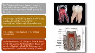
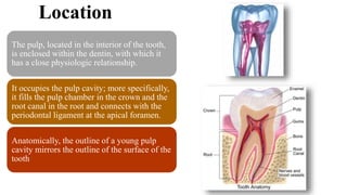
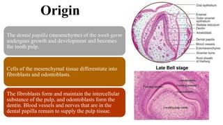
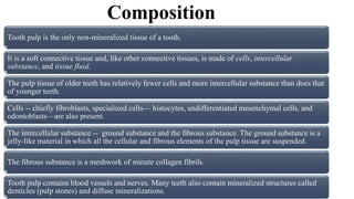
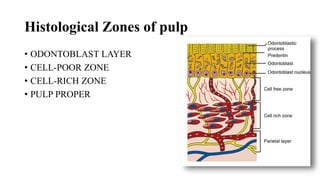
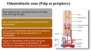
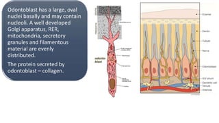
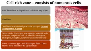
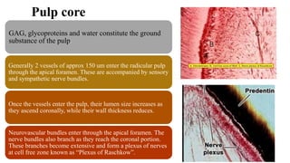
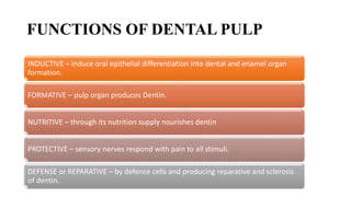
Pulp
DR DINRAJ KULKARNI
BDS, MDS, PGDCR, PGDNT, FICD,
PhD
The Pulp is a soft mesenchymal connective
tissue derived from ectomesenchyme &
occupies pulp cavity in the central portion of
the tooth.
It is enclosed all around by dentin except at the
apical portion of the root, where it
communicates with the apical periodontium.
It is a special organ because of the unique
environment.
Pulp is responsible for vitality of tooth & more
specifically dentin. Anything affects dentin may
have its effects on pulp & the disturbance of
pulp may affect quality and quantity of dentin
During the 8th week of IUL, there is condensation of the
mesenchyme under the enamel organ-Dental papilla.
The enamel organ enlarge and enclose the dental papilla in their
central portion.
Dental papilla controls the morphology & type of tooth to be
formed.
Dental papilla shows : extensive proliferation of cells & high
vascularity.
Following the differentiation of the IEE into ameloblasts,
odontoblast differentiate from the peripheral cells of dental
papilla
Well organized capillaries are found at beginning of
dentinogenesis
GENERALFEATURES
 Total of 52 pulp organs 32 in the permanent and20 in the primary teeth.
 Total pulp volume in permanent teeth is 0.38cc with mean being 0.02CC.
 Eachof these organs hasa shape that conforms to that of the respective tooth.
 Hasability to formdentin throughout life.
 Average life time of primary pulp in oral cavity is – 8.3 years & maximum
life of the deciduous pulp is – 9.6 years.
 Pulp responds to all stimuli by pain because it has only – free nerve
endings
The pulp cavity is divided
into
1. Coronal pulp
2. Radicular pulp
Coronal pulp
It is the pulp occupying the pulp
chamber of the crown of the tooth
In young teeth it resembles the shape of
the outer dentin
It has six surfaces : occlusal, mesial,
distal, buccal, lingual and floor.
Pulp horns are projections into the cusp
This pulp constricts at the cervical region
where it continues as the radicular pulp
Radicular pulp
It is the pulp occupying the pulp canals of the
root of the tooth
In the anterior tooth it is single and in the
posterior teeth it is multiple
The radicular portions of the pulp is
continuous with the periapical tissues through
apical foramen
As age advances the width of the radicular
pulp is reduced, and so is the apical foramen.
Pulp cavity terminates at root apex as small opening
called apical foramen
Radicular pulp continuous with connective tissue of
the periodontium through this foramen.
Diameter in an adult- maxillary teeth-0.4mm &
mandibular teeth-0.3mm
Wide open during development of root.
Sometimes apical opening is found on the lateral side
of the apex
There may be 2-3 foramina split by cementum or
dentin- APICAL DELTA
Accessory canals
Leading laterally from the radicular pulp into the
periodontal tissue.
Present in the apical third of the root sheath cells
Formed due to premature loss of HERS or when
developing root encounters a blood vessel.
Overall occurrence is 33%
May also be present at the furcation region
Location
The pulp, located in the interior of the tooth,
is enclosed within the dentin, with which it
has a close physiologic relationship.
It occupies the pulp cavity; more specifically,
it fills the pulp chamber in the crown and the
root canal in the root and connects with the
periodontal ligament at the apical foramen.
Anatomically, the outline of a young pulp
cavity mirrors the outline of the surface of the
tooth
Origin
The dental papilla (mesenchyme) of the tooth germ
undergoes growth and development and becomes
the tooth pulp.
Cells of the mesenchymal tissue differentiate into
fibroblasts and odontoblasts.
The fibroblasts form and maintain the intercellular
substance of the pulp, and odontoblasts form the
dentin. Blood vessels and nerves that are in the
dental papilla remain to supply the pulp tissue.
Composition
Tooth pulp is the only non-mineralized tissue of a tooth.
It is a soft connective tissue and, like other connective tissues, is made of cells, intercellular
substance, and tissue fluid.
The pulp tissue of older teeth has relatively fewer cells and more intercellular substance than does that
of younger teeth.
Cells -- chiefly fibroblasts, specialized cells— histocytes, undifferentiated mesenchymal cells, and
odontoblasts—are also present.
The intercellular substance -- ground substance and the fibrous substance. The ground substance is a
jelly-like material in which all the cellular and fibrous elements of the pulp tissue are suspended.
The fibrous substance is a meshwork of minute collagen fibrils.
Tooth pulp contains blood vessels and nerves. Many teeth also contain mineralized structures called
denticles (pulp stones) and diffuse mineralizations.
Histological Zones of pulp
• ODONTOBLAST LAYER
• CELL-POOR ZONE
• CELL-RICH ZONE
• PULP PROPER
Odontoblastic zone (Pulp at periphery)
Most characteristic, specialized and non dividing
cells with long life span.
They form peripheral portion of the pulp and lines
the predentin.
Life cycle of odontoblasts starts from the peripheral
Dental papilla cells  preodontoblasts 
odontoblasts.
Stages of odontoblasts reflects their functional
activity i.e. from secretory odontoblasts 
transitional odontoblasts  quiescent odontoblasts.
Types of functional odontoblast
15
Ready for synthesis of dentin
Secretory Odontoblast
Intermediate stage between 2
Transitional Odontoblast
Resting Odontoblast
In the coronal pulp – odontoblasts are columnar, arranged
in a palisaded pattern, and have pseudostratified or
multilayered appearance though only one layer actually
exists. This is due to the different length of the cell bodies
with their nuclei located at different layers.
Odontoblasts extend their cytoplasmic process into the dentin
 each dentinal tubule contains only one odontoblastic
process.
In the coronal pulp at cervical portion they are tall columnar 
in the mid root level are cuboidal or pyramidal in shape  at
the apical portion they are short & flattened.
Number of odontoblasts per unit area in coronal pulp is more.
Cell bodies of of the odontoblasts are connected to each other
& to the cells of cell rich zone & to the cells of pulp core by
cell junctions and complexes (tight, adhering and gap
junctions).
Odontoblast has a large, oval
nuclei basally and may contain
nucleoli. A well developed
Golgi apparatus, RER,
mitochondria, secretory
granules and filamentous
material are evenly
distributed.
The protein secreted by
odontoblast – collagen.
Zone of Weil or cell free zone
This zone appears as space between odontoblastic zone
and cell rich zone. There are no cells in this zone.
Represents the space into which odontoblasts move
during tooth development
40 microns wide &relatively free of cells.
Traversed by,
Blood vessels, unmyelinated nerves, cytoplasmic
process of fibroblasts. The diameter of largest arterial
vessels in the pulp range from – 50-60 um.
Some researcher feel that appearance of cell free zone
is an artefact
Cell rich zone – consists of numerous cells
Zone formed due to migration of cells from pulp proper
Fibroblasts
Undifferentiated mesenchymal cells, pericytes (around
the capillaries of pulp)
Defence cells (histiocytes, macrophages, dendritic cells,
mast cells and plasma cells. In addition, neutrophils,
eosinophils, basophils, lymphocytes and monocytes).
Fibers – contains type I and III collagen fibers. These
become more thicken as the age advances
Pulp core
GAG, glycoproteins and water constitute the ground
substance of the pulp.
Generally 2 vessels of approx 150 um enter the radicular pulp
through the apical foramen. These are accompanied by sensory
and sympathetic nerve bundles.
Once the vessels enter the pulp, their lumen size increases as
they ascend coronally, while their wall thickness reduces.
Neurovascular bundles enter through the apical foramen. The
nerve bundles also branch as they reach the coronal portion.
These branches become extensive and form a plexus of nerves
at cell free zone known as “Plexus of Raschkow”.
Types of nerve fibers
Above the cell free zone, myelinated fibers begin
to lose their myelin sheath.
In the cell free zone, they form a rich network
responsible for pain. Many of these fibers pass
between the odontoblastic zone.
Unmyelinated nerve fibers of pulp are – associated with
blood vessels and are sympathetic in nature.
FUNCTIONS OF DENTAL PULP
INDUCTIVE – induce oral epithelial differentiation into dental and enamel organ
formation.
FORMATIVE – pulp organ produces Dentin.
NUTRITIVE – through its nutrition supply nourishes dentin
PROTECTIVE – sensory nerves respond with pain to all stimuli.
DEFENSE or REPARATIVE – by defence cells and producing reparative and sclerosis
of dentin.
Pulp stones or
Denticles
Appearing in either or both coronal
and root portions of the pulp organ
Develop in teeth that appear to be
normal in other respects.
Asymptomatic unless they impinge
on nerves (or)blood vessels
Seen in functional as well as
embedded or unerupted teeth.
Based on presence or
absence of dentinal
tubules,
True
denticles
False
denticles
Diffuse
calcifications
Based on their
location in the tooth
surface or near pulpal
area
Free
denticles
Attaches
denticles
Embedded
denticles
Age changes
Formation of secondary dentin through out life, reduces the size of the pulp chamber and root
canals
Decrease in cellularity and their size. The intracellular organelles also decrease in number.
Odontoblast decrease in size & number, & may disappear in certain areas. Especially on pulpal
floor over bifurcation & trifurcation.
Increase in number & thickness in collagen fibers, i.e. pulp fibrosis. The fibrosis may be
longitudinal in the root and more diffuse in coronal area
Reduction in the nerve fibers & blood vessels, leads to complete obliteration of root canals.
Atherosclerotic plaque formation is seen in the vessel walls of the pulp tissue.
Dystrophic calcification may be seen in root canals.
4.pulp.pptx

4.pulp.pptx

  • 1.
    Pulp DR DINRAJ KULKARNI BDS,MDS, PGDCR, PGDNT, FICD, PhD
  • 2.
    The Pulp isa soft mesenchymal connective tissue derived from ectomesenchyme & occupies pulp cavity in the central portion of the tooth. It is enclosed all around by dentin except at the apical portion of the root, where it communicates with the apical periodontium. It is a special organ because of the unique environment. Pulp is responsible for vitality of tooth & more specifically dentin. Anything affects dentin may have its effects on pulp & the disturbance of pulp may affect quality and quantity of dentin
  • 3.
    During the 8thweek of IUL, there is condensation of the mesenchyme under the enamel organ-Dental papilla. The enamel organ enlarge and enclose the dental papilla in their central portion. Dental papilla controls the morphology & type of tooth to be formed. Dental papilla shows : extensive proliferation of cells & high vascularity. Following the differentiation of the IEE into ameloblasts, odontoblast differentiate from the peripheral cells of dental papilla Well organized capillaries are found at beginning of dentinogenesis
  • 4.
    GENERALFEATURES  Total of52 pulp organs 32 in the permanent and20 in the primary teeth.  Total pulp volume in permanent teeth is 0.38cc with mean being 0.02CC.  Eachof these organs hasa shape that conforms to that of the respective tooth.  Hasability to formdentin throughout life.  Average life time of primary pulp in oral cavity is – 8.3 years & maximum life of the deciduous pulp is – 9.6 years.  Pulp responds to all stimuli by pain because it has only – free nerve endings
  • 5.
    The pulp cavityis divided into 1. Coronal pulp 2. Radicular pulp
  • 6.
    Coronal pulp It isthe pulp occupying the pulp chamber of the crown of the tooth In young teeth it resembles the shape of the outer dentin It has six surfaces : occlusal, mesial, distal, buccal, lingual and floor. Pulp horns are projections into the cusp This pulp constricts at the cervical region where it continues as the radicular pulp
  • 7.
    Radicular pulp It isthe pulp occupying the pulp canals of the root of the tooth In the anterior tooth it is single and in the posterior teeth it is multiple The radicular portions of the pulp is continuous with the periapical tissues through apical foramen As age advances the width of the radicular pulp is reduced, and so is the apical foramen.
  • 8.
    Pulp cavity terminatesat root apex as small opening called apical foramen Radicular pulp continuous with connective tissue of the periodontium through this foramen. Diameter in an adult- maxillary teeth-0.4mm & mandibular teeth-0.3mm Wide open during development of root. Sometimes apical opening is found on the lateral side of the apex There may be 2-3 foramina split by cementum or dentin- APICAL DELTA
  • 9.
    Accessory canals Leading laterallyfrom the radicular pulp into the periodontal tissue. Present in the apical third of the root sheath cells Formed due to premature loss of HERS or when developing root encounters a blood vessel. Overall occurrence is 33% May also be present at the furcation region
  • 10.
    Location The pulp, locatedin the interior of the tooth, is enclosed within the dentin, with which it has a close physiologic relationship. It occupies the pulp cavity; more specifically, it fills the pulp chamber in the crown and the root canal in the root and connects with the periodontal ligament at the apical foramen. Anatomically, the outline of a young pulp cavity mirrors the outline of the surface of the tooth
  • 11.
    Origin The dental papilla(mesenchyme) of the tooth germ undergoes growth and development and becomes the tooth pulp. Cells of the mesenchymal tissue differentiate into fibroblasts and odontoblasts. The fibroblasts form and maintain the intercellular substance of the pulp, and odontoblasts form the dentin. Blood vessels and nerves that are in the dental papilla remain to supply the pulp tissue.
  • 12.
    Composition Tooth pulp isthe only non-mineralized tissue of a tooth. It is a soft connective tissue and, like other connective tissues, is made of cells, intercellular substance, and tissue fluid. The pulp tissue of older teeth has relatively fewer cells and more intercellular substance than does that of younger teeth. Cells -- chiefly fibroblasts, specialized cells— histocytes, undifferentiated mesenchymal cells, and odontoblasts—are also present. The intercellular substance -- ground substance and the fibrous substance. The ground substance is a jelly-like material in which all the cellular and fibrous elements of the pulp tissue are suspended. The fibrous substance is a meshwork of minute collagen fibrils. Tooth pulp contains blood vessels and nerves. Many teeth also contain mineralized structures called denticles (pulp stones) and diffuse mineralizations.
  • 13.
    Histological Zones ofpulp • ODONTOBLAST LAYER • CELL-POOR ZONE • CELL-RICH ZONE • PULP PROPER
  • 14.
    Odontoblastic zone (Pulpat periphery) Most characteristic, specialized and non dividing cells with long life span. They form peripheral portion of the pulp and lines the predentin. Life cycle of odontoblasts starts from the peripheral Dental papilla cells  preodontoblasts  odontoblasts. Stages of odontoblasts reflects their functional activity i.e. from secretory odontoblasts  transitional odontoblasts  quiescent odontoblasts.
  • 15.
    Types of functionalodontoblast 15 Ready for synthesis of dentin Secretory Odontoblast Intermediate stage between 2 Transitional Odontoblast Resting Odontoblast
  • 16.
    In the coronalpulp – odontoblasts are columnar, arranged in a palisaded pattern, and have pseudostratified or multilayered appearance though only one layer actually exists. This is due to the different length of the cell bodies with their nuclei located at different layers. Odontoblasts extend their cytoplasmic process into the dentin  each dentinal tubule contains only one odontoblastic process. In the coronal pulp at cervical portion they are tall columnar  in the mid root level are cuboidal or pyramidal in shape  at the apical portion they are short & flattened. Number of odontoblasts per unit area in coronal pulp is more. Cell bodies of of the odontoblasts are connected to each other & to the cells of cell rich zone & to the cells of pulp core by cell junctions and complexes (tight, adhering and gap junctions).
  • 17.
    Odontoblast has alarge, oval nuclei basally and may contain nucleoli. A well developed Golgi apparatus, RER, mitochondria, secretory granules and filamentous material are evenly distributed. The protein secreted by odontoblast – collagen.
  • 18.
    Zone of Weilor cell free zone This zone appears as space between odontoblastic zone and cell rich zone. There are no cells in this zone. Represents the space into which odontoblasts move during tooth development 40 microns wide &relatively free of cells. Traversed by, Blood vessels, unmyelinated nerves, cytoplasmic process of fibroblasts. The diameter of largest arterial vessels in the pulp range from – 50-60 um. Some researcher feel that appearance of cell free zone is an artefact
  • 19.
    Cell rich zone– consists of numerous cells Zone formed due to migration of cells from pulp proper Fibroblasts Undifferentiated mesenchymal cells, pericytes (around the capillaries of pulp) Defence cells (histiocytes, macrophages, dendritic cells, mast cells and plasma cells. In addition, neutrophils, eosinophils, basophils, lymphocytes and monocytes). Fibers – contains type I and III collagen fibers. These become more thicken as the age advances
  • 20.
    Pulp core GAG, glycoproteinsand water constitute the ground substance of the pulp. Generally 2 vessels of approx 150 um enter the radicular pulp through the apical foramen. These are accompanied by sensory and sympathetic nerve bundles. Once the vessels enter the pulp, their lumen size increases as they ascend coronally, while their wall thickness reduces. Neurovascular bundles enter through the apical foramen. The nerve bundles also branch as they reach the coronal portion. These branches become extensive and form a plexus of nerves at cell free zone known as “Plexus of Raschkow”.
  • 21.
    Types of nervefibers Above the cell free zone, myelinated fibers begin to lose their myelin sheath. In the cell free zone, they form a rich network responsible for pain. Many of these fibers pass between the odontoblastic zone.
  • 22.
    Unmyelinated nerve fibersof pulp are – associated with blood vessels and are sympathetic in nature.
  • 23.
    FUNCTIONS OF DENTALPULP INDUCTIVE – induce oral epithelial differentiation into dental and enamel organ formation. FORMATIVE – pulp organ produces Dentin. NUTRITIVE – through its nutrition supply nourishes dentin PROTECTIVE – sensory nerves respond with pain to all stimuli. DEFENSE or REPARATIVE – by defence cells and producing reparative and sclerosis of dentin.
  • 24.
    Pulp stones or Denticles Appearingin either or both coronal and root portions of the pulp organ Develop in teeth that appear to be normal in other respects. Asymptomatic unless they impinge on nerves (or)blood vessels Seen in functional as well as embedded or unerupted teeth.
  • 25.
    Based on presenceor absence of dentinal tubules, True denticles False denticles Diffuse calcifications Based on their location in the tooth surface or near pulpal area Free denticles Attaches denticles Embedded denticles
  • 26.
    Age changes Formation ofsecondary dentin through out life, reduces the size of the pulp chamber and root canals Decrease in cellularity and their size. The intracellular organelles also decrease in number. Odontoblast decrease in size & number, & may disappear in certain areas. Especially on pulpal floor over bifurcation & trifurcation. Increase in number & thickness in collagen fibers, i.e. pulp fibrosis. The fibrosis may be longitudinal in the root and more diffuse in coronal area Reduction in the nerve fibers & blood vessels, leads to complete obliteration of root canals. Atherosclerotic plaque formation is seen in the vessel walls of the pulp tissue. Dystrophic calcification may be seen in root canals.