The dental pulp is a soft connective tissue located within the tooth. It supports the dentin and provides nutrition for tooth development. The pulp is divided into the coronal pulp in the crown and the radicular pulp in the roots. The coronal pulp contains pulp horns that extend into tooth cusps while the radicular pulp tapers toward the apical foramen. The pulp contains cells like odontoblasts, fibroblasts, and immune cells and has functions like tooth development, nutrition, sensation, repair, and defense against pathogens. Changes with aging include decreased cell size and number as well as increased fibrosis and calcification. Clinical considerations for the pulp include its shape, size changes with age, and the effects of
This document discusses the stages of amelogenesis, the formation of enamel. It describes 6 stages: 1) morphogenic, 2) differentiating, 3) secretory, 4) maturative, 5) protective, and 6) desmolytic. During the secretory stage, ameloblasts secrete enamel matrix proteins and form Tomes' processes to deposit the matrix along the developing enamel surface. In the maturative stage, ameloblasts engulf the matrix and facilitate its mineralization into mature enamel. The protective stage involves deposition of an enamel cuticle, while in the desmolytic stage, the reduced enamel epithelium aids in tooth eruption.
PHYSICAL PROPERTIES
CHEMICAL PROPERTIES
STRUCTURE OF ENAMEL
DEVELOPMENT OF ENAMEL
EPITHELIAL ENAMEL ORGAN
AMELOGENESIS
LIFE CYCLE OF AMELOBLASTS
AGE CHANGES IN ENAMEL
DEFECTS OF AMELOGENESIS
CLINICAL IMPLICATIONS
The document describes the life cycle of ameloblasts and the process of amelogenesis. There are seven stages in the life cycle of ameloblasts: 1) morphogenic, 2) differentiation, 3) secretory, 4) transitional, 5) maturative, 6) protective, and 7) desmolytic. Amelogenesis involves two processes - formation of the enamel matrix through protein secretion and mineralization through hydroxyapatite deposition and maturation. Defects in amelogenesis can result from conditions like febrile diseases or tetracycline intake, producing malformed or discolored enamel. Enamel undergoes age-related changes like attrition, discoloration, and increased hardness due
Amelogenesis is the process of enamel formation. It occurs in two steps - organic matrix formation and mineralization. During the secretory stage, cells called ameloblasts secrete enamel matrix proteins that make up the organic portion of enamel. In the maturation stage, ameloblasts facilitate the removal of water and organic material from enamel while depositing minerals, resulting in fully mineralized enamel. Ameloblasts undergo morphological and functional changes throughout their lifespan to facilitate the different stages of enamel formation.
The document discusses the peridontium and its components, which include the gingiva, periodontal ligament, cementum, and alveolar bone. It focuses on cementum, describing it as a hard connective tissue that covers tooth roots and provides attachment for collagen fibers. Cementum begins forming at the cementoenamel junction and continues to the root apex. It contains cementoblasts and cementocytes that aid in its formation and structure. Cementum comes in cellular and acellular varieties and demonstrates incremental lines from its continuous deposition over time.
The dental pulp is loose connective tissue located within the tooth. It can be divided into the coronal pulp within the crown and radicular pulp within the root. The pulp contains cellular elements like odontoblasts, fibroblasts, and defensive cells, as well as neurovascular elements. With age, the size of the pulp decreases as secondary dentin is deposited. The number of cells and vascularity also decrease with age. Accessory canals may form due to developmental processes or resorption of tissue during aging. The pulp provides nutrients and defenses to the tooth.
The document summarizes the process of dentinogenesis or dentin formation. It involves differentiation of odontoblasts from dental papilla cells, secretion of an organic matrix, and mineralization of the matrix. Odontoblasts secrete collagen fibers and matrix vesicles that initiate mineralization. Dentin is formed in mantle dentin near enamel and circumpulpal dentin further inside via continuous mineralization. Root dentin formation begins after crown completion, guided by Hertwig's epithelial root sheath.
This document summarizes the structure and formation of enamel. It begins by describing enamel as the hardest substance in the body, composed primarily of hydroxyapatite crystals. It then discusses the composition, structure, and organization of enamel rods and crystals. Hunter-Schreger bands and incremental lines are described as well. The lifecycle of ameloblasts and stages of amelogenesis - morphogenic, organizing, formative, maturative, and protective - are summarized.
The document summarizes the structure and composition of dentin. It discusses the different types of dentin - primary, secondary, tertiary - and their locations and functions. It also describes odontoblasts, the cells responsible for dentin formation, and dentinal tubules, the structures that span the thickness of dentin.
The pulp is a soft connective tissue located within the tooth. It has several unique features, including being surrounded by rigid dentin walls and susceptible to changes in pressure. The pulp contains odontoblasts, fibroblasts, undifferentiated cells, and defense cells. It is highly vascularized and innervated. During development, dental papilla forms the pulp through proliferation and differentiation of cells. The pulp cavity is divided into coronal and radicular regions. Nerves and blood vessels enter through the apical foramen, supplying the pulp.
This document discusses tooth shedding, or the process by which primary teeth are replaced by permanent teeth. It defines shedding as the physiological process by which deciduous teeth are resorbed and lost to make way for successor teeth. Key points covered include the factors affecting shedding like pressure from erupting permanent teeth and genetic factors; the histology of shedding involving resorption of dental hard and soft tissues; the typical pattern of shedding from anterior to posterior teeth; and potential abnormalities in shedding like retained, submerged, or residual primary teeth.
The dental pulp originates from cranial neural crest cells that migrate into the developing tooth germ. During tooth development, these cells form the dental papilla which becomes the dental pulp. The pulp contains odontoblasts, fibroblasts, undifferentiated mesenchymal cells, and macrophages. It has a cell-rich zone containing blood vessels and a cell-free zone near the odontoblasts. The pulp shapes change from development to maturity as the root forms and remodels. It is divided into coronal and radicular portions, connected through the apical foramen.
This document provides information on dentin, including its composition, formation, and types. Some key points:
- Dentin makes up the bulk of the tooth and is composed of 65% inorganic material (mainly hydroxyapatite) and 35% organic material (collagen and proteoglycans).
- Odontoblasts are cells responsible for dentin formation. Their processes extend into dentinal tubules that permeate the dentin.
- Dentin formation begins with predentin, which mineralizes to become circumpulpal dentin. Mantle dentin forms the outer layer near the enamel.
- Dentinal tubules contain peritubular dentin and connect the
The document provides an overview of the pulp-dentin complex, including dentin and pulp. It discusses the physical and chemical properties of dentin, its structure including dentinal tubules and types of dentin. Dentinogenesis and age-related changes are also covered. The morphology, development, zones and cell types in pulp are summarized. The document establishes that dentin and pulp are embryologically, histologically and functionally the same tissue and should be considered as a complex.
The dental pulp contains zones including the odontoblastic zone, cell-free zone, and cell-rich zone. Principal cells include odontoblasts that synthesize dentin, fibroblasts that form the pulp matrix, and immune cells. Blood vessels enter the pulp and branches form capillaries. Nerves form the Raschkow plexus near the odontoblasts. The pulp provides nutrients and sensation to the tooth.
Ameloblast are the enamel forming cells. Understanding of life cycle of ameloblast aids in the understanding of various developmetal anomalies in particular and various other oral pathologies.
The document discusses the anatomy and features of dental pulp. It describes the pulp as a soft connective tissue occupying the pulp cavity at the center of teeth. The pulp is divided into coronal and radicular pulp. The coronal pulp is in the pulp chamber while the radicular pulp occupies the root canals. The document outlines the cell types found in pulp, including odontoblasts, fibroblasts, and immune cells. It also discusses the structural organization and development of pulp.
Enamel is the hardest tissue in the body and is composed primarily of hydroxyapatite crystals. It is formed through the process of amelogenesis by ameloblasts, which transition between secretory and maturation stages as they lay down the enamel matrix and facilitate mineralization. The unique structure of enamel, including enamel rods and interrod enamel, provides hardness but requires the underlying dentin to compensate for enamel's brittleness. The complex life cycle of ameloblasts and specialized enamel proteins are required for this intricate mineralization process and formation of enamel's highly mineralized structure.
The oral cavity is lined by oral mucosa, which is continuous with the skin and pharyngeal mucosa. The oral mucosa consists of three types - masticatory, lining, and specialized mucosa. Masticatory mucosa covers areas involved in chewing and is keratinized, while lining mucosa covers other soft tissue areas and is non-keratinized. Specialized mucosa covers the dorsal tongue and contains papillae involved in taste. The oral mucosa provides protection, sensation, secretion, permeability and thermal regulation functions.
Dentin is the hard tissue that forms the bulk of the tooth beneath enamel. It consists of a bone-like matrix with dentinal tubules that contain odontoblast processes and nerves. Dentin is less mineralized than enamel but provides strength and protects the pulp. The three main theories of dentin hypersensitivity are direct neural stimulation, transduction, and the most accepted hydrodynamic theory, which proposes that fluid movement in the dentinal tubules causes mechanical stimulation of intratubular nerves when exposed dentin is subjected to stimuli.
This document provides information about the maxillary second molar tooth. It discusses the chronology of development, anatomical features including cusp and root aspects, common variations, and pulp cavity morphology. The maxillary second molar has similar buccal and mesial aspects to the first molar but with less divergent roots. It typically has a rhomboidal occlusal outline and four main cusps. The document also notes that difficulty accessing the maxillary second molar is a common dental problem.
Amelogenesis is the process of enamel formation and involves four main stages: secretory, transition, maturation, and protective. During the secretory stage, ameloblasts secrete enamel matrix proteins that form an organic scaffold for enamel mineralization. In the transition stage, ameloblasts transition from a secretory to resorptive phenotype. During maturation, ameloblasts facilitate enamel crystal growth and remove enamel proteins. Finally, in the protective stage, ameloblasts protect mature enamel but have a reduced metabolic activity.
Permanent Mandibular First Premolar and Differences between First and Second ...Dr Monika Negi
This document provides information about the permanent mandibular first premolar and differences between the mandibular first and second premolars. It discusses the general features of premolars, including their transitional location between canines and molars. For the mandibular first premolar, it describes the chronology, number of roots, crown and root outlines, contact areas, and surface anatomy from various aspects. It then compares the mandibular first and second premolars, noting differences in their geometric outlines, facial outlines and surface anatomy, lingual outlines, proximal outlines, occlusal aspects, and pulp cavities.
The dental pulp is a soft connective tissue located within the pulp chamber and root canal of teeth. It develops from the dental papilla early in tooth formation. The pulp contains odontoblasts along its periphery, as well as fibroblasts, macrophages, blood vessels and nerves throughout its connective tissue matrix. It is responsible for dentin formation and sensation in teeth. The pulp narrows as teeth mature and the root forms, with branches extending into the root canal system and exiting through the apical foramen. The pulp plays an important role in tooth function but is also vulnerable to damage.
The pulp is a soft connective tissue found within the tooth that develops from the dental papilla. It occupies the pulp chamber in the crown and root canals in the root. The pulp is responsible for dentin formation and tooth sensitivity. It contains cells like odontoblasts and fibroblasts, nerves, blood vessels, and ground substance. The odontoblastic zone lines the pulp periphery and contains columnar odontoblasts that secrete dentin. With age, secondary dentin formation reduces the pulp size and mineral deposits may form within it.
Indian Dental Academy: will be one of the most relevant and exciting training center with best faculty and flexible training programs for dental professionals who wish to advance in their dental practice,Offers certified courses in Dental implants,Orthodontics,Endodontics,Cosmetic Dentistry, Prosthetic Dentistry, Periodontics and General Dentistry.
The document discusses the dental pulp, including its development, structure, cells, and features. It notes that the dental pulp develops from the dental papilla during tooth formation. The pulp contains coronial and radicular regions, with the radicular pulp terminating at the apical foramen. The pulp has histological zones including the odontoblastic layer and cell-rich and cell-poor zones. Key cells include odontoblasts, fibroblasts, and defense cells. Odontoblasts are responsible for dentin formation and are arranged in palisades along the pulp periphery.
The dental pulp occupies the center of each tooth and consists of soft connective tissue. The pulp has two main parts - the coronal pulp in the crown and the radicular pulp in the root. The coronal pulp contains pulp horns that extend into tooth cusps, while the radicular pulp extends from the crown down the root canal. Blood vessels and nerves enter the pulp through the apical foramen at the root tip. With age, dentin deposition causes the pulp to shrink. Accessory canals may also form connections between the pulp and periodontium. The pulp contains fibroblasts, odontoblasts that form dentin, undifferentiated cells, and defense cells. It functions in tooth development
Indian Dental Academy: will be one of the most relevant and exciting training center with best faculty and flexible training programs for dental professionals who wish to advance in their dental practice,Offers certified courses in Dental implants,Orthodontics,Endodontics,Cosmetic Dentistry, Prosthetic Dentistry, Periodontics and General Dentistry.
The dental pulp is a soft connective tissue located within the tooth. It develops from the dental papilla during tooth formation. The pulp has four zones - the odontoblastic zone containing cells that form dentin, the cell-free zone, cell-rich zone containing many cells, and a central zone with large blood vessels and nerves. The pulp receives blood vessels through the apical foramen and contains many cell types including odontoblasts, fibroblasts, immune cells, and undifferentiated cells. It is highly innervated with sensory fibers that detect pain and sympathetic fibers that control blood flow. The pulp plays key roles in tooth development, defense against infection, and sensitivity.
This document discusses the anatomy, histology, and functions of the dental pulp. It begins by describing the development of the dental papilla and enamel organ during embryogenesis. It then details the four distinct histological zones of the pulp, including the odontoblastic, cell-free, cell-rich, and pulp core zones. Other topics covered include the pulp's vascular, nervous, and lymphatic supply, as well as the composition of its intercellular substance and cells such as odontoblasts, fibroblasts, and defense cells. The document concludes by discussing regressive changes in the aging pulp and applied aspects of pulp preservation during restorative procedures.
The dental pulp is the soft tissue contained within the pulp chamber and root canals of teeth. It is composed of loose connective tissue and nerves that provide sensation and nourishment to the tooth. The pulp contains cells such as odontoblasts, fibroblasts, macrophages, and stem cells embedded within an extracellular matrix. Odontoblasts are responsible for dentin formation and maintenance. With age, the pulp undergoes regressive changes like fibrosis, calcification in the form of pulp stones, and decreased cellularity. Diseases like caries, trauma, and chemical irritation can lead to inflammation of the pulp tissue.
The document discusses the dental pulp. It describes the pulp as the soft connective tissue contained within the pulp chamber and root canals that supports the dentin. The pulp is divided into zones including the odontoblastic zone, cell-free zone, cell-rich zone, and pulp core. The pulp provides inductive, formative, nutritive, sensory, and protective functions to the tooth. Age-related changes in the pulp include reductions in cell number and activity, fibrosis, and formation of pulp stones.
The dental pulp is loose connective tissue located in the center of the tooth that contains blood vessels, nerves, and cells like odontoblasts. It has formative, nutritive, protective, and defensive functions. Over time, the pulp undergoes changes like decreased size and vascularity, increased calcification in the form of pulp stones or diffuse deposits, and reduced vitality.
The document summarizes key information about dental pulp:
1. Dental pulp is the mesenchyme tissue inside the pulp cavity and surrounds the dentin. It contains blood vessels, nerves, fibroblasts, macrophages, and odontoblasts.
2. There are a total of 52 pulp organs in the adult dentition. Molar pulps are 3-4 times larger than incisor pulps. Pulp develops from the dental papilla and is surrounded by dentin except at openings.
3. Histologically, pulp contains four zones - the odontoblastic zone, cell-free zone of Weil, cell-rich zone, and pulp core. Odontoblasts
PULP
• FUNCTIONS OF THE PULP
• Formative: Elaboration of dentin to form the tooth
• Protective: Protection against and repairing of the effectsof noxious stimuli
• Nutritive: Preserving the vitality of all the cellular elements
• Sensory: Perception of stimuli
• ZONES OF PULP
• Starting at the periphery, the pulp is divided into four zones:
• Odontoblastic zone, which surrounds the periphery of
• the pulp
• Cell-free zone
• Cell-rich zone
• Central zone
ODONTOBLASTIC ZONE
• The primary function of the odontoblasts throughout the life of the pulp is the production and deposition of dentin.
• The crowded arrangement of the coronal odontoblasts is due to the rapid reduction of the pulp chamber by the deposition of dentin, which compresses the existing cells to a stratified layer.
• This crowding of odontoblasts produces more cells per unit area and, therefore, more dentinal tubules (45,000/mm2) in the pulpal side than in the enamel side (20,000/mm2).
• The unmyelinated nerves for sensory perception are also found in the pulpal end of the periodontoblastic space of the dentinal tubules
• The incremental lines represent rest periods in dentinogenesis, whereas the interglobular dentin and the granular layer of Tomes probably represent a defect in matrix formation.
• The accentuated incremental line that occurs at birth is called the neonatal line.
• In some areas in the mature dentin, the matrix has not calcified or is hypocalcified. These areas are called interglobular dentin.
• One also sees spaces in the root dentin near the cementodentinal junction called the granular layer of Tomes.
• The dentinal tubules extend from the predentin border to the dentinoenamel and the dentinocemental junctions.
• Dentinal Tubules are conical in shape, with a 2.5 μm mean diameter in the pulpal wall and a 0.9 μm mean diameter in the dentinoenamel or dentinocemental junctions because of the deposition of the peritubular dentin.
• The continuous deposition of peritubular dentinformation of the sclerotic dentin, which has a glassy appearance under transmitted light.
• Primary dentin is elaborated before the teeth erupt and is divided into mantle and circumpulpal dentin.
• Mantle dentin, the first calcified layer of the dentin deposited against the enamel, forms the dentinal side of the dentinoenamel junction.
• Circumpulpal dentin is the dentin formed after the layer of mantle dentin.
• Secondary dentin is elaborated after eruption of the teeth. It can be differentiated from primary dentin by the sharpbending of the tubules producing a line of demarcation.
• secondary dentin is deposited in greater quantities in the floor and roof of the pulp chamber than on the walls.
• Tertiary Dentin
• Two types of tertiary dentin are recognized:
• Tertiary dentin formed by primary odontoblasts following a mild stimulus is called reactionary dentin.
• Tertiary dentin formed by newly differentiated or secondary odontoblasts is termed reparative dentin.
• Reparative dentin,
This document provides an overview of gingival anatomy and histology. It discusses the 3 parts of the gingiva - marginal, attached, and interdental gingiva. Microscopically, it describes the stratified squamous epithelium and underlying connective tissue. The epithelium consists of basal, spinous, granular, and corneal layers. The connective tissue contains collagen fibers, fibroblasts, and ground substance. Gingival fibers attach the gingiva to the tooth cementum. Blood supply and innervation is also summarized.
The dental pulp is the soft connective tissue inside teeth that supports dentin. It has four zones - the odontoblastic zone near the dentin, a cell-free zone beneath it, a cell-rich zone with many cells, and a pulp core containing major blood vessels and nerves. Odontoblasts are distinctive cells that form dentin, with about 60,000 per square millimeter. The pulp also contains fibroblasts that form collagen matrix, undifferentiated cells that give rise to connective tissues, macrophages that eliminate dead cells, lymphocytes, dendritic cells, nerves, and occasionally calcified pulp stones.
The document discusses the structure and development of the dental pulp. It begins by describing the pulp as a soft tissue composed of mesenchymal cells and specialized odontoblasts. The close relationship between odontoblasts and dentin results in the pulp-dentin complex. The primary role of the pulp is to produce dentin, but it also functions as a sensory organ. The document then proceeds to discuss pulp embryology and development, pulp anatomy and histology, innervation and sensitivity, aging changes, and other related topics.
The periodontal ligament is a dense connective tissue located between the cementum and alveolar bone. It contains principal fiber bundles (alveolar, horizontal, oblique, apical, interradicular), fibroblasts, cementoblasts, osteoblasts, osteoclasts, cementoclasts, progenitor cells, epithelial rests of Malassez, macrophages, lymphocytes, plasma cells and nerves and blood vessels. The principal fibers resist various forces on teeth and help maintain them in the alveolar bone.
How to Control Your Asthma Tips by gokuldas hospital.Gokuldas Hospital
Respiratory issues like asthma are the most sensitive issue that is affecting millions worldwide. It hampers the daily activities leaving the body tired and breathless.
The key to a good grip on asthma is proper knowledge and management strategies. Understanding the patient-specific symptoms and carving out an effective treatment likewise is the best way to keep asthma under control.
Summer is a time for fun in the sun, but the heat and humidity can also wreak havoc on your skin. From itchy rashes to unwanted pigmentation, several skin conditions become more prevalent during these warmer months.
STUDIES IN SUPPORT OF SPECIAL POPULATIONS: GERIATRICS E7shruti jagirdar
Unit 4: MRA 103T Regulatory affairs
This guideline is directed principally toward new Molecular Entities that are
likely to have significant use in the elderly, either because the disease intended
to be treated is characteristically a disease of aging ( e.g., Alzheimer's disease) or
because the population to be treated is known to include substantial numbers of
geriatric patients (e.g., hypertension).
5-hydroxytryptamine or 5-HT or Serotonin is a neurotransmitter that serves a range of roles in the human body. It is sometimes referred to as the happy chemical since it promotes overall well-being and happiness.
It is mostly found in the brain, intestines, and blood platelets.
5-HT is utilised to transport messages between nerve cells, is known to be involved in smooth muscle contraction, and adds to overall well-being and pleasure, among other benefits. 5-HT regulates the body's sleep-wake cycles and internal clock by acting as a precursor to melatonin.
It is hypothesised to regulate hunger, emotions, motor, cognitive, and autonomic processes.
Nano-gold for Cancer Therapy chemistry investigatory projectSIVAVINAYAKPK
chemistry investigatory project
The development of nanogold-based cancer therapy could revolutionize oncology by providing a more targeted, less invasive treatment option. This project contributes to the growing body of research aimed at harnessing nanotechnology for medical applications, paving the way for future clinical trials and potential commercial applications.
Cancer remains one of the leading causes of death worldwide, prompting the need for innovative treatment methods. Nanotechnology offers promising new approaches, including the use of gold nanoparticles (nanogold) for targeted cancer therapy. Nanogold particles possess unique physical and chemical properties that make them suitable for drug delivery, imaging, and photothermal therapy.
Travel Clinic Cardiff: Health Advice for International TravelersNX Healthcare
Travel Clinic Cardiff offers comprehensive travel health services, including vaccinations, travel advice, and preventive care for international travelers. Our expert team ensures you are well-prepared and protected for your journey, providing personalized consultations tailored to your destination. Conveniently located in Cardiff, we help you travel with confidence and peace of mind. Visit us: www.nxhealthcare.co.uk
Discover the benefits of homeopathic medicine for irregular periods with our guide on 5 common remedies. Learn how these natural treatments can help regulate menstrual cycles and improve overall menstrual health.
Visit Us: https://drdeepikashomeopathy.com/service/irregular-periods-treatment/
2. INTRODUCTION
• Pulp is a soft
mesenchymal connective
issue that occupies pulp
cavity in the central part
of the teeth
• It supports the dentin and
is unique because of its
unique environment.
• initially it was dental
papilla with increased
amount of capillaries
which provide nutrition
during amelogenesis and
dentinogenesis.
DR.AMANI MAHAJAN
3. Total of 52 pulp organs: 32 in the permanent
and 20 in the primary teeth.
Total pulp volume in permanent teeth is
0.38cc with mean being 0.02CC
Each of these organs has a shape that
conforms to that of the respective tooth.
DR.AMANI MAHAJAN
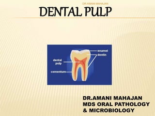
4. The pulp cavity is divided into
1. Coronal pulp
2. Radicular pulp
DR.AMANI MAHAJAN
5. It is located centrally in the crowns.
It has six surfaces: occlusal, mesial, distal, buccal,
lingual and floor.
It has pulp horns, which are protrusions that extend into the cusps of
each tooth. The number of these pulp horns thus depends upon the
number of cusps.
The cervical region of the pulp organs constricts as does the contour
of the crown. At this point coronal pulp joins with the radicular pulp.
Coronal pulp
DR.AMANI MAHAJAN
6. It is present in the roots.
It extends from the cervical region to the root apex.
They are not always straight and vary in size, shape and number.
The dentinal wall taper and shape of the radicular pulp is tubular.
During root formation the root end is wide opening limited by an
epithelial diaphragm.
As growth proceeds, more dentin is formed so that as the tooth
matures, the radicular pulp is narrower.
Apical pulp canal becomes smaller also because of apical
cementum deposition.
Radicular pulp
DR.AMANI MAHAJAN
7. The average size of apical foramen in maxillary teeth in adults is
0.4mm.
In mandibular teeth , it is about 0.3mm .
The location and shape of the apical foramen may undergo changes
as a result of functional influences on the teeth.
Tooth may be tipped from horizontal pressure or it may migrate
mesially. Under such conditions tissue entering pulp through the
apical foramen may exert pressure on the wall of the foramen
causing resorption.
APICAL FORAMEN
DR.AMANI MAHAJAN
8. At the same time cementum is laid down on the opposite
side of the apical root canal resulting in relocation of the
apical foramen.
Sometimes apical foramen may be found on the lateral side
of the apex although the root itself is not curved.
There may be presence of two or more foramina separated
by a portion of dentin and cementum or only by cementum
DR.AMANI MAHAJAN
9. From the radicular pulp they run laterally through the root dentin
to the PDL tissue. They occur in areas where
• there is premature loss of root sheath cells
• Where the developing root encounters a blood vessel.
They act as a route for transmission of infection from the pulpal
tissue to periodontal tissues and vice a versa .Branching pattern of
the small accessory canals and minor foramina seen at the tip or
apex of some tooth roots form the apical delta
ACCESSORY CANALS
DR.AMANI MAHAJAN
10. STRUCTURALORGANIZATION OF PULP
Central region of both radicular and
coronal pulp contains large nerve
trunks and blood vessels.
Peripherally, the pulp is
circumscribed by specialized
odontogenic region composed of:
1. Odontoblasts
2. The cell free zone (of Weil’s)
3. The cell rich zone and
4. The pulp core which is
characterized by major vessels
and nerves
DR.AMANI MAHAJAN
11. ODONTOBLAST LAYER
A layer of odontoblasts are found along
the pulp periphery.
They are dentin forming cells.
DR.AMANI MAHAJAN
12. Immediately subjacent to odontoblastic zone in coronal
pulp there is a narrow zone, approximately 40µm in
width that is relatively free of cells and hence called Cell
free zone of Weils.
It is traversed by blood vessels, unmyelinated nerve
fibers and slender cytoplasmic processes of fibroblasts. It
may not be apparent in young pulp where dentin forms
rapidly or in older pulps where reparative dentin is being
produced.
Cell free zone
DR.AMANI MAHAJAN
13. Below cell free zone a layer containing relatively high
proportions of fibroblasts compared with more central
region of pulp.
it is more prominent in coronal pulp than in radicular
pulp.
It also include large number of macrophages, dendritic
cells and undifferentiated mesenchymal cells.
CELL RICH ZONE
DR.AMANI MAHAJAN
14. The pulp proper is the central mass of pulp.it contains
the large vessels and nerves.
The connective tissue cells in this zone consist of
fibroblasts or pulpal cells
Pulp proper
DR.AMANI MAHAJAN
16. FIBROBLASTS
Fibroblasts are the most numerous cells in the pulp.
They form the collagen fibers, through out the pulp during the life
of the tooth.
The fibroblasts are stellate shaped cells having extensive processes
that contact and are joined by intercellular junctions to the processes
of other fibroblast
DR.AMANI MAHAJAN
17. Young pulp - Fibroblasts have abudant
cytoplasm having numerous cell
organells.
Older pulp - Fibroblasts appear round
or spindle shaped posses short
processes having few cytoplasmic
organelles such cells are called
fibrocytes
DR.AMANI MAHAJAN
18. Dual function :
a) It has capability of ingesting and
degrading the organic matrix.
b) Pathway of both synthesis and degreadation
in the same cell.
DR.AMANI MAHAJAN
19. Primary cells in the very young pulp.
They are polyhedral in shape with peripheral
processes and a large oval nuclei.
They are Totipotent cells i.e. when the need arises
they can be converted into macrophages, odontoblast,
fibroblast etc.
UNDIFFERENTIATED MESENCHYMAL CELLS
DR.AMANI MAHAJAN
20. macrophages, dendritic cells, plasma cells, mast cells, neutrophils,
eosinophils, basophils, lymphocytes, monocytes are present in the
pulp.
Macrophages are Irregularly shaped cell with short blunt processes.
Macrophages appear as large oval or sometimes elongated cells that
exhibit a dark-stained nucleus.
They act as scavengers ,removing extravasated red blood cells, dead
cells and foreign bodies from the tissue. Ingested material is destroyed
by lysosomes.
DEFENSE CELLS
DR.AMANI MAHAJAN
21. Dendritic cells are Antigen-presenting cells. These cells
participate in immunosurveillance and increase in number in
carious teeth, where they infiltrate the odontoblast layer
Plasma cells function in the production of antibodies
Mast cells are widely distributed. Seldom found in normal pulp
tissue although they are routinely found in inflamed pulp
DR.AMANI MAHAJAN
22. ODONTOBLASTS
2nd most prominent cells in the pulp.
A Peripheral area of the pulp where the
odontoblasts reside is termed odontogenic
zone.
Arranged in Palisading pattern cells are tall
columnar forming a layer of 3 to 5 cells in
depth.
Shape may vary
cornal pulp- columnar
midportion - cuboidal
DR.AMANI MAHAJAN
23. These cells have large process extending into
Dentin.
Odontoblasts in the crown are larger than in the
root.
DR.AMANI MAHAJAN
24. Shape of the odontoblasts also reflect the functional
activity of the cell.
During active phase, cells show increase in endoplasmic
reticulum golgi appartus and secretory vesicles.
Resting (or) Non active phase cells are flattened little
cytoplasm condensed chromatin and decrease no of ER
DR.AMANI MAHAJAN
27. INDUCTIVE: Induces oral epithelial differentiation into dental lamina &
enamel organ. Interacts with Enamel organ and determines tooth
morphology
FORMATIVE: pulpal odontoblasts produce dentin which surrounds &
protects it
NUTRITIVE: nourishes dentin through odontoblast by means of blood
vascular system of pulp
PROTECTIVE: when any stimuli like heat, cold, pressure, chemicals is
applied nerves in pulp gets stimulated and produce pain
DEFENSE OR REPARATIVE: responds to irritation by producing
reparative dentin and mineralizing any affected dentinal tubules.
DR.AMANI MAHAJAN
28. REGRESSIVE CHANGES (AGING)
1)CELL CHANGES: The cells are characterized by a decrease in
size and a number of cytoplasmic organelles.
2) FIBROSIS: There is increase in fibers.
3)VASCULAR CHANGES: Atherosclerotic plaques may appear in
pulpal vessels. Calcifications may be seen. Blood flow decreases
with age
4) PULP STONES
DR.AMANI MAHAJAN
29. PULP STONES
Pulp stones are nodular, calcified masses appearing in either or both
the coronal or root portion of the pulp organ.
TRUE DENTICLES
FALSE DENTICLE
DIFFUSE CALCIFICATION
DR.AMANI MAHAJAN
30. TRUE DENTICLES-Structure similar to dentin. Comparatively
rare and usually located close to the apical foramen. Development
is due to inclusion of remnants of epithelial root sheath within the
pulp
FALSE DENTICLES-Do not exhibit dentinal tubules but appear
as concentric layers of calcified tissue
DIFFUSE CALCIFICATIONS-Appear as irregular calcific
deposits in the pulp tissue, usually following collagenous fiber
bundles or blood vessels. Usually found in root canal and less
often in coronal area
DR.AMANI MAHAJAN
31. Pulp stones are also classified according to their location in relation
to the surrounding dentinal wall:
1.Free denticles –present in the pulp
2.Attached denticles-present in between pulp and dentin
3.Embedded denticles-present in the dentin
DR.AMANI MAHAJAN
32. CLINICAL CONSIDERATIONS
Shape of the pulp chamber and its extensions into the
cusps pulpal horns is important.The pulpal horns project
high into the cusps exposure of pulp can occur.
With the advancing age the size of pulp chamber
decreases whereas in young age pulp horns are wider &
high.
Cavity preparation: speed, heat, pressure & coolant may
all cause pulp irritation.
Thickness & nature of remaining dentine may affect pulp
response to dental material. Remaining dentin thickness:
2 mm.
DR.AMANI MAHAJAN
33. CLINICAL CONSIDERATIONS
Shape of the apical foramen and its location may play an
important part in treatment of root canals. Accessory canals
& multiple canals are rarely seen in IOPA.
Irreversible changes occur at temperatures higher than 45
degrees centigrade. It has been noticed that at a
temperature lower than -2 degrees centigrade the pulpal
necrosis can occur.
DR.AMANI MAHAJAN