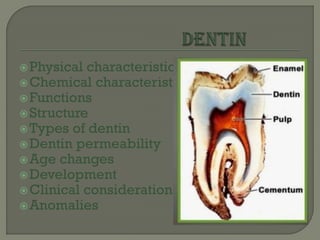
2. Dentin provide the bulk and general
form of the tooth and is characterised as
a hard tissue with tubules throughout its
thickness.
Thickness- 3-10 mm or more
3. Color- light yellowish, becomes darker
with age.
Dentin is viscoelastic and subject to
slight deformation.
Compressive strength- 297 Mpa.
Tensile strength- 52 Mpa.
Elastic modulus- 18 Gpa.
Knoop hardness number- 68.
dentin is somewhat harder in its central
part than near the pulp or its periphery.
4. The lower content of mineral salts in
dentin renders it more radiolucent than
enamel.
Composition: 1. organic matter (35%)
2. inorganic matter(65%)
3. water
7. Provide support to the enamel.
Responsible for transmitting impulses
from enamel or root to the dental pulp.
8. Dentinal tubules-
The course of the dentinal tubules follow a
gentle curve in the crown, less so in the root
where it resembles a gentle S(sigmoid)
shape- primary curvatures.
These tubules end perpendicular to the
dentinoenamel and dentinocementum
junction
branches of dentinal tubules near the
terminals are called terminal branches
which is profuse in root dentin.
9. Dentinal tubules have lateral branches
throughout dentin which are termed
canaliculi or microtubules.
Tubules are larger in diameter near pulpal
cavity(3-4 um) and smaller at their outer ends
(1um).
10.
11. Peritubular dentin:
The dentin that
immediately
surrounds the dentinal
tubules is termed as
peritubular dentin.
Forms walls of tubules.
It is more highly
mineralized.
it is also termed as
intratubular dentin.
12. Between the odontoblastic process and
peritubular dentin, a space-
peridodontoblastic space
This space contain dentinal fluid.
The normal flow of the fluid is outwards
from the pulp.
Dentinal fluid has higher K+ and lower
Na+ content.
Movement of this fluid explains dentin
sensitivity.
13. Intertubular Dentin:
The main body of dentin is
composed of intertubular
dentin.
It is located between the
dental tubules or more
specifically between the
zones of peritubular
dentin.
14. predentin:
The predentin is located
adjacent to the pulp
tissue and is 2 to 6 um
wide.
It is first formed dentin
and is not mineralized
as the collagen fibers
undergo mineralization
at the predentin-dentin
junction , the predentin
becomes dentin and a
new layer of predentin
forms circumpulpally.
15. Odontoblast process:
Odontoblast processes are the cytoplasmic
extensions of the odontoblast.
The odontoblast cells reside in the
peripheral pulp as the pulp predentin
border and their processes extend into the
dentinal tubules.
The processes are largest in diameter near
the pulp(3-4 um) and taper to approx 1um
further into the dentin.
They are 7um in diameter and 40 um in
length.
16.
17. Interglobular dentin:
Sometimes mineralization of dentin begins
in small globular areas that fail to coalesce
into a homogenous mass.This results in
zones of hypomineralization between the
globules.
This dentin forms in crowns of teeth in the
circumpulpal dentin just below mantle
dentin.
It separates mantle dentin from
circumpulpal dentin.
18.
19. Sclerotic dentin:
Occurs in caries,
attrition, abrasion
conditions.
By blocking the
tubules, it reduces
dentin permeability
and thus prolongs the
pulp vitality.
Also called
transparent dentin.
Observed in older
individuals, mostly in
roots.
20. Tome’s granular layer:
When dry ground
sections of root dentin are
visualised in transmitted
light, a zone adjacent to
cementum appears
granular.
Increases in amount from
CEJ to apex.
Formed by coalescing
and looping of dentinal
tubules.
22. 1. Primary dentin: a)Mantle dentin:
Mantle dentin is the name of the first formed
dentin in the crown underlying the
dentinoenamel junction
Provides cushioning effect to the tooth.
It is about 20 um thick.
The largest diameter collagen fibers (0.1-
0.2 um) are argyrophillic.- VON KORFF’s
FIBERS.
Contain type III collagen.
Less mineralized.
Undergoes globular mineralization.
23. 1.Primary dentin: b)Circumpulpal dentin:
Circumpulpal dentin forms the remaining
primary dentin or bulk of the tooth.
The collagen fibrils in circumpulpal dentin
are much smaller in diameter and are more
closely packed together compared to the
mantle.
The circumpulpal dentin contain slightly
more mineral than mantle dentin.
Mineralizes either by globular or linear
pattern.
24. 2.Secondary dentin:
It is a narrow band of dentin bordering
the pulp and representing that dentin
formed after root completion.
Contains fewer tubules.
Due to regular arrangement of dentinal
tubules it is known as regular secondary
dentin.
It grows much slower than primary
dentin.
25. 3.Tertiary dentin:
It is formed as a reaction to external insult
such as caries.The dentin is formed from a
pre-existing odontoblasts.
Also known as reactive dentin
reparative dentin
irritation dentin
replacement dentin
adventitious dentin
defense dentin
26. Incremental lines of von Ebner:
Appear s fine lines or striations in dentin.
They run at right angles to dentin.
The distance between lines varies from
4-8um in crown and closely packed in
roots.
The course of lines show growth pattern
of dentin.
27.
28. Contour lines of Owen:
Some incremental lines
are accentuated
because of
disturbances in the
matrix and
mineralization process.
Such lines are seen in
ground sections.
These lines show
hypocalcified bands.
29. Depends upon patency of dentinal
tubules.
Reduction in dentin permeability would
lessen the sensitivity of dentin.
31. Vitality of dentin – Since the odontoblast and its
process are an integral part of dentin.Dentin is a
vital tissue. Dentin is laid down throughout life,
although after the teeth have erupted and have
been functioning for a short time, dentinogenesis
slows and further dentin formation is at much
slower rate.
Pathologic effect of dental caries, abrasion,
attrition or the cutting of operative procedure
cause changes in dentin.
These are described as the development of dead
tracts , sclerosis and addition of reparative dentin
32. Reparative Dentin – If by extensive abrasion, erosion,
caries or operative procedures the odontoblast
processes are exposed or cut, the odontoblast die or
if they live, deposit reparative dentin.
The majority of odontoblast in this situation
degenerate but a few many continue to form dentin.
Reparative dentin is characterised as having fewer or
more twisted tubules than normal dentin.
It is believed that bacteria , living or dead or their
toxic products as well as chemical substances from
reparative materials, migrate down the pulp and
stimulate pulpal response leading to the reparative
dentin formation.
33. Dead Tracts – In dried ground sections of normal dentin the
odontoblast processes disintegrate,and the empty tubules
are filled with air.They appear black in transmitted and
white in reflected light.
Loss of odontoblast processes may also occur in teeth
containing vital pulp as a result of caries,attrition,abrasion,
cavity,preparation,or erosion.
their degeneration is often observed in the area of narrow
pulpal horns because of crowding of odontoblast.
Again,where reparative dentin seal dentin tubules at their
pulpal ends dentinal tubules fill with fluid or gaseous
subtsances.
Dentin areas characterized by degenerated odontoblast
process give rise to dead tracts.
Dead tracts are probably the initial step in the formation of
sclerotic dentin
34.
35. Lifecycle of odontoblast:
1.Odontoblast Differentiation (Pre-
odontoblasts).
2.Formative (secretory) stage:
a. Mantle dentin formation.
b. Odontoblastic process appearance.
3. Quiescent (resting) stage.
36.
37.
38.
39. Dentinogenesis:
Formation of dentin begins when the tooth germ reach the
bell stage.The dental papilla is the formative organ of
dentin,formed of ectomesenchymal spindle shaped cells in
loose intercellular substance, separated from the inner
dental epithelium by cell free zone.
Dentin is formed by cells called odontoblasts that
differentiate from ectomesenchymal cells of the dental
papilla following an organizing effect (induction) that comes
from the inner dental epithelium.
the dental papilla is the formative organ of dentin and
eventually becomes the pulp of the tooth,a change in
terminology generally associated with the moment of dentin
formation beginning.
40. Dentinogenesis begins at the cusp-tips after the
odontoblast have differentiate and begin
collagen production.As the odontoblast
differentiate , they change and form an ovoid
columnar shape and their nucleus become
basally oriented in this early stage of
development.
As the matrix formation continues the
odontoblast process lengthens as does the
dentinal tubules.Initially,daily increment of
approximately 4 um of dentin are formed
41. 1. Formation of predentin:
The first indication of predentin
formation is the development of bundles
of fibrils among the fully differentiated
odontoblast.These bundles were known
as Von Korff’s fibers, that form the major
component of the first formed thickness
of dentin and are attached to the
basement membrane of the inner dental
epithelium.
42. Once the secretory vesicles secreted
outside the cell, the procollagen molecules
aggregated as large fibers of type I
collagen fibers in ground substance which
is the product of odontoblasts incorporated
with some of pre-existing substance of the
cell free zone to form Mantle dentin.The
large collagen fibrils are 0.1-0.2 µm in
diameter; these fibrils are aligned at right
angles to the basement membrane, while in
the mantle dentin of the root, they are
parallel to it.
43. •Formation of circumpulpal dentin:
Once the layer of mantle dentin is formed, dentinogenesis
continue in a slightly different manner to form circumpulpal
dentin which is the basic structure of dentin and forms its
bulk.The odontoblasts increase in size obliterating the
intercellular spaces with extensive junctional complexes
develops to form distinct row of odontoblasts.
As the matrix is formed,the odontoblasts begin to move
towards the pulp.The plasma membrane of the odontoblasts
adjacent to the inner dental epithelium pushes out several
short processes called Odontoblastic Process (Tome’s
Fiber).Occasionally,one of them may penetrate the
basement membrane and interpose itself between the cells
of the inner dental epithelium to form Enamel Spindle.
44. Mineralization:
First crystal deposition is in form of very fine
plates of hydroxyapatite on the surfaces of
collagen fibrils and in the ground substance.
The crystals associated with the collagen fibrils
are arranged in an orderly fashion,with their
long axes paralleling the fibril long axes and in
rows conforming to 64 nm (640 A degree)
Within the globular islands of mineralization,
crystal deposition appears to take place radially
from common centres so called spherulite form.
45. These are the first sites of calcification of
dentin.
The general calcification process is gradual,
but the peritubular region becomes highly
mineralized at a very early stage.
Dentin sialoprotein present in mineralizing
dentin affects the rate of mineral deposition
while other proteoglycans present more in
predentin, inhibit calcification to prevent
premature calcification of predentin.
46. Dentin has a tubular structure.
Tubules are enlarged by action of microorganism,
alteration of fluid and cellular content of tubules
cause stimulation of nerve ending in contact with
these cell. Caries can spread along DEJ.
the sensitivity of dentin is a important clinical
consideration after placement of restoration that
conduct heat or cold. dentin responds to such
stimuli by changes in the dentin tubules underlying
the restoration.
sensitivity of the tooth will diminish after a few
weeks because of these changes
47. Dentinal tubules increase in size by loss of
intratubular and peritubular dentin.this
dentin is subject to decalcification by caries
or acid cleansing of the cavity, which
removes the smear layer, this dentin is about
40% more calcified than the remainder
dentin
Dentin is permeable hard tissue with
tubules leading from the dentinoenamel
junction to pulp.Therefore, in cavity
preparation sealing of the dentinal tubules
is a requisite of effective resoration.