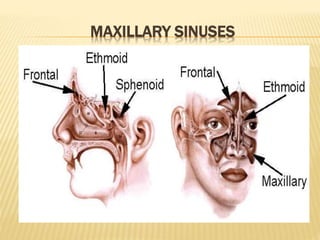
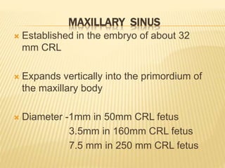
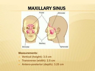
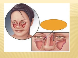
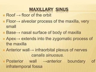
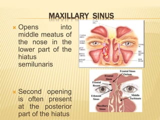
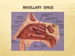
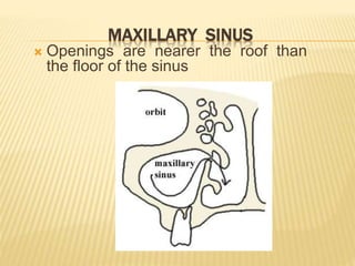
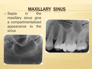
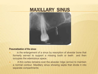
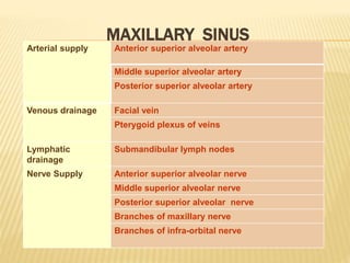
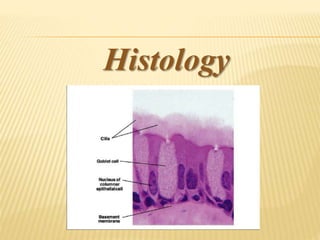
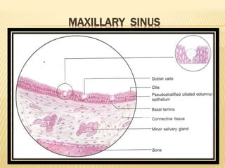
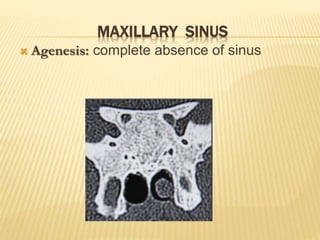
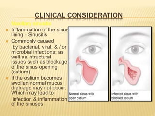
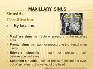
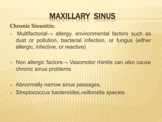
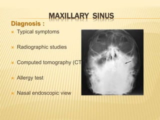
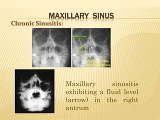
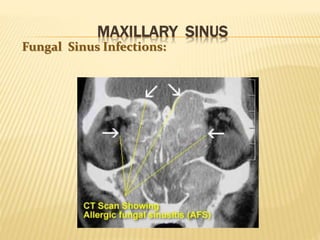
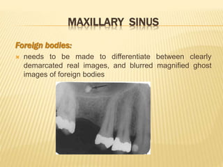
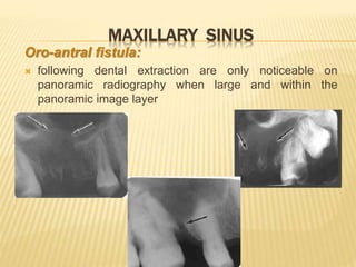
The maxillary sinus is one of the paranasal sinuses located within the body of the maxilla bone. It develops during embryological development and enlarges throughout life through resorption of surrounding bone. The maxillary sinus is lined with ciliated pseudostratified columnar epithelium and can be affected by developmental anomalies, infections like acute or chronic sinusitis, and fungal infections. Maxillary sinusitis presents with pain and tenderness and is diagnosed through symptoms, imaging, and endoscopy.
This document provides an overview of the periodontal ligament (PDL), including its development, cells, extracellular components, fiber groups, and structures. The PDL is a specialized connective tissue that attaches teeth to alveolar bone. It contains fibroblasts that secrete collagen fibers, along with blood vessels, nerves, and progenitor cells. The principal fiber groups resist various forces on teeth. The PDL allows teeth to withstand chewing forces through its extracellular matrix and continual remodeling by synthetic and resorptive cells.
The document discusses the periodontal ligament (PDL), which is the soft connective tissue that surrounds tooth roots and attaches cementum to alveolar bone. It defines PDL and describes its extent, average width, development from the dental follicle, orientation of collagen fibers, cellular elements including fibroblasts, cementoblasts, osteoblasts, and epithelial rests of Mallassez. The document also covers the biochemical composition and ground substance of PDL, as well as its blood supply, nerve supply, age-related changes, and role in healing after periodontal surgery.
The maxillary sinus is the largest of the paranasal sinuses. It is located within the body of the maxilla and has a pyramidal shape. The maxillary sinus develops during fetal development from the maxillary process and reaches its maximum size by age 18. It is important for functions like voice resonance and warming inhaled air. Disease processes like sinusitis, cysts, tumors or dental infections can involve the maxillary sinus. Radiographs are important for evaluating the sinus floor and its relationship to tooth roots. Surgical procedures may be needed to treat conditions like oroantral fistulas or remove foreign bodies from the sinus.
Here are some suggested du'as before and after studying, and during exams:
Before studying:
اللهم أعني على ذكرك وشكرك وحسن عبادتك
O Allah, help me remember You, be grateful to You and worship You in the best way.
اللهم بارك لي في علمي وزدني من فضلك وانفعني بما علمت
O Allah, bless me in my knowledge, increase me in Your bounty and benefit me with what I
Aging causes irreversible changes to the dental hard tissues over time. The three main tissues - enamel, dentin, and cementum - all undergo changes as part of the aging process. Enamel becomes less permeable and more discolored with age. Dentin develops more dead tracts and sclerotic dentin. Cementum may experience hypercementosis and the formation of cementicles. The alveolar bone also undergoes resorption, decreasing in height and width over time. These morphological and functional changes to the dental tissues are a natural part of the biological aging process.
The document discusses the peridontium and its components, which include the gingiva, periodontal ligament, cementum, and alveolar bone. It focuses on cementum, describing it as a hard connective tissue that covers tooth roots and provides attachment for collagen fibers. Cementum begins forming at the cementoenamel junction and continues to the root apex. It contains cementoblasts and cementocytes that aid in its formation and structure. Cementum comes in cellular and acellular varieties and demonstrates incremental lines from its continuous deposition over time.
This document provides information on cementum, including its definition, physical characteristics, chemical composition, formation (cementogenesis), classification, functions, anomalies, and clinical considerations. Cementum is the mineralized tissue covering tooth roots. It is softer than dentin and lacks enamel's luster. Cementum formation involves acellular and cellular stages. Cementum attaches the periodontal ligament fibers to the tooth root and allows for tooth repair. Abnormalities include hypercementosis, ankylosis, and cementomas. Cementum is an important part of the periodontium that aids in tooth attachment and repair.
Dentinogenesis is the formation of dentin, which begins before enamel formation. Dentin is formed by odontoblast cells in two phases: first the formation of an organic collagen matrix, followed by deposition of hydroxyapatite crystals. As dentinogenesis begins, odontoblasts elongate and secrete an unmineralized collagen matrix called predentin. Over time, predentin adjacent to the pulp mineralizes and forms dentin while new predentin is deposited, resulting in incremental dentin growth of approximately 4 micrometers per day. Dentinogenesis continues throughout life but slows after eruption.
The maxillary sinus is an air-filled pyramidal cavity within the body of the maxilla. It develops from evaginations of the nasal cavity epithelium beginning around 12 weeks of gestation. The sinus is lined by ciliated pseudostratified columnar epithelium and contains seromucous glands. Mucociliary flow helps clear secretions from the sinus into the nasal cavity. Clinical considerations involving the maxillary sinus include developmental anomalies, infections, orofacial fistulas involving tooth extraction, and malignant lesions such as various carcinomas.
The pulp is a soft connective tissue located within the tooth. It has several unique features, including being surrounded by rigid dentin walls and susceptible to changes in pressure. The pulp contains odontoblasts, fibroblasts, undifferentiated cells, and defense cells. It is highly vascularized and innervated. During development, dental papilla forms the pulp through proliferation and differentiation of cells. The pulp cavity is divided into coronal and radicular regions. Nerves and blood vessels enter through the apical foramen, supplying the pulp.
This document provides an overview of cementum, including:
- Its physical characteristics, composition, classification, and formation process (cementogenesis).
- The cells involved in cementum formation and maintenance, including cementoblasts and cementocytes.
- Its locations and junctions with other tissues like enamel and dentin.
- The functions of cementum in anchoring teeth, adaptation, and repair.
- Some developmental anomalies and abnormalities that can affect cementum.
This document discusses important anatomical landmarks for complete dentures in the maxilla and mandible. It describes 14 maxillary landmarks including the labial and buccal frenums, vestibules, alveolar ridge, tuberosity, hamular notch, hard palate features, and rugae. It also describes 9 mandibular landmarks like the labial and lingual frenums and vestibules, buccal shelf area, retromolar pad, and pear shaped pad. Understanding these landmarks is essential for proper denture fit and function as well as preservation of underlying tissues.
Coronal and radicular pulp
Apical foramen
Accessory canal
Functions of dental pulp
Components of dental pulp
Functions of pulpal extracellular matrix
Organization of cells in the pulp
The principle cells of the pulp
The pathways of collagen synthesis
Matrix and ground substances
Vasculature and lymphatic supply
Innervation of Dentin- pulp complex
Disorders of the dental pulp
Advances in pulp vitality testing
The document discusses the properties and development of dentin. It begins by introducing dentin and its role in tooth structure. Then it covers the physical and chemical properties of dentin, including its composition, hardness, thickness and density. The stages of dentin development and mineralization are described. Histologically, the key features of dentin are dentinal tubules, peritubular dentin, intertubular dentin and predentin. Structural lines like the dentinoenamel junction and Tome's granular layer are also outlined. Finally, the document notes different types of dentin like mantle dentin.
The periodontal ligament is a connective tissue that connects the tooth to the alveolar bone. It contains collagen fibers, fibroblasts, cementoblasts, osteoblasts and other cells. The principal collagen fibers of the periodontal ligament originate on the cementum and insert into the alveolar bone in different orientations to provide structural support to the tooth and resist various forces. The periodontal ligament is essential for functions such as tooth eruption and maintains the space between the tooth and bone.
The document provides an overview of the maxillary sinus, including its:
1) Definition as the largest air-containing cavity in the maxilla that opens into the nasal cavity.
2) Anatomy as a four-sided pyramid with walls related to surrounding structures like the facial surface of the maxilla.
3) Functions such as warming inhaled air and enhancing facial growth.
It discusses the sinus's histology, blood supply, drainage through the ostium into the nasal cavity, relationships to nearby teeth, and clinical considerations regarding infections.
This document provides an overview of the anatomy, histology, development and clinical implications of alveolar bone. It describes the components and cellular makeup of bone, including osteoblasts, osteocytes and osteoclasts. It explains that the alveolar process develops with tooth eruption and is resorbed after tooth loss. Factors that regulate bone formation and resorption are discussed. The document also outlines how alveolar bone is affected by tooth loss, orthodontic forces and non-functioning teeth.
The dental pulp is loose connective tissue located within the tooth. It can be divided into the coronal pulp within the crown and radicular pulp within the root. The pulp contains cellular elements like odontoblasts, fibroblasts, and defensive cells, as well as neurovascular elements. With age, the size of the pulp decreases as secondary dentin is deposited. The number of cells and vascularity also decrease with age. Accessory canals may form due to developmental processes or resorption of tissue during aging. The pulp provides nutrients and defenses to the tooth.
Cementum is the mineralized connective tissue covering tooth roots. It has several functions including anchoring collagen fibers from the periodontal ligament to provide attachment between the tooth and bone. Cementum can be classified based on its location, cellularity, fiber content, and other characteristics. It plays roles in adaptation, repair, and maintaining the periodontium. The cemento-enamel junction describes the interface between cementum and enamel at the cervical portion of the tooth root.
Maxillary sinus is the largest of the paranasal sinuses. It develops from a shallow groove in the maxilla and reaches its maximum size by age 18. It has multiple walls and communicates with the nasal cavity via the osteum. Maxillary sinusitis can result from dental issues like periapical abscesses, cysts, foreign bodies or trauma. Odontogenic tumors and cysts can also involve the maxillary sinus. Care must be taken during dental procedures near the maxillary sinus to prevent oroantral communications.
The maxillary sinus is the largest of the paranasal sinuses. It is located within the body of the maxilla bone and communicates with the nasal cavity via an opening called the osteum. The maxillary sinus develops during fetal development and reaches its maximum size around 18 years of age. It has thin walls that are in close proximity to important structures like the orbit and teeth. Conditions like sinusitis or cysts can develop within the maxillary sinus and spread to surrounding areas due to its anatomical relationships. Precise knowledge of the maxillary sinus anatomy is important for dental surgeons to avoid complications during procedures involving nearby teeth.
Indian Dental Academy: will be one of the most relevant and exciting training center with best faculty and flexible training programs for dental professionals who wish to advance in their dental practice,Offers certified courses in Dental implants,Orthodontics,Endodontics,Cosmetic Dentistry, Prosthetic Dentistry, Periodontics and General Dentistry.
The development of teeth occurs through a series of stages beginning with the tooth bud. The tooth bud develops into a tooth germ containing three components - the enamel organ, dental papilla, and dental follicle. The tooth germ progresses through bud, cap, and bell stages as the enamel organ invaginates and the dental papilla becomes enclosed. During the bell stage, hard tissues like enamel and dentin begin to form. Root development also occurs during the bell stage directed by Hertwig's epithelial root sheath, forming the periodontal ligament, cementum, and roots.
The document summarizes the oral mucosa and its components. It describes that the oral mucosa lines the oral cavity and provides protection, sensation and secretion. It is made up of epithelium, lamina propria, submucosa and periosteum/bone. The document further describes the different types of oral epithelium and the layers that make up keratinized and non-keratinized oral epithelium. It also summarizes the different types of oral mucosa including lining mucosa and masticatory mucosa, as well as the structures they line.
Alveolar bone is the specialized bone that forms the sockets for teeth in the maxilla and mandible. It consists of alveolar bone proper surrounding the tooth root, supporting alveolar bone made of cortical plates and spongy bone, and bundle bone where periodontal ligament fibers insert. Osteoblasts build bone matrix while osteoclasts resorb it, allowing remodeling. With age, alveolar bone thins with wider marrow spaces and more fragile trabeculae, leading the alveolar crest to slope down distally as teeth tilt mesially.
This document provides information about the maxillary air sinus (antrum). It discusses the embryology, anatomy, functions, clinical importance and diseases of the maxillary sinus. The maxillary sinus begins developing in the fourth month of gestation and reaches adult size by age 18. It is pyramidal in shape with thin walls. Diseases discussed include acute and chronic sinusitis, polyps, cysts and tumors. Surgical procedures for treating maxillary sinus diseases like antral lavage and Caldwell-Luc operation are also summarized.
This document provides an overview of the maxillary sinus, including its anatomy, development, functions, clinical examination, common infections, and considerations for dental implants. The maxillary sinus is the largest paranasal sinus located within the maxilla. It is pyramidal in shape and has boundaries of the orbital floor, lateral nasal wall, and alveolar process. The sinus develops during gestation and pneumatizes after tooth loss. Examination involves transillumination and radiography to identify infections or anatomical variations. Maxillary sinus augmentation may be needed to place implants when bone quantity is insufficient.
The document provides an overview of the maxillary sinus, including its anatomy, development, functions, and relationship to maxillary sinusitis. Key points include:
- The maxillary sinus is an air-filled cavity located within the body of the maxilla.
- It is innervated by branches of the maxillary nerve and supplied by branches of the maxillary artery.
- The maxillary sinus develops after 3 months of gestation and increases in size through childhood and adolescence.
- Maxillary sinusitis can be acute or chronic and is usually caused by infection, allergy, or dental issues like an infected tooth root extending into the sinus.
Maxillary sinus diseases are presented by Dr. Vishal Modha. The maxillary sinus is the largest paranasal sinus located within the maxilla bone. It develops embryologically from the lateral nasal wall and grows postnatally. The maxillary sinus anatomy includes thin bony walls that are vulnerable to trauma and contain important structures. Mucociliary drainage flows from the maxillary sinus ostium to the nasal cavity. Common maxillary sinus diseases include acute or chronic sinusitis, which can result from dental infections, trauma, or nasal obstruction and cause symptoms like facial pain and nasal congestion. Radiographs and CT scans may reveal mucosal thickening or opacification in sinusitis. Treatment involves antibiotics,
1. The maxillary sinus is an air-filled space within the body of the maxilla bone that communicates with the nasal cavity.
2. It develops from the 4th month of gestation and reaches its maximum size by 18 years of age.
3. Common pathologies of the maxillary sinus include acute or chronic sinusitis, odontogenic cysts such as dentigerous or radicular cysts, and benign or malignant tumors.
4. Diagnostic evaluation of sinus disease involves medical history, clinical examination including transillumination, and radiographic imaging like panoramic radiograph, CT scan, or MRI.
The document discusses the paranasal sinuses. There are four pairs of paranasal sinuses located around the nasal cavity that develop from invaginations of the nasal cavity mucosa into the bones. The maxillary sinus is the largest sinus and most clinically relevant as it is close to the teeth. Sinusitis is inflammation of the sinuses which can be acute or chronic and is usually caused by infection, trauma, dental issues or tumors. Anatomical relationships between the sinuses and structures like the orbit and teeth are important for dentists to understand.
This document provides an overview of the maxillary sinus, including its discovery, anatomy, development, functions, and associated pathologies. Some key points:
- The maxillary sinus was first discovered and illustrated by Leonardo da Vinci, but was described in detail by Nathaniel Highmore in 1651.
- It is a pyramid-shaped air space within the body of the maxilla. It is bounded by the zygomatic process, nasal surface, orbital surface, and alveolar process.
- Development begins in the newborn as a tubular structure, becoming ovoid in childhood and pyramidal in adults.
- Pathologies associated with the maxillary sinus include sinusitis, cyst
The maxillary sinus is the largest of the paranasal sinuses. It develops within the body of the maxilla and communicates with the nasal cavity via the ostium in the middle meatus. The maxillary sinus has important anatomical relationships with surrounding structures like the orbit, teeth roots, and nerves. Diagnostic evaluation of the maxillary sinus involves medical history, clinical examination including transillumination, and imaging modalities like radiography, CT, MRI, ultrasound, and endoscopy.
The maxillary sinus is the largest paranasal air sinus located in the body of the maxilla. It has a pyramidal shape with its base facing medially and apex extending laterally. The roof is formed by the orbital floor and the thin floor corresponds to the level of the ala of the nose, with tooth roots sometimes projecting into it. Drainage occurs through the hiatus semilunaris in the middle meatus. Issues like sinusitis, oroantral fistula, and reduced alveolar ridge volume after tooth extraction are important clinical considerations related to the maxillary sinus.
The document discusses the maxillary sinus, including its functions, structure, development, vascular and nerve supply, diseases, and sinusitis. The maxillary sinus (also called the antrum of Highmore) is the largest of the paranasal sinuses. It is pyramidal in shape and located behind the cheekbone. Sinusitis occurs when the sinuses become blocked and inflamed, usually due to a viral or bacterial infection. Symptoms include nasal congestion, facial pain, and headaches. Diagnosis involves imaging like CT scans. Treatment involves medications like antibiotics, decongestants, corticosteroids, and surgery if medications are not effective.
The document summarizes the anatomy and physiology of the nose. It describes the three main parts of the nose - the external nose, nasal cavity, and nasal sinuses. It discusses the bones, cartilages, walls, openings and drainage pathways of the nose. It also covers the vascular, nerve and lymphatic supply, functions, mucociliary clearance and disorders like allergic rhinitis.
The document provides an overview of the anatomy of the nose and paranasal sinuses. It describes the external structures of the nose including the nasal bones, cartilages, and openings. Internally, it details the nasal cavity including its walls, floors, meatuses and contents. The paranasal sinuses are then outlined, specifically the maxillary, frontal, sphenoid and ethmoid sinuses. Finally, common clinical conditions involving the nose and sinuses like sinusitis, nosebleeds and rhinitis are briefly discussed.
The maxillary sinus is the largest of the paranasal sinuses. It is located below the cheeks and above the teeth. Maxillary sinusitis is usually caused by infection, trauma, or dental issues like cysts or displaced roots. Symptoms include pain, nasal discharge, and tenderness of teeth. Diagnosis involves examination, transillumination, and radiography. Treatment may include antibiotics, surgery like Caldwell-Luc to drain pus, or repair of oroantral fistulas caused by tooth extractions. Complications can arise if fistulas are not properly closed.
1) Sinusitis is inflammation of the paranasal sinuses, the air-filled cavities around the nose. The four main sinus cavities are the maxillary, frontal, ethmoid, and sphenoid sinuses.
2) Acute sinusitis is caused by viral or bacterial infection following a cold or allergy. Symptoms include facial pain, nasal congestion, and discolored nasal discharge. Maxillary sinusitis commonly involves pain in the cheek.
3) Treatment involves antibiotics, nasal decongestants, pain relievers, and surgery if symptoms persist. Surgical treatments include antral puncture to drain pus from the maxillary sinus or functional endoscopic sinus surgery. Comp
The document summarizes key information about dental pulp:
1. Dental pulp is the mesenchyme tissue inside the pulp cavity and surrounds the dentin. It contains blood vessels, nerves, fibroblasts, macrophages, and odontoblasts.
2. There are a total of 52 pulp organs in the adult dentition. Molar pulps are 3-4 times larger than incisor pulps. Pulp develops from the dental papilla and is surrounded by dentin except at openings.
3. Histologically, pulp contains four zones - the odontoblastic zone, cell-free zone of Weil, cell-rich zone, and pulp core. Odontoblasts
MANDIBULAR CENTRAL AND LATERAL INCISOR.pptsmithanaik1980
The document describes the anatomy and features of the mandibular central and lateral incisors. It discusses that the mandibular central incisor is the smallest tooth and is bilaterally symmetrical, while the lateral incisor is slightly larger with an incisal edge that declines distally. Key distinguishing features between the two incisors include the lateral having a lower distal contact point, distally tipped crown, and distolingual twist of the incisal edge. Both assist in biting and cutting food from the front view appearing as narrow teeth with a straight incisal edge and tapering sides.
The document describes the anatomical features of human canine teeth. It notes that canines have a thick, conical crown with a long, flattened root. There are two canines in each jaw, located between the lateral incisors and first premolars. Canines are the longest teeth in the dental arch and have a single cusp. The document then describes the anatomical features of both the maxillary and mandibular canines in detail.
The oral mucosa is the moist lining of the oral cavity that continues with the skin and esophagus. It has three main functions - protection, sensation, and secretion. It protects the deeper tissues from mechanical forces and toxins, senses stimuli like temperature and pain, and secretes saliva through minor salivary glands. The oral mucosa varies between keratinized mucosa covering areas like the gingiva and hard palate, non-keratinized mucosa in areas like the floor of the mouth and cheeks, and specialized mucosa bearing taste buds on the tongue.
The dental pulp is the soft tissue contained within the pulp chamber and root canals of teeth. It is composed of loose connective tissue and nerves that provide sensation and nourishment to the tooth. The pulp contains cells such as odontoblasts, fibroblasts, macrophages, and stem cells embedded within an extracellular matrix. Odontoblasts are responsible for dentin formation and maintenance. With age, the pulp undergoes regressive changes like fibrosis, calcification in the form of pulp stones, and decreased cellularity. Diseases like caries, trauma, and chemical irritation can lead to inflammation of the pulp tissue.
The document describes the development of teeth from the embryonic stage through eruption. It discusses the key stages of odontogenesis including the initiation of primary tooth buds between 6-8 weeks in utero, and permanent teeth beginning at 20 weeks. The primary stages of development discussed are the bud stage, cap stage, and bell stage. The bud stage is characterized by thickening of the dental lamina. In the cap stage the enamel organ takes on a cap shape with the formation of the dental papilla and follicle inside. In the bell stage the shape of the tooth crown is defined and cells differentiate into ameloblasts and odontoblasts.
share - Lions, tigers, AI and health misinformation, oh my!.pptxTina Purnat
• Pitfalls and pivots needed to use AI effectively in public health
• Evidence-based strategies to address health misinformation effectively
• Building trust with communities online and offline
• Equipping health professionals to address questions, concerns and health misinformation
• Assessing risk and mitigating harm from adverse health narratives in communities, health workforce and health system
Rasamanikya is a excellent preparation in the field of Rasashastra, it is used in various Kushtha Roga, Shwasa, Vicharchika, Bhagandara, Vatarakta, and Phiranga Roga. In this article Preparation& Comparative analytical profile for both Formulationon i.e Rasamanikya prepared by Kushmanda swarasa & Churnodhaka Shodita Haratala. The study aims to provide insights into the comparative efficacy and analytical aspects of these formulations for enhanced therapeutic outcomes.
Recomendações da OMS sobre cuidados maternos e neonatais para uma experiência pós-natal positiva.
Em consonância com os ODS – Objetivos do Desenvolvimento Sustentável e a Estratégia Global para a Saúde das Mulheres, Crianças e Adolescentes, e aplicando uma abordagem baseada nos direitos humanos, os esforços de cuidados pós-natais devem expandir-se para além da cobertura e da simples sobrevivência, de modo a incluir cuidados de qualidade.
Estas diretrizes visam melhorar a qualidade dos cuidados pós-natais essenciais e de rotina prestados às mulheres e aos recém-nascidos, com o objetivo final de melhorar a saúde e o bem-estar materno e neonatal.
Uma “experiência pós-natal positiva” é um resultado importante para todas as mulheres que dão à luz e para os seus recém-nascidos, estabelecendo as bases para a melhoria da saúde e do bem-estar a curto e longo prazo. Uma experiência pós-natal positiva é definida como aquela em que as mulheres, pessoas que gestam, os recém-nascidos, os casais, os pais, os cuidadores e as famílias recebem informação consistente, garantia e apoio de profissionais de saúde motivados; e onde um sistema de saúde flexível e com recursos reconheça as necessidades das mulheres e dos bebês e respeite o seu contexto cultural.
Estas diretrizes consolidadas apresentam algumas recomendações novas e já bem fundamentadas sobre cuidados pós-natais de rotina para mulheres e neonatos que recebem cuidados no pós-parto em unidades de saúde ou na comunidade, independentemente dos recursos disponíveis.
É fornecido um conjunto abrangente de recomendações para cuidados durante o período puerperal, com ênfase nos cuidados essenciais que todas as mulheres e recém-nascidos devem receber, e com a devida atenção à qualidade dos cuidados; isto é, a entrega e a experiência do cuidado recebido. Estas diretrizes atualizam e ampliam as recomendações da OMS de 2014 sobre cuidados pós-natais da mãe e do recém-nascido e complementam as atuais diretrizes da OMS sobre a gestão de complicações pós-natais.
O estabelecimento da amamentação e o manejo das principais intercorrências é contemplada.
Recomendamos muito.
Vamos discutir essas recomendações no nosso curso de pós-graduação em Aleitamento no Instituto Ciclos.
Esta publicação só está disponível em inglês até o momento.
Prof. Marcus Renato de Carvalho
www.agostodourado.com
Basavarajeeyam is a Sreshta Sangraha grantha (Compiled book ), written by Neelkanta kotturu Basavaraja Virachita. It contains 25 Prakaranas, First 24 Chapters related to Rogas& 25th to Rasadravyas.
These lecture slides, by Dr Sidra Arshad, offer a quick overview of the physiological basis of a normal electrocardiogram.
Learning objectives:
1. Define an electrocardiogram (ECG) and electrocardiography
2. Describe how dipoles generated by the heart produce the waveforms of the ECG
3. Describe the components of a normal electrocardiogram of a typical bipolar lead (limb II)
4. Differentiate between intervals and segments
5. Enlist some common indications for obtaining an ECG
6. Describe the flow of current around the heart during the cardiac cycle
7. Discuss the placement and polarity of the leads of electrocardiograph
8. Describe the normal electrocardiograms recorded from the limb leads and explain the physiological basis of the different records that are obtained
9. Define mean electrical vector (axis) of the heart and give the normal range
10. Define the mean QRS vector
11. Describe the axes of leads (hexagonal reference system)
12. Comprehend the vectorial analysis of the normal ECG
13. Determine the mean electrical axis of the ventricular QRS and appreciate the mean axis deviation
14. Explain the concepts of current of injury, J point, and their significance
Study Resources:
1. Chapter 11, Guyton and Hall Textbook of Medical Physiology, 14th edition
2. Chapter 9, Human Physiology - From Cells to Systems, Lauralee Sherwood, 9th edition
3. Chapter 29, Ganong’s Review of Medical Physiology, 26th edition
4. Electrocardiogram, StatPearls - https://www.ncbi.nlm.nih.gov/books/NBK549803/
5. ECG in Medical Practice by ABM Abdullah, 4th edition
6. Chapter 3, Cardiology Explained, https://www.ncbi.nlm.nih.gov/books/NBK2214/
7. ECG Basics, http://www.nataliescasebook.com/tag/e-c-g-basics
Cell Therapy Expansion and Challenges in Autoimmune DiseaseHealth Advances
There is increasing confidence that cell therapies will soon play a role in the treatment of autoimmune disorders, but the extent of this impact remains to be seen. Early readouts on autologous CAR-Ts in lupus are encouraging, but manufacturing and cost limitations are likely to restrict access to highly refractory patients. Allogeneic CAR-Ts have the potential to broaden access to earlier lines of treatment due to their inherent cost benefits, however they will need to demonstrate comparable or improved efficacy to established modalities.
In addition to infrastructure and capacity constraints, CAR-Ts face a very different risk-benefit dynamic in autoimmune compared to oncology, highlighting the need for tolerable therapies with low adverse event risk. CAR-NK and Treg-based therapies are also being developed in certain autoimmune disorders and may demonstrate favorable safety profiles. Several novel non-cell therapies such as bispecific antibodies, nanobodies, and RNAi drugs, may also offer future alternative competitive solutions with variable value propositions.
Widespread adoption of cell therapies will not only require strong efficacy and safety data, but also adapted pricing and access strategies. At oncology-based price points, CAR-Ts are unlikely to achieve broad market access in autoimmune disorders, with eligible patient populations that are potentially orders of magnitude greater than the number of currently addressable cancer patients. Developers have made strides towards reducing cell therapy COGS while improving manufacturing efficiency, but payors will inevitably restrict access until more sustainable pricing is achieved.
Despite these headwinds, industry leaders and investors remain confident that cell therapies are poised to address significant unmet need in patients suffering from autoimmune disorders. However, the extent of this impact on the treatment landscape remains to be seen, as the industry rapidly approaches an inflection point.
6. PARANASAL SINUSES
Arranged in pairs
Except ethmoidal sinuses-3 groups of air-filled
spaces
All of them open into the nasal cavity through its
lateral wall
Lined by mucous membrane consisting of
ciliated columnar epithelium
7. PARANASAL SINUSES
Pneumatic diverticula from the nasal cavity
Process begins prenatally
Continues through the course of an organism's lifetime
At birth→ rudimentary/ absent. Enlarge rapidly →6-7
years. (during time of eruption of permanent tooth and
then after puberty)
Birth to adult life, the growth→ resorption of the
surrounding cancellous bone
8. MAXILLARY SINUS
Also k/a ANTRUM OF HIGHMORE.
The pneumatic space that is lodged inside the body
of the maxilla and that communicates with the
environment by way of the middle nasal meatus and
the nasal vestibule
11. MAXILLARY SINUS
Horizontal shift of palatal shelves
Fusion of the 2 shelves with one
another
Fusion with the nasal septum
Nasal septum separate the secondary
oral cavity from 2 secondary nasal
chambers
Folding of lateral nasal wall→ 3 nasal
conchae & 3 nasal meatus
13. MAXILLARY SINUS
Established in the embryo of about 32
mm CRL
Expands vertically into the primordium of
the maxillary body
Diameter -1mm in 50mm CRL fetus
3.5mm in 160mm CRL fetus
7.5 mm in 250 mm CRL fetus
17. MAXILLARY SINUS
Roof → floor of the orbit
Floor→ alveolar process of the maxilla, very
small
Base→ nasal surface of body of maxilla
Apex→ extends into the zygomatic process of
the maxilla
Anterior wall→ infraorbital plexus of nerves
canalis sinuosus.
Posterior wall →anterior boundary of
infratemporal fossa
18. MAXILLARY SINUS
Opens into
middle meatus of
the nose in the
lower part of the
hiatus
semilunaris
Second opening
is often present
at the posterior
part of the hiatus
21. MAXILLARY SINUS
Septa in the
maxillary sinus give
a compartmentalized
appearance to the
sinus
22. MAXILLARY SINUS
Pneumatization of the sinus:
is the enlargement of a sinus by resorption of alveolar bone that
formerly served to support a missing tooth or teeth and then
occupies the edentulous space.
A thin cortex remains over the alveolar ridge (arrow) to maintain
a normal contour. Maxillary sinus showing septa that divide it into
separate compartments
23. MAXILLARY SINUS
Arterial supply Anterior superior alveolar artery
Middle superior alveolar artery
Posterior superior alveolar artery
Venous drainage Facial vein
Pterygoid plexus of veins
Lymphatic
drainage
Submandibular lymph nodes
Nerve Supply Anterior superior alveolar nerve
Middle superior alveolar nerve
Posterior superior alveolar nerve
Branches of maxillary nerve
Branches of infra-orbital nerve
26. HISTOLOGY:
3 microscopically distinct layers surround the
space of the maxillary sinus:
Epithelial layer
Basal lamina
Sub epithelial layer- including the periosteum
27. HISTOLOGY
. It is lined by pseudo stratified ciliated
columnar epithelium.
.
The epithelial layer is mainly composed of
ciliated columnar cells. Along with these,
there are non ciliated columnar cells, basal
cells and goblet cells.
29. GOBLET CELL
Goblet cells are unicellular secretary organ
which are goblet shaped with a basally
placed nucleus and apical cytoplasm filled
with secretory products, producing mucous.
In a hematoxylyn and eosin stained section
cytoplasm of goblet cells appear empty. Cilia
of the lining epithelium help to move the
secretions.
30. HISTOLOGY
This epithelium is separated from sub
epithelial connective tissue by a basal
lamina.
Sub epithelial connective tissue layer has
collagen fibers and fibroblasts and also minor
salivary glands which include both serous
and mucous glands. This layer is attached to
the periosteum of the bone of the maxilla.
31. MAXILLARY SINUS
Important Histologic features:
1. Pseudo stratified ciliated columnar
epithelium
2. Goblet cells
3. Both serous & mucous minor salivary
glands
33. MAXILLARY SINUS
Accessory space to nasal cavity
Humidifying & heating of inhaled air
Protects internal structures like brain, &
eyeball against exposure to cold air
Resonance of voice
Lightening of skull weight
Resistance to mechanical shock
38. MAXILLARY SINUS
Aplasia/ hypoplasia:
altered/ under development –alone or in
association with other anomalies like-
cleft palate, high palate, septal deformity,
absence of concha, mandibulofacial
dysostosis
39. CLINICAL CONSIDERATION
Maxillary sinusitis
Inflammation of the sinus
lining - Sinusitis
Commonly caused
by bacterial, viral, & / or
microbial infections; as
well as, structural
issues such as blockage
of the sinus opening
(ostium).
If the ostium becomes
swollen normal mucus
drainage may not occur.
Which may lead to
infection & inflammation
of the sinuses
40. It causes headache with thick purulent
persistent discharge from nose
Diagnosed by:- transillumination &
radiograph
In chronic cases:- superior alveolar nerve
is involoved & may cause pain.
(Examination of maxillary sinus &
maxillary teeth is required—due to
pathology)
41. RELATIONSHIP OF MAXILLARY SINUS &
MAXILLARY MOLAR
The roots of the
maxillary first and
second molars are in
intimate relation to the
floor of the maxillary
sinus .
42. MAXILLARY SINUS
Infections:
Sinusitis:
an inflammation of the paranasal sinuses, which may or may not
be as a result of infection from bacterial, fungal, viral, allergic or
autoimmune issues
-(Wikipedia, the free encyclopedia)
Rhinosinusitis:
Newer classifications of sinusitis refer to it as rhinosinusitis
Taking into account the thought that inflammation of the
sinuses cannot occur without some inflammation of the nose as
well (rhinitis)
(-Ramadan H, Sanclement J, Thomas J (2005). “Chronic rhinosinusitis and
biofilms.". Otolaryngol Head Neck Surg 132 (3): 414–7)
44. MAXILLARY SINUS
Sinusitis:
Classification:
A. By location:
Maxillary sinusitis - pain or pressure in the maxillary
area
Frontal sinusitis - pain or pressure in the frontal sinus
cavity
Ethmoid sinusitis - pain or pressure pain
between/behind eyes
Sphenoid sinusitis - pain or pressure behind the eyes,
but often refers to the vertex of the head
45. MAXILLARY SINUS
Sinusitis:
Classification:
B. By duration:
Acute: going on less than four weeks
Subacute: between 4-12 weeks
Chronic: going on for 12 weeks or more
(-Harrison's Manual of Medicine 16/e)
46. MAXILLARY SINUS
Acute Sinusitis:
Precipitated by an earlier URT infection→ viral
Most common: Haemophilus influenzae, Streptococcus
pneumoniae, Moraxella catarrhalis, Staphylococcus
aureus
Others: dental problems, fungal invasion, diabetes or
other immune deficiencies
Blocking of openings to the sinuses, allergies
Maxillary sinusitis caused by apical
infection and extensive periodontal
lesions involving the molars and
premolar
47. MAXILLARY SINUS
Symptoms :
Pain, tenderness, and swelling
Yellow or green pus
Fever & chills
Change in vision or swelling around the eye→
very serious condition
48. CONTD……
Histologic feautures:
Acute inflammatory infiltrate with edema of
the connective tissue
Squamous metaplasia of the specialized
ciliated columnar epithelium
49. MAXILLARY SINUS
Acute Sinusitis:
Diagnosis :
Typical symptoms
Radiographic studies
Computed tomography (CT)
Check for dental abscesses
Endoscopic view
Left-sided maxillar sinusitis
(Absence of the air transparency
of left maxillar sinus)
50. MAXILLARY SINUS
Management:
Antihistamine along with decongestant or pain
reliever
Antibiotics
Rest
Nasal irrigation/sparys
Hot drinks
Most cases of acute sinusitis, antibiotics and
nasal corticosteriods work no better than a
placebo”
Pulsating nasal irrigator
(Ian G. Williamson et al. "Antibiotics and Topical Nasal Steroid for
Treatment of Acute Maxillary Sinusitis". JAMA 2007; 298: 2487–249)
51. MAXILLARY SINUS
Chronic Sinusitis:
Multifactorial→ allergy, environmental factors such as
dust or pollution, bacterial infection, or fungus (either
allergic, infective, or reactive)
Non allergic factors→ Vasomotor rhinitis can also cause
chronic sinus problems
Abnormally narrow sinus passages,
Streptococcus bacteroides,veillonella species
54. MAXILLARY SINUS
Fungal Sinus Infections:
Fungi can cause significant nasal and sinus
inflammation
“Triggers” & incite an inflammatory response in
susceptible individuals
Fungus balls are an overgrowth of fungi in
otherwise healthy people
Symptoms: sinus pain, pressure, nasal
congestion, drainage of fluids, chronic infections
56. MAXILLARY SINUS
Fungal Sinus Infections:
Allergic fungal sinusitis:
Fungi cause a reaction characterized by marked
nasal congestion and the formation of nasal and
sinus polyps
Polyps obstruct the nose and the openings to
the sinuses and produce chronic inflammation
Involve only one side of the nose
Management: Surgery/corticosteroids/
antibiotics/ antifungal drugs
57. MAXILLARY SINUS
Fungal Sinus Infections:
Invasive fungal sinusitis:
Very serious disorder
In those immune system is impaired by
chemotherapy or by diseases such as poorly
controlled diabetes, leukemia, lymphoma, multiple
myeloma, or AIDS
Symptoms: pain, fever, and discharge of pus from
the nose
The fungus may spread to the eye socket, causing a
bulging of the affected eye (proptosis) and blindness
Management: Biopsy, surgery, antifungal drugs
62. MAXILLARY SINUS
Antrolith:
•Antroliths are calcified
masses found in the
maxillary sinus
•They are formed by
deposition of calcific material
on a nidus such as a root
fragment, bone chip, foreign
object, or a mass of stagnant
mucus in sites of previous
inflammation
63. MAXILLARY SINUS
Foreign bodies:
needs to be made to differentiate between clearly
demarcated real images, and blurred magnified ghost
images of foreign bodies
64. MAXILLARY SINUS
Oro-antral fistula:
following dental extraction are only noticeable on
panoramic radiography when large and within the
panoramic image layer
66. MAXILLARY SINUS
The growth of tumors within the maxilla is not
concentric
Hence, the site of origin is not necessarily the
epicenter of the lesion
The early detection of insidious maxillary sinus
disease can be very important for the patient’
prognosis, especially in the case of malignant
neoplasia