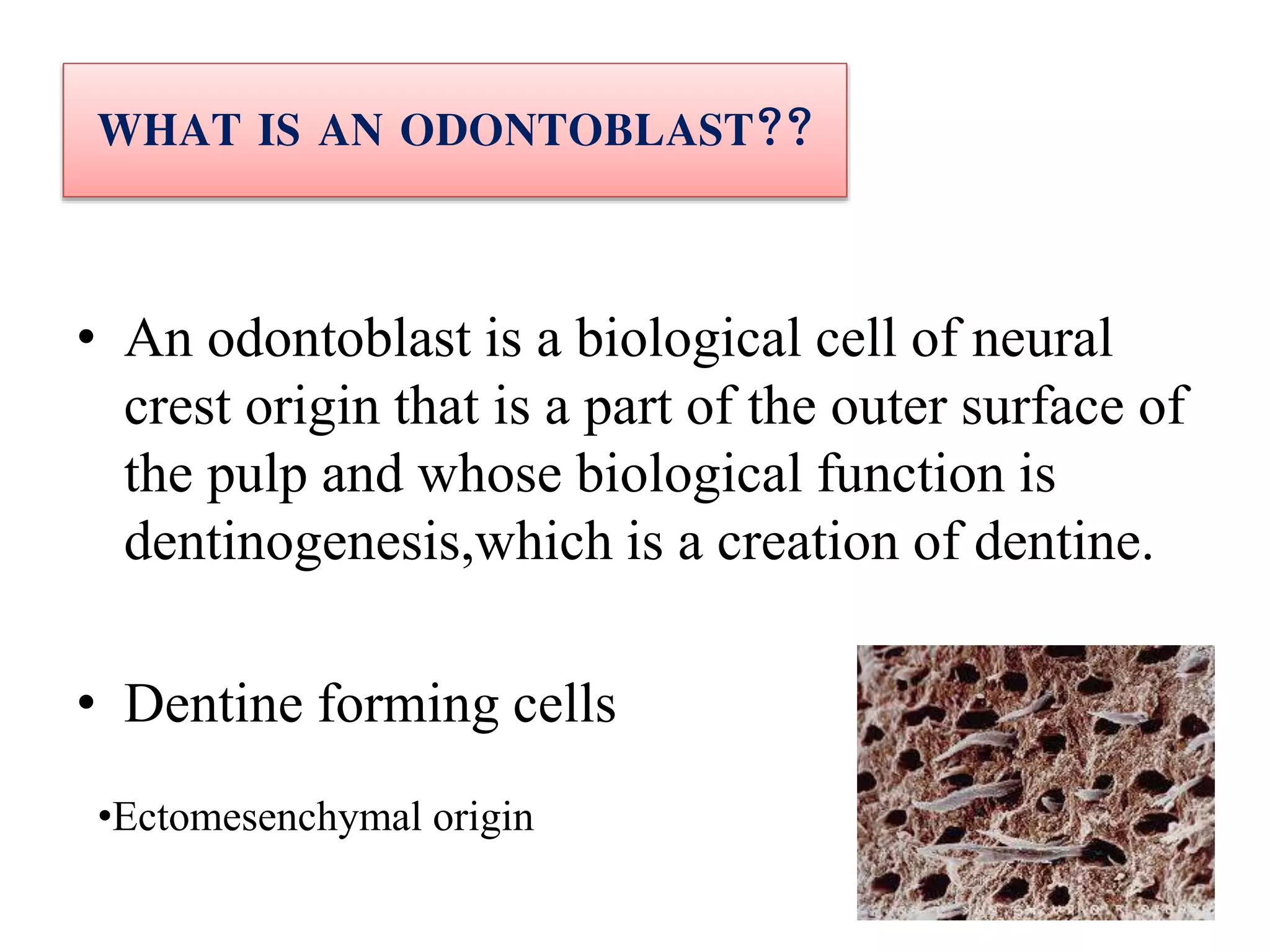
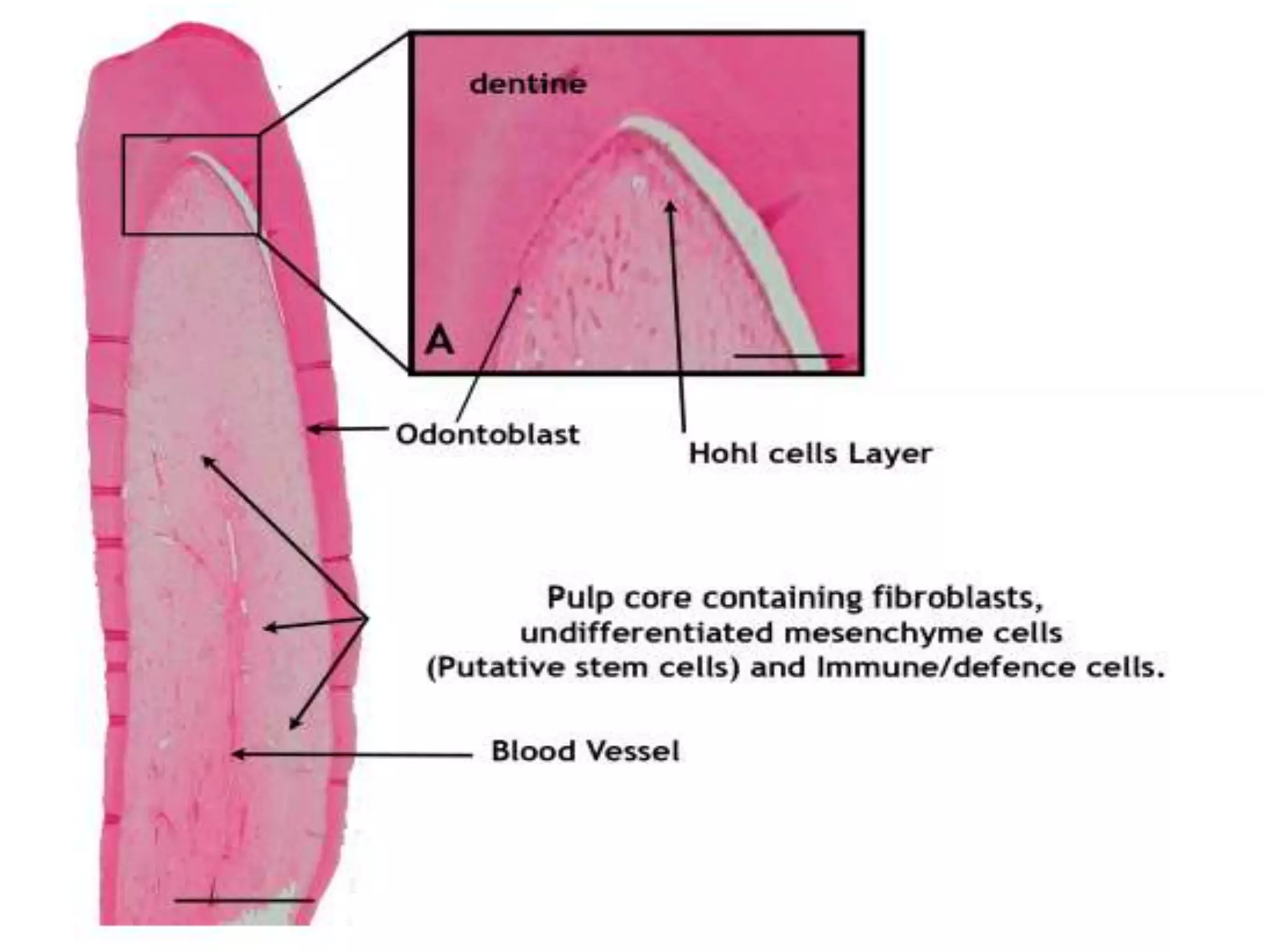
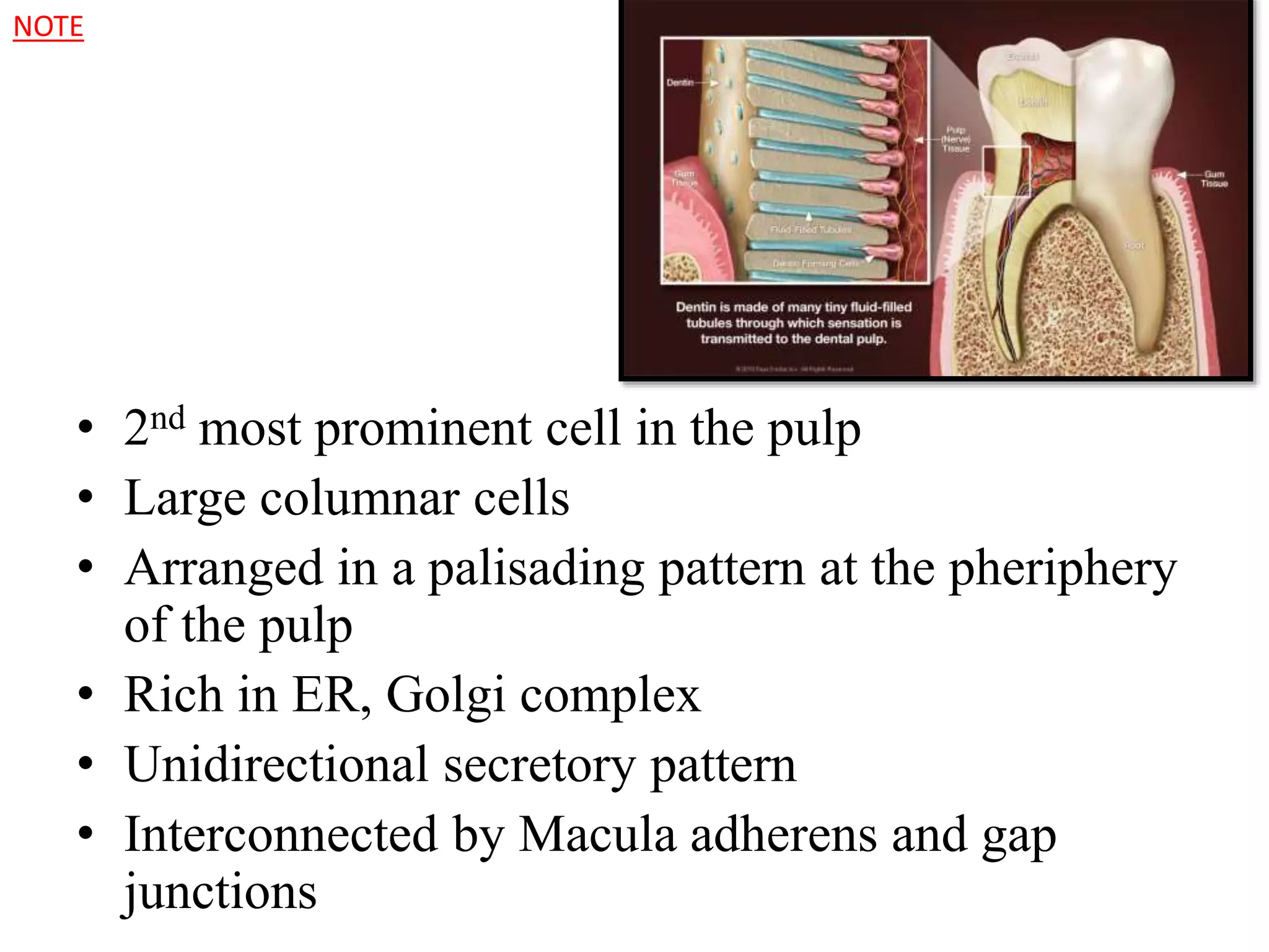
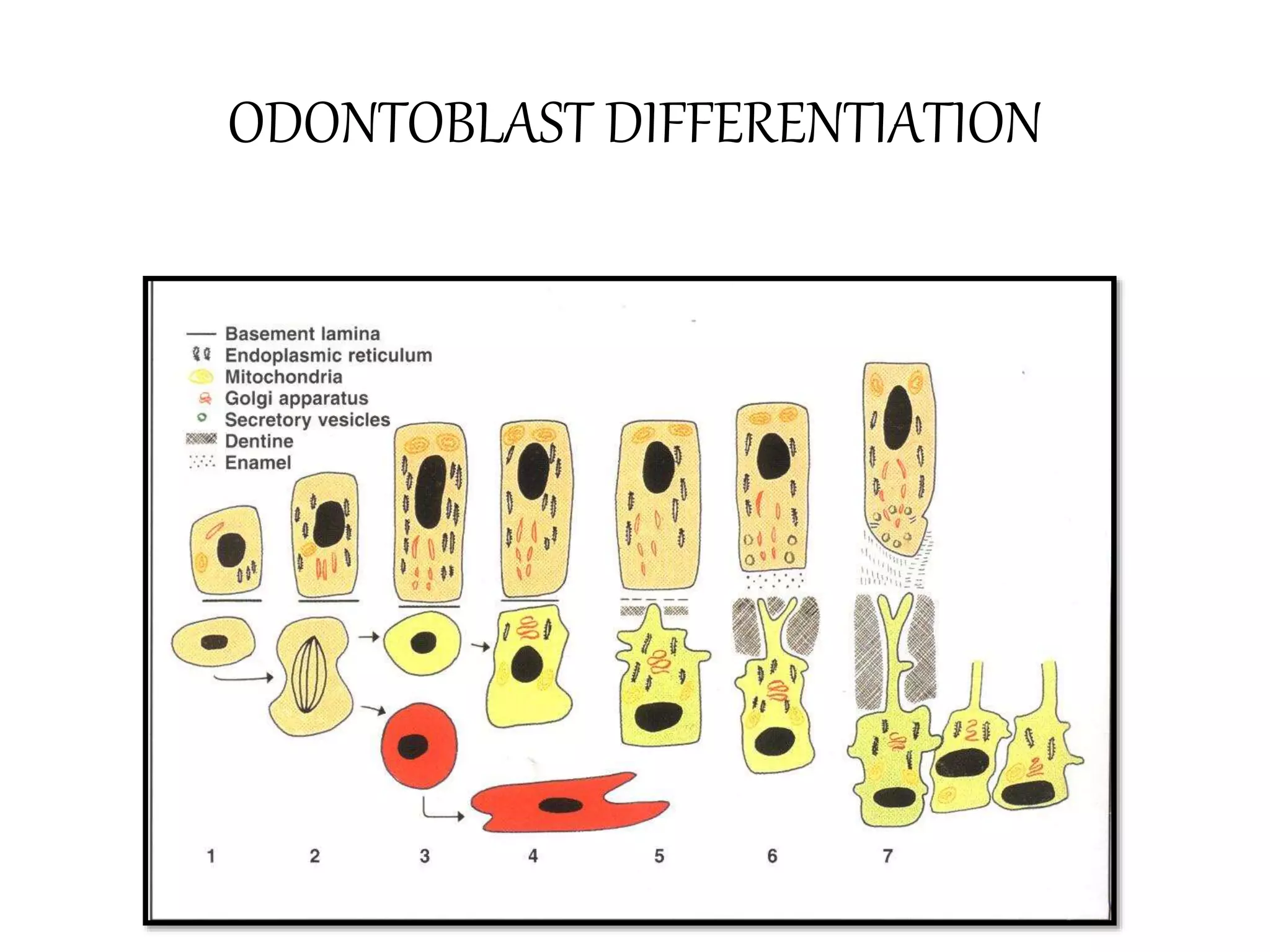
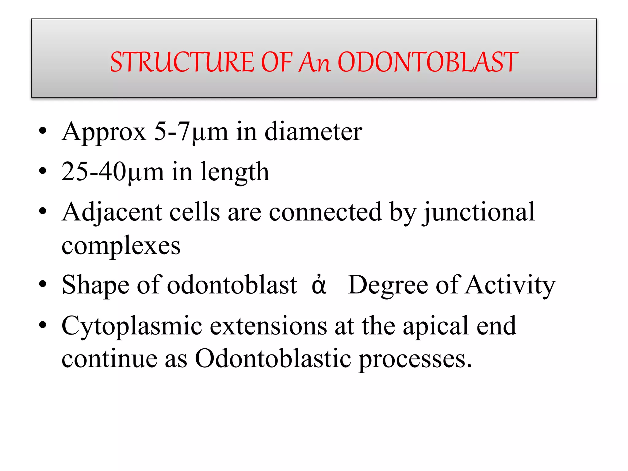
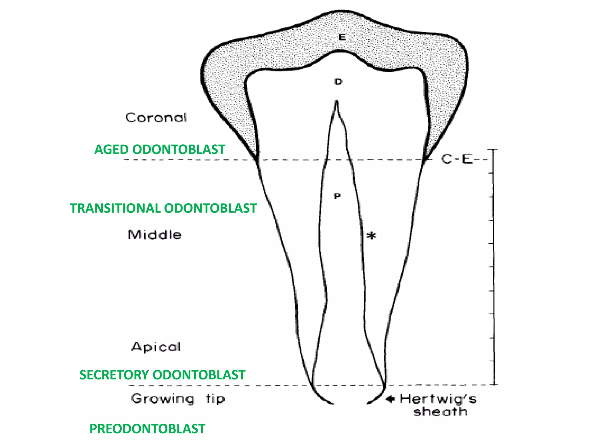
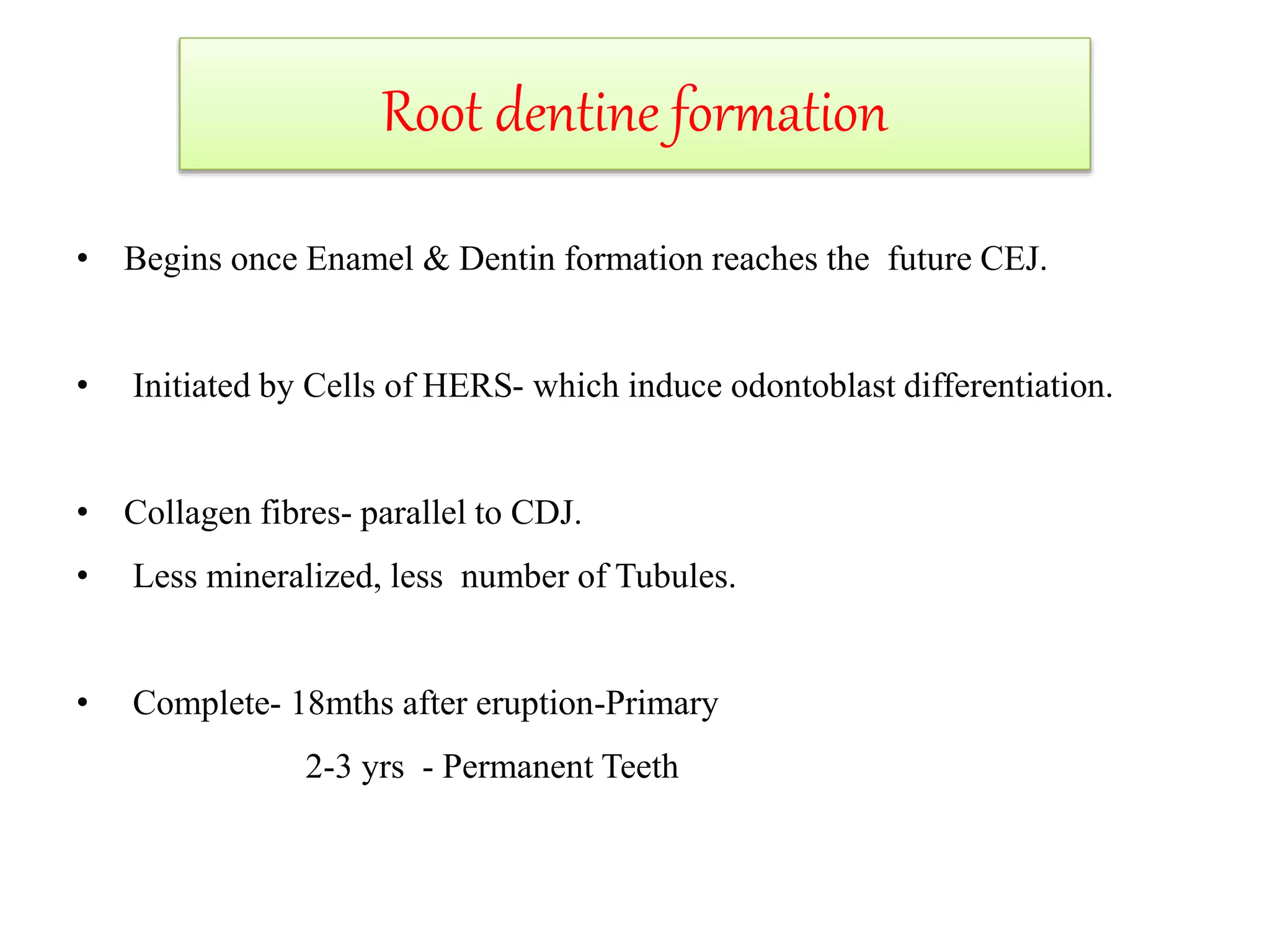
ODONTOBLAST
SHRAVYA.M
CONTENTS
 INTRODUCTION
 ORIGIN
 DEVELOPMENT
 LIFE CYCLE
 STRUCTURE OF
ODONTOBLAST
GENES
ODONTOBLAST
&
DENTINOGENESIS
ROOT DENTINE
 BLOOD SUPPLY
 FATE OF ODONTOBLAST
 ULTRASTRUCTURE
 HISTOCHEMICAL STAINS
 IHC MARKERS
 CLINICAL CONSIDERATION
 REFERENCES
WHAT IS AN ODONTOBLAST??
• An odontoblast is a biological cell of neural
crest origin that is a part of the outer surface of
the pulp and whose biological function is
dentinogenesis,which is a creation of dentine.
• Dentine forming cells
•Ectomesenchymal origin
• 2nd most prominent cell in the pulp
• Large columnar cells
• Arranged in a palisading pattern at the pheriphery
of the pulp
• Rich in ER, Golgi complex
• Unidirectional secretory pattern
• Interconnected by Macula adherens and gap
junctions
NOTE
• No. of odontoblast = no.of dentinal tubules
• 59,000 -76,000/sqmm
• Deposits 4µm of dentine everyday.
• Morphologic variations of odontoblasts :
-Tall columnar cells in the crown of the tooth
-Low columnar cells in the middle of the tooth
-Flattened cells at the apex
NOTE
STAGES OF TOOTH
DEVELOPMENT
• BUD STAGE
•CAP STAGE
•EARLY BELL
NOTE:
Pre Ameloblasts
Are First Formed
Which Then
Influences The
Formation Of Pre
Odontoblasts
•ADVANCED BELL STAGE
LIFE CYCLE
PRE-ODONTOBLAST STAGE
SYNTHETIC
ODONTOBLAST STAGE
TRANSITIONAL
ODONTOBLAST STAGE
RESTING STAGE
ODONTOCYTES
Life cycle of odontoblast
• PRE-ODONTOBLAST STAGE:
-Preodontoblasts have a small ovoid cells with small
nucleus
- few strands of dense fibrillar material radiating from the
fibrillar center.
-↑nuclear-cytoplasmic ratio
- few orgenells
-The cells are located around the growing tip of the root
-BMP ,GDf11 , BSP genes
`
• SYNTHETIC / ACTIVE STAGE:
-Tall columnar cells
-Large basal nucleus with a basophilic cytoplasm
-Numerous endoplasmic reticulum, mitochondria
-Well developed golgi complex
- Abundant synthetic orgenelles
-Numerous secretory granules.
• TRANSITIONAL / INTERMEDIATE STAGE:
-Shows features of synthetic cells but orgenells are less in
number and less prominent.
-Nucleus shows condensation of chromatin with orgenells
distributed around the nucleus.
-↓secretory granules
-Autophagic vacuoles
• RESTING / AGED ODONTOBLAST STAGE:
-Stubby cells
-Scanty cytoplasm
-Dark close faced nucleus
-↓Orgenelles
-Secretory granules are absent
STRUCTURal DEVELOPMENT OF ODONTOBLASTS
PREODONTOBLASTS - ↑NUCLEUS-CYTOPLASMIC
RATIO & few cisterns of rough endoplasmic
reticulum , small golgi apparatus and few
mitochondria.
1ST SIGN OF ODONTOBLAST DIFFERENTIATION- apical
point of dental papilla during early bell stage.
↑Aperiodic fibrils ̃15-20nm in diameter and 0.5-
1µm long are perpendicular to basal lamina.
PREODONTOBLASTS
• Cytoplasmic Processes Of The Adjascent
Preodontoblasts Become Closely Associated
With The Aperiodic Fibrils And Contact The
Basal Lamina.
• Simultaneously polarisation of preodontoblasts
begin.
NOTE
• It has been suggested that receptor-ligand
interactions involving substrate adhesion
molecules among the aperiodic fibrils serve to
orient and immobilize the preodontoblasts,
inducing them to begin differentiation.
• AND ODONTOBLASTS ARE THUS FORMED.
NOTE
GENES REGULATING THE ODONTOBLAST DIFFERENTIATIONBMP (BONE MORPHOGENETIC PROTEINS)
ACTIVIN
FGF(FIBROBLAST GROWTH FACTOR)
WNT (Wingless homologue in vertebrates)
Lhx6 ,Lhx7 (Lim-homeobox domain gene)
Barx1 (BarH like homologue in vertebrates)
Msx , Msx2 (Msh-like genes in vertebrates)
Dix1, Dix2 (Distaless homologue in vertebrates)
Pax9 (Paired box homeotic genes)
Gli1 ,Gli2 , Gli3 (Glioma associated oncogene homologe )
Lef1 (Lymphoid enchancer – binding factor)
ODONTOBLAST DIFFERENTIATION
Migration
Golgi apparatus,RER basement membrane
Nucleus proximal end
↑number of RER
↑size of Golgi complex
↑number of mitochondria
Numerous matrix vesicles
Thus Odontoblasts exhibit cylindrical shape.
CELLULAR CHANGES
STRUCTURE OF An ODONTOBLAST
• Approx 5-7µm in diameter
• 25-40µm in length
• Adjacent cells are connected by junctional
complexes
• Shape of odontoblast ἀ Degree of Activity
• Cytoplasmic extensions at the apical end
continue as Odontoblastic processes.
NUCLEUS
SECRETORY VESICLES
STRUCTURAL POLARITY
MITOCHONDRIA
GOLGI COMPLEX
RER
ENDOPLASMIC RETICULUM
• The endoplasmic reticulum is a membrane-
bound structure that is a part of every
eukaryotic cell.
• FUNCTIONS:
-Synthesis of various proteins and carbohydrates
-Transports the vesicles to the golgi complex
• As the proteins travel through the organelle, they create small
pockets.
-These pockets pinch off from the organelle and form vesicles.
-The vesicles transport the proteins to the Golgi complex, where
they are processed and distributed to different cells.
• Their number increases from from pre-odontoblast stage to
active stage and further reduces as the cells reach quinesence.
GOLGI COMPLEX
• The golgi complex appears as an aggregation of
smooth walled vesicles and cisternal profiles
organised into distinct and separate groups
• Functions:
*Synthesis of complex carbohydrates
*Sulfation of proteins.
Matrix vesicles
• Site of initial mineralization.
• They are small membrane bound structure that
buds off from the cell to form an independent unit.
*They contain alkaline phosphatase,
calcium adenosinetriphosphatase,
metalloproteinases,
proteoglycans , sulfur and calcium.
.
• *Microanalysis of Matrix vesicles indicates
CALCIUM and SULFUR PEAK of matrix
vesicles are higher than the odontoblast
cytoplasm
ODONTOBLAST PROCESSES
• Tomes fibres
• Is the secretory pole of odontoblast
• Form the major content of dentinal tubules
• 3-4 microns in diameter at pulpal end and taper to
1 micron near the periphery.
• Cytoplasmic orgenells are also present close to
the cell body.
• Their extension into the dentinal tubules varies
• Enamel spindles
•They play a
major role in
predentine
deposition
•Also the
secretory
granules on these
processes are the
precursors for
peri-tubular
dentine.
• Coated vesicles and Coated pits reflect
pinocytotic activity along the odontoblastic
process at the level of predentine close to cell
body.
Cytoskeleton
• A well-developed system of microtubules is present both
within the cell body and the odontoblast process.
• Each microtubule is about 25 nm wide with a wall
approximately 7-8 nm thick
• Functions of microtubules-
- transportation of intracellular substances
- stabilises cellular shape
• These cytoplasmic filaments are usually more highly
concentrated at the cell periphery, forming distinct bands just
beneath the cell membrane.
Nucleus
• Nuclear changes in various stages of
odontoblast differentiation:
• a) Pre-odontoblast stage:
*These cells present moderately reticulated
nucleoli that tend to be circular in shape.
*The nucleolar area is 0.55µm
*Few strands of dense fibrillar material radiating
from the fibrillar center
*Located at the growing tips
FC
Fg
d NUCLEAR
BODY
I
• b) Secretory Stage :
*characterized by a large, irregular, and
reticulated nucleolus.
*Size 1.24µm
*71% is occupied by dense fibrillar component.
*2 fibrillar centres.
*Prominent interstitial space
*Chromatin clumps
*Located at the apical region
• c) Transitional stage:
*Seen only under electron microscope
*Nucleus is displaced from the basal extremity
*Nucleolus is reduced in size
*Size- 0.54µm
*Granular layer is clearly seggregated
*Highly condensed chromatin
*Located at the middle region
• d) Aged Odontoblast stage
• Small, compact haematoxyphilic nucleolus
• 0.39µm
• Segregated components
• Large lipid filled vacuoles in the cytoplasm
• Located at the coronal region.
• The fibrillar center remains connected on one side
to the dense chromatin that encloses the
nucleolus.
AGED ODONTOBLAST
TRANSITIONAL ODONTOBLAST
SECRETORY ODONTOBLAST
PREODONTOBLAST
• They possess junctional structures constituted
by – Gap
- Tight junctions
-Desmosomes.
• Gap junctions and Tight junctions of focal or
macular type appear on early odontoblasts
during differentiation.
JUNCTIONS
GJ
GAP JUNCTIONS
-Occur on the lateral
surface of the cells at
the Base.
-They can
form,dissolve &
reform as the function
dictates.
TIGHT JUNCTIONS:
*Freeze fracture image
of a tight junction
*Extensive and
branched rows of
zipperlike particles.
ODONTOBLAST & DENTINOGENESIS
• After the differentiation of odontoblasts, the odontoblast
starts functiong by depositing Von Korffs fibres (Type III
collagen)
• They extend towards the IEE and fan out on the ground
substance.
• As the odontoblasts increase in size they deposit Type I
collagen that orient parallel to the future DEJ.
• Thus a layer of mantle dentine (15-20mm thick) is formed
by odontoblasts.
Odontoblast depositing coronal mantle predentine
• MATRIX VESICLES contain Alkaline Phosphatase -↑
concentration of phosphates + Calcium →Hydroxyapatite
Crystals.
Crystals- grow rapidly, rupture the matrix vesicles
The process continues …
Spread -clusters of crystallites → fuse with adjacent clusters to
form a continuous layer of mineralized matrix
Initially- on the surface of the collagen fibrils and ground substance,
later within the fibrils- aligned with collagen.
Various Matrix Proteins Influence Mineralization:
• DPP- Binds to Ca, Controls Growth of H.A Crystals
• Osteonectin- Inhibits growth of H.A crystals, promotes their
Binding to Collagen
• Gla-proteins, Phospholipids- Act as nucleators to concentrate
calcium.
• Proteoglycans- inhibit premature mineralization seen in
predentin.
COLLAGEN SYNTHESIS BY ODONTOBLASTS
Hydroxylation of procollagen with N- and C-
terminal extensions.
Prolylhydroxylase Lysylhydroxylase.
Triple helix assembly
Triple helix formation
Golgi complex
Glycosylation
Transported
Procollagen
COLLAGEN
+n of O-linked
galactose residues
Trans- face
Pathway of
Collagen
synthesis in
odontoblast
SECONDARY AND TERTIARY DENTIN
 Secondary dentin is deposited after root formation is completed and
is formed by the same odontoblasts that formed primary dentin.
 Tertiary dentin is deposited at specific site in response to injury by
damaged odontoblasts/replacement cells from pulp.
Root dentine formation
• Begins once Enamel & Dentin formation reaches the future CEJ.
• Initiated by Cells of HERS- which induce odontoblast differentiation.
• Collagen fibres- parallel to CDJ.
• Less mineralized, less number of Tubules.
• Complete- 18mths after eruption-Primary
2-3 yrs - Permanent Teeth
BLOOD SUPPLY
Capillary supply in
the
subodontoblastic
layer
They migrate
between the
odontoblasts and
regress.
FATE OF ODONTOBLASTS
• Life span of odontoblasts is equal to that of a viable tooth
because once differentiated they cannot undergo further cell
division.
• Resting Odontoblasts involved in secondary dentinogenesis
is renamed as “ODONTOCYTES” because their function
and properties are similar to osteocytes.
• These odontocytes may participate during reactionary
dentinogenesis
• Gene DMP1 is involved in the differention of secretory
odontoblasts into odontocytes
Ultrastructure
Histochemical Stains
• The Phosphophoryn content of the odontoblast are
retained in the sections during the specimen preparation
and stained selectively in situ.
• Various stains for dentine are:
Haematoxylin & Eosin
Methylene blue
Alcian blue
PAS
Modified Gallego stain
Ruthenium red
IHC MARKERS
• Murine Dentin Matrix Protein 1 (Dmp1)
• Dentin Sialophosphoprotein (DSPP)
• S35
• P33
• 3H-Serine
• 3H-Proline
• Actin
• Tubulin
• Vimentin
Clinical significance
1)Pathological differences in the functional life
cyle of odontoblasts – DENTINOGENESIS
IMPERFECTA
2)Pre-odontoblasts do not differentiate into odontoblasts
– Shell/ Thistle-tube teeth
3)Odontoblasts do not differentiate into Osteocytes –
Pulpal obliteration
4) Outward resorption of dentinal tubules by
odontoclasts results in pulpal tissue appearing pink
through the thin enamel – Pink Tooth
TO SUMMARIZE:
REFERENCES
• Nanci. A: Ten cates oral histology, 8th ed, Elsevier
• Kumar. G S : Orban’s Oral histology &
embryology, 13th ed , Elsevier
• Berkovitz. B K B: Oral anatomy , Histology and
Embryology,3rd ed, Mosby
• Jose.M: Essentials of Oral Biology, CBS
• Sasaki T, Garant P “Structure An Organization Of Odontoblasts” ,
The Anatomical Record245:235-249(1996)
• Couve E “Morphometric Analysis Of The Nucleolus During The Life
Cycle Of Human Odontoblasts”, The Anatomical Record 213:215-
224(1985)
• A. Nanci, M. Fortin, And L. GHITESCU “Endocytotic Functions Of
Ameloblasts And Odontoblasts:lmmunocytochemical And Tracer
Studies On The Uptake Of Plasma Proteins” , THE ANATOMICAL
RECORD 245219-234 (1996)
• Ushiyama J, “Gap Junctions Between Odontoblasts Revealed By
Transjunctional Flux Of Fluroscent Tracers”, Cell Tissues Res 258-
611-616(1989)
• Arana VE , Katchburian E, “Development Of Tight Junctions
Between Odontoblasts Innearly Dentinogenesis As Revealed By
Freeze-fracture” . The Anatomical Record 248:332-338(1997)
• Sigal M J , Pitaru S, Aubin JE,Cate T, “A Combined Scanning
Electron Microscopy And Immunofluorescence Study
Demonstrating That The Odontoblast Process Extends To
Dentinoenamel Junction In Human Teeth.” The Anatomical
Record 210:453-462(1984)
Corrections
• Add Odontoblast sensitivity –fluid dynamic
theory
• Difference between tomes granular layer &
granular process

ODONTOBLAST

  • 1.
  • 2.
    CONTENTS  INTRODUCTION  ORIGIN DEVELOPMENT  LIFE CYCLE  STRUCTURE OF ODONTOBLAST GENES ODONTOBLAST & DENTINOGENESIS ROOT DENTINE
  • 3.
     BLOOD SUPPLY FATE OF ODONTOBLAST  ULTRASTRUCTURE  HISTOCHEMICAL STAINS  IHC MARKERS  CLINICAL CONSIDERATION  REFERENCES
  • 4.
    WHAT IS ANODONTOBLAST?? • An odontoblast is a biological cell of neural crest origin that is a part of the outer surface of the pulp and whose biological function is dentinogenesis,which is a creation of dentine. • Dentine forming cells •Ectomesenchymal origin
  • 7.
    • 2nd mostprominent cell in the pulp • Large columnar cells • Arranged in a palisading pattern at the pheriphery of the pulp • Rich in ER, Golgi complex • Unidirectional secretory pattern • Interconnected by Macula adherens and gap junctions NOTE
  • 8.
    • No. ofodontoblast = no.of dentinal tubules • 59,000 -76,000/sqmm • Deposits 4µm of dentine everyday. • Morphologic variations of odontoblasts : -Tall columnar cells in the crown of the tooth -Low columnar cells in the middle of the tooth -Flattened cells at the apex NOTE
  • 9.
  • 10.
  • 11.
    •EARLY BELL NOTE: Pre Ameloblasts AreFirst Formed Which Then Influences The Formation Of Pre Odontoblasts
  • 12.
  • 13.
    LIFE CYCLE PRE-ODONTOBLAST STAGE SYNTHETIC ODONTOBLASTSTAGE TRANSITIONAL ODONTOBLAST STAGE RESTING STAGE ODONTOCYTES
  • 14.
    Life cycle ofodontoblast • PRE-ODONTOBLAST STAGE: -Preodontoblasts have a small ovoid cells with small nucleus - few strands of dense fibrillar material radiating from the fibrillar center. -↑nuclear-cytoplasmic ratio - few orgenells -The cells are located around the growing tip of the root -BMP ,GDf11 , BSP genes
  • 15.
  • 16.
    • SYNTHETIC /ACTIVE STAGE: -Tall columnar cells -Large basal nucleus with a basophilic cytoplasm -Numerous endoplasmic reticulum, mitochondria -Well developed golgi complex - Abundant synthetic orgenelles -Numerous secretory granules. • TRANSITIONAL / INTERMEDIATE STAGE: -Shows features of synthetic cells but orgenells are less in number and less prominent. -Nucleus shows condensation of chromatin with orgenells distributed around the nucleus. -↓secretory granules -Autophagic vacuoles
  • 17.
    • RESTING /AGED ODONTOBLAST STAGE: -Stubby cells -Scanty cytoplasm -Dark close faced nucleus -↓Orgenelles -Secretory granules are absent
  • 20.
    STRUCTURal DEVELOPMENT OFODONTOBLASTS PREODONTOBLASTS - ↑NUCLEUS-CYTOPLASMIC RATIO & few cisterns of rough endoplasmic reticulum , small golgi apparatus and few mitochondria. 1ST SIGN OF ODONTOBLAST DIFFERENTIATION- apical point of dental papilla during early bell stage. ↑Aperiodic fibrils ̃15-20nm in diameter and 0.5- 1µm long are perpendicular to basal lamina.
  • 21.
  • 22.
    • Cytoplasmic ProcessesOf The Adjascent Preodontoblasts Become Closely Associated With The Aperiodic Fibrils And Contact The Basal Lamina. • Simultaneously polarisation of preodontoblasts begin. NOTE
  • 23.
    • It hasbeen suggested that receptor-ligand interactions involving substrate adhesion molecules among the aperiodic fibrils serve to orient and immobilize the preodontoblasts, inducing them to begin differentiation. • AND ODONTOBLASTS ARE THUS FORMED. NOTE
  • 24.
    GENES REGULATING THEODONTOBLAST DIFFERENTIATIONBMP (BONE MORPHOGENETIC PROTEINS) ACTIVIN FGF(FIBROBLAST GROWTH FACTOR) WNT (Wingless homologue in vertebrates) Lhx6 ,Lhx7 (Lim-homeobox domain gene) Barx1 (BarH like homologue in vertebrates) Msx , Msx2 (Msh-like genes in vertebrates) Dix1, Dix2 (Distaless homologue in vertebrates) Pax9 (Paired box homeotic genes) Gli1 ,Gli2 , Gli3 (Glioma associated oncogene homologe ) Lef1 (Lymphoid enchancer – binding factor)
  • 25.
  • 26.
  • 27.
    Migration Golgi apparatus,RER basementmembrane Nucleus proximal end ↑number of RER ↑size of Golgi complex ↑number of mitochondria Numerous matrix vesicles Thus Odontoblasts exhibit cylindrical shape. CELLULAR CHANGES
  • 28.
    STRUCTURE OF AnODONTOBLAST • Approx 5-7µm in diameter • 25-40µm in length • Adjacent cells are connected by junctional complexes • Shape of odontoblast ἀ Degree of Activity • Cytoplasmic extensions at the apical end continue as Odontoblastic processes.
  • 29.
  • 30.
    ENDOPLASMIC RETICULUM • Theendoplasmic reticulum is a membrane- bound structure that is a part of every eukaryotic cell. • FUNCTIONS: -Synthesis of various proteins and carbohydrates -Transports the vesicles to the golgi complex
  • 31.
    • As theproteins travel through the organelle, they create small pockets. -These pockets pinch off from the organelle and form vesicles. -The vesicles transport the proteins to the Golgi complex, where they are processed and distributed to different cells. • Their number increases from from pre-odontoblast stage to active stage and further reduces as the cells reach quinesence.
  • 32.
    GOLGI COMPLEX • Thegolgi complex appears as an aggregation of smooth walled vesicles and cisternal profiles organised into distinct and separate groups • Functions: *Synthesis of complex carbohydrates *Sulfation of proteins.
  • 33.
    Matrix vesicles • Siteof initial mineralization. • They are small membrane bound structure that buds off from the cell to form an independent unit. *They contain alkaline phosphatase, calcium adenosinetriphosphatase, metalloproteinases, proteoglycans , sulfur and calcium. .
  • 34.
    • *Microanalysis ofMatrix vesicles indicates CALCIUM and SULFUR PEAK of matrix vesicles are higher than the odontoblast cytoplasm
  • 35.
    ODONTOBLAST PROCESSES • Tomesfibres • Is the secretory pole of odontoblast • Form the major content of dentinal tubules • 3-4 microns in diameter at pulpal end and taper to 1 micron near the periphery. • Cytoplasmic orgenells are also present close to the cell body. • Their extension into the dentinal tubules varies • Enamel spindles
  • 36.
    •They play a majorrole in predentine deposition •Also the secretory granules on these processes are the precursors for peri-tubular dentine.
  • 37.
    • Coated vesiclesand Coated pits reflect pinocytotic activity along the odontoblastic process at the level of predentine close to cell body.
  • 38.
    Cytoskeleton • A well-developedsystem of microtubules is present both within the cell body and the odontoblast process. • Each microtubule is about 25 nm wide with a wall approximately 7-8 nm thick • Functions of microtubules- - transportation of intracellular substances - stabilises cellular shape • These cytoplasmic filaments are usually more highly concentrated at the cell periphery, forming distinct bands just beneath the cell membrane.
  • 39.
    Nucleus • Nuclear changesin various stages of odontoblast differentiation: • a) Pre-odontoblast stage: *These cells present moderately reticulated nucleoli that tend to be circular in shape. *The nucleolar area is 0.55µm *Few strands of dense fibrillar material radiating from the fibrillar center *Located at the growing tips
  • 40.
  • 41.
    • b) SecretoryStage : *characterized by a large, irregular, and reticulated nucleolus. *Size 1.24µm *71% is occupied by dense fibrillar component. *2 fibrillar centres. *Prominent interstitial space *Chromatin clumps *Located at the apical region
  • 43.
    • c) Transitionalstage: *Seen only under electron microscope *Nucleus is displaced from the basal extremity *Nucleolus is reduced in size *Size- 0.54µm *Granular layer is clearly seggregated *Highly condensed chromatin *Located at the middle region
  • 45.
    • d) AgedOdontoblast stage • Small, compact haematoxyphilic nucleolus • 0.39µm • Segregated components • Large lipid filled vacuoles in the cytoplasm • Located at the coronal region. • The fibrillar center remains connected on one side to the dense chromatin that encloses the nucleolus.
  • 48.
  • 49.
    • They possessjunctional structures constituted by – Gap - Tight junctions -Desmosomes. • Gap junctions and Tight junctions of focal or macular type appear on early odontoblasts during differentiation. JUNCTIONS
  • 50.
    GJ GAP JUNCTIONS -Occur onthe lateral surface of the cells at the Base. -They can form,dissolve & reform as the function dictates.
  • 51.
    TIGHT JUNCTIONS: *Freeze fractureimage of a tight junction *Extensive and branched rows of zipperlike particles.
  • 52.
    ODONTOBLAST & DENTINOGENESIS •After the differentiation of odontoblasts, the odontoblast starts functiong by depositing Von Korffs fibres (Type III collagen) • They extend towards the IEE and fan out on the ground substance. • As the odontoblasts increase in size they deposit Type I collagen that orient parallel to the future DEJ. • Thus a layer of mantle dentine (15-20mm thick) is formed by odontoblasts.
  • 53.
  • 54.
    • MATRIX VESICLEScontain Alkaline Phosphatase -↑ concentration of phosphates + Calcium →Hydroxyapatite Crystals. Crystals- grow rapidly, rupture the matrix vesicles The process continues … Spread -clusters of crystallites → fuse with adjacent clusters to form a continuous layer of mineralized matrix Initially- on the surface of the collagen fibrils and ground substance, later within the fibrils- aligned with collagen.
  • 56.
    Various Matrix ProteinsInfluence Mineralization: • DPP- Binds to Ca, Controls Growth of H.A Crystals • Osteonectin- Inhibits growth of H.A crystals, promotes their Binding to Collagen • Gla-proteins, Phospholipids- Act as nucleators to concentrate calcium. • Proteoglycans- inhibit premature mineralization seen in predentin.
  • 57.
    COLLAGEN SYNTHESIS BYODONTOBLASTS Hydroxylation of procollagen with N- and C- terminal extensions. Prolylhydroxylase Lysylhydroxylase. Triple helix assembly Triple helix formation Golgi complex Glycosylation Transported Procollagen COLLAGEN +n of O-linked galactose residues Trans- face
  • 58.
  • 59.
    SECONDARY AND TERTIARYDENTIN  Secondary dentin is deposited after root formation is completed and is formed by the same odontoblasts that formed primary dentin.  Tertiary dentin is deposited at specific site in response to injury by damaged odontoblasts/replacement cells from pulp.
  • 60.
    Root dentine formation •Begins once Enamel & Dentin formation reaches the future CEJ. • Initiated by Cells of HERS- which induce odontoblast differentiation. • Collagen fibres- parallel to CDJ. • Less mineralized, less number of Tubules. • Complete- 18mths after eruption-Primary 2-3 yrs - Permanent Teeth
  • 61.
    BLOOD SUPPLY Capillary supplyin the subodontoblastic layer They migrate between the odontoblasts and regress.
  • 62.
    FATE OF ODONTOBLASTS •Life span of odontoblasts is equal to that of a viable tooth because once differentiated they cannot undergo further cell division. • Resting Odontoblasts involved in secondary dentinogenesis is renamed as “ODONTOCYTES” because their function and properties are similar to osteocytes. • These odontocytes may participate during reactionary dentinogenesis • Gene DMP1 is involved in the differention of secretory odontoblasts into odontocytes
  • 63.
  • 69.
    Histochemical Stains • ThePhosphophoryn content of the odontoblast are retained in the sections during the specimen preparation and stained selectively in situ. • Various stains for dentine are: Haematoxylin & Eosin Methylene blue Alcian blue PAS Modified Gallego stain Ruthenium red
  • 70.
    IHC MARKERS • MurineDentin Matrix Protein 1 (Dmp1) • Dentin Sialophosphoprotein (DSPP) • S35 • P33 • 3H-Serine • 3H-Proline • Actin • Tubulin • Vimentin
  • 71.
    Clinical significance 1)Pathological differencesin the functional life cyle of odontoblasts – DENTINOGENESIS IMPERFECTA
  • 72.
    2)Pre-odontoblasts do notdifferentiate into odontoblasts – Shell/ Thistle-tube teeth 3)Odontoblasts do not differentiate into Osteocytes – Pulpal obliteration 4) Outward resorption of dentinal tubules by odontoclasts results in pulpal tissue appearing pink through the thin enamel – Pink Tooth
  • 73.
  • 75.
    REFERENCES • Nanci. A:Ten cates oral histology, 8th ed, Elsevier • Kumar. G S : Orban’s Oral histology & embryology, 13th ed , Elsevier • Berkovitz. B K B: Oral anatomy , Histology and Embryology,3rd ed, Mosby • Jose.M: Essentials of Oral Biology, CBS
  • 76.
    • Sasaki T,Garant P “Structure An Organization Of Odontoblasts” , The Anatomical Record245:235-249(1996) • Couve E “Morphometric Analysis Of The Nucleolus During The Life Cycle Of Human Odontoblasts”, The Anatomical Record 213:215- 224(1985) • A. Nanci, M. Fortin, And L. GHITESCU “Endocytotic Functions Of Ameloblasts And Odontoblasts:lmmunocytochemical And Tracer Studies On The Uptake Of Plasma Proteins” , THE ANATOMICAL RECORD 245219-234 (1996) • Ushiyama J, “Gap Junctions Between Odontoblasts Revealed By Transjunctional Flux Of Fluroscent Tracers”, Cell Tissues Res 258- 611-616(1989)
  • 77.
    • Arana VE, Katchburian E, “Development Of Tight Junctions Between Odontoblasts Innearly Dentinogenesis As Revealed By Freeze-fracture” . The Anatomical Record 248:332-338(1997) • Sigal M J , Pitaru S, Aubin JE,Cate T, “A Combined Scanning Electron Microscopy And Immunofluorescence Study Demonstrating That The Odontoblast Process Extends To Dentinoenamel Junction In Human Teeth.” The Anatomical Record 210:453-462(1984)
  • 79.
    Corrections • Add Odontoblastsensitivity –fluid dynamic theory • Difference between tomes granular layer & granular process

Editor's Notes

  • #7 Intermediate forms between fibroblasts and odontoblasts in d subodontoblastic layer .elongated bipolar cells that form matrix during reparative dentine formn.
  • #35 A)Matrix vesicles as seen in electron microscope B)Freeze fracture showing intramembranous particle c)Histochemical demonstration of Ca-Adenosinetriphosphatase activity
  • #41 FC – Fibrillar centre ………….. FG- Firillogranular material ………D- Fibrillar component…………. I – interstitial space