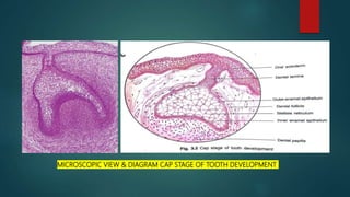
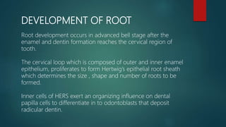
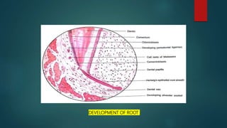
PHYSICAL PROPERTIES
CHEMICAL PROPERTIES
STRUCTURE OF ENAMEL
DEVELOPMENT OF ENAMEL
EPITHELIAL ENAMEL ORGAN
AMELOGENESIS
LIFE CYCLE OF AMELOBLASTS
AGE CHANGES IN ENAMEL
DEFECTS OF AMELOGENESIS
CLINICAL IMPLICATIONS
An odontoblast is a biological cell of neural crest origin whose main function is formation of dentin.
This slide gives a detailed explanation of the same.
Upload By : Ahmed Ali Abbas
Babylon University College of Dentistry
download this file from Website on google theoptimalsmile.wix.com/dentistry
Oral histology
Coronal and radicular pulp
Apical foramen
Accessory canal
Functions of dental pulp
Components of dental pulp
Functions of pulpal extracellular matrix
Organization of cells in the pulp
The principle cells of the pulp
The pathways of collagen synthesis
Matrix and ground substances
Vasculature and lymphatic supply
Innervation of Dentin- pulp complex
Disorders of the dental pulp
Advances in pulp vitality testing
I prepared this presentation during the first year of my MDS. This will give you a basic idea and necessary information about the pulp of the teeth and its histology. Hope you guys find it useful.
The early development of tooth from six week of prenatal life. Description of different stages- bud,cap and bell stage and amelogenesis, dentinogenesis. Description of root development.
The initiation of tooth development begins at 37 days of development
with formation of a continuous horseshoe-band of thickened epithelium
in the location of upper and lower jaws – Primary Epithelial Band
Dental lamina appears as a thickening
of the oral epithelium adjacent to
condensation of ectomesenchyme
20 areas of enlargement or knobs
appear, which will form tooth buds
for the 20 primary teeth
Not all will appear at the same time.
The first to develop are those of the
anterior mandible region
At this early stage the tooth buds
have already determined their crown morphology
Successional lamina: lamina from
which permanent teeth develop
The dental lamina begins to function
at 6th prenatal week and continues to
15th year of birth (3rd molar)
Tooth development is a continuous process, however can be
divided into 3 stages:
1. Bud Stage
2. Cap Stage
3. Bell Stage
4. Hertwigs epithelial root sheath and root formation
The bud stage is represented by the first epithelial incursion into the ectomesenchyme of the jaw.
The epithelial cells show little if any change in shape or function.
The supporting ectomesenchymal cells are packed closely beneath and around the epithelial bud. As the epithelial bud continues to proliferate into the ectomesenchyme, cellular density increases immediately adjacent to the epithelial outgrowth.
This process is classically referred to as a condensation of the ectomesenchyme.
The epithelium of the dental lamina separated from the underlying ectomesenchyme by basement membrane.
Bud stage is characterized by rounded, localized growth of
epithelium surrounded by proliferating mesenchymal cells,which are packed closely beneath and around the epithelial buds
The transition from bud to cap marks the onset of morphologic differences between tooth germs that give rise to different types of teeth.
Differential cellular division in the epithelial bud initiates a change in shape so that now the epithelial outgrowth assumes a more complex outline with a flattened internal portion along which the mesenchymal condensation densifies.
As the tooth bud grows larger, it drags along with it part of the dental lamina; thus from that point on, the developing tooth is tethered to the dental lamina by an extension called the lateral lamina.
At this early stage of tooth development, identifying the formative elements of the tooth and its supporting tissues is already possible.
The epithelial outgrowth, which superficially resembles a cap sitting on a ball of condensed ectomesenchyme , is still referred to widely as the dental organ but actually should be called the enamel organ, because it eventually will form the enamel of the tooth. Henceforth, the term enamel organ is used.
Condensation of the ectomesenchyme immediately subjacent to the tooth bud caused by lack of extracellular matrix secretion by the cells thus preventing separation.
A Brief Description about the development of teeth. Understanding the process of tooth development is of particular importance for the dentist; as developmental disturbances may occur at any stage of this process resulting in different types of tooth anomalies.
Tooth development can be classified either based on morphology or histology
Morphological stages:
Bud stage
Cap stage
Bell stage:
* Early
* Advanced
Physiological stages:
Initiation
Proliferation
Histodifferentiation
Morphodifferentiation
Apposition
BE UPDATE TO IT,, AS IT IS 3 years back from 2017
Kindly mail me if you feel, needy of this presentation
you can find my mail id @ slide share,,, if not mail me @
sukesh3567@gmail.com.
Good luck
Upload By : Ahmed Ali Abbas
Babylon University College of Dentistry
download this file from Website on google theoptimalsmile.wix.com/dentistry
Oral histology
Indian Dental Academy: will be one of the most relevant and exciting training center with best faculty and flexible training programs for dental professionals who wish to advance in their dental practice,Offers certified courses in Dental implants,Orthodontics,Endodontics,Cosmetic Dentistry, Prosthetic Dentistry, Periodontics and General Dentistry.
Acetabularia Information For Class 9 .docxvaibhavrinwa19
Acetabularia acetabulum is a single-celled green alga that in its vegetative state is morphologically differentiated into a basal rhizoid and an axially elongated stalk, which bears whorls of branching hairs. The single diploid nucleus resides in the rhizoid.
Model Attribute Check Company Auto PropertyCeline George
In Odoo, the multi-company feature allows you to manage multiple companies within a single Odoo database instance. Each company can have its own configurations while still sharing common resources such as products, customers, and suppliers.
June 3, 2024 Anti-Semitism Letter Sent to MIT President Kornbluth and MIT Cor...Levi Shapiro
Letter from the Congress of the United States regarding Anti-Semitism sent June 3rd to MIT President Sally Kornbluth, MIT Corp Chair, Mark Gorenberg
Dear Dr. Kornbluth and Mr. Gorenberg,
The US House of Representatives is deeply concerned by ongoing and pervasive acts of antisemitic
harassment and intimidation at the Massachusetts Institute of Technology (MIT). Failing to act decisively to ensure a safe learning environment for all students would be a grave dereliction of your responsibilities as President of MIT and Chair of the MIT Corporation.
This Congress will not stand idly by and allow an environment hostile to Jewish students to persist. The House believes that your institution is in violation of Title VI of the Civil Rights Act, and the inability or
unwillingness to rectify this violation through action requires accountability.
Postsecondary education is a unique opportunity for students to learn and have their ideas and beliefs challenged. However, universities receiving hundreds of millions of federal funds annually have denied
students that opportunity and have been hijacked to become venues for the promotion of terrorism, antisemitic harassment and intimidation, unlawful encampments, and in some cases, assaults and riots.
The House of Representatives will not countenance the use of federal funds to indoctrinate students into hateful, antisemitic, anti-American supporters of terrorism. Investigations into campus antisemitism by the Committee on Education and the Workforce and the Committee on Ways and Means have been expanded into a Congress-wide probe across all relevant jurisdictions to address this national crisis. The undersigned Committees will conduct oversight into the use of federal funds at MIT and its learning environment under authorities granted to each Committee.
• The Committee on Education and the Workforce has been investigating your institution since December 7, 2023. The Committee has broad jurisdiction over postsecondary education, including its compliance with Title VI of the Civil Rights Act, campus safety concerns over disruptions to the learning environment, and the awarding of federal student aid under the Higher Education Act.
• The Committee on Oversight and Accountability is investigating the sources of funding and other support flowing to groups espousing pro-Hamas propaganda and engaged in antisemitic harassment and intimidation of students. The Committee on Oversight and Accountability is the principal oversight committee of the US House of Representatives and has broad authority to investigate “any matter” at “any time” under House Rule X.
• The Committee on Ways and Means has been investigating several universities since November 15, 2023, when the Committee held a hearing entitled From Ivory Towers to Dark Corners: Investigating the Nexus Between Antisemitism, Tax-Exempt Universities, and Terror Financing. The Committee followed the hearing with letters to those institutions on January 10, 202
The French Revolution, which began in 1789, was a period of radical social and political upheaval in France. It marked the decline of absolute monarchies, the rise of secular and democratic republics, and the eventual rise of Napoleon Bonaparte. This revolutionary period is crucial in understanding the transition from feudalism to modernity in Europe.
For more information, visit-www.vavaclasses.com
The Roman Empire A Historical Colossus.pdfkaushalkr1407
The Roman Empire, a vast and enduring power, stands as one of history's most remarkable civilizations, leaving an indelible imprint on the world. It emerged from the Roman Republic, transitioning into an imperial powerhouse under the leadership of Augustus Caesar in 27 BCE. This transformation marked the beginning of an era defined by unprecedented territorial expansion, architectural marvels, and profound cultural influence.
The empire's roots lie in the city of Rome, founded, according to legend, by Romulus in 753 BCE. Over centuries, Rome evolved from a small settlement to a formidable republic, characterized by a complex political system with elected officials and checks on power. However, internal strife, class conflicts, and military ambitions paved the way for the end of the Republic. Julius Caesar’s dictatorship and subsequent assassination in 44 BCE created a power vacuum, leading to a civil war. Octavian, later Augustus, emerged victorious, heralding the Roman Empire’s birth.
Under Augustus, the empire experienced the Pax Romana, a 200-year period of relative peace and stability. Augustus reformed the military, established efficient administrative systems, and initiated grand construction projects. The empire's borders expanded, encompassing territories from Britain to Egypt and from Spain to the Euphrates. Roman legions, renowned for their discipline and engineering prowess, secured and maintained these vast territories, building roads, fortifications, and cities that facilitated control and integration.
The Roman Empire’s society was hierarchical, with a rigid class system. At the top were the patricians, wealthy elites who held significant political power. Below them were the plebeians, free citizens with limited political influence, and the vast numbers of slaves who formed the backbone of the economy. The family unit was central, governed by the paterfamilias, the male head who held absolute authority.
Culturally, the Romans were eclectic, absorbing and adapting elements from the civilizations they encountered, particularly the Greeks. Roman art, literature, and philosophy reflected this synthesis, creating a rich cultural tapestry. Latin, the Roman language, became the lingua franca of the Western world, influencing numerous modern languages.
Roman architecture and engineering achievements were monumental. They perfected the arch, vault, and dome, constructing enduring structures like the Colosseum, Pantheon, and aqueducts. These engineering marvels not only showcased Roman ingenuity but also served practical purposes, from public entertainment to water supply.
Instructions for Submissions thorugh G- Classroom.pptxJheel Barad
This presentation provides a briefing on how to upload submissions and documents in Google Classroom. It was prepared as part of an orientation for new Sainik School in-service teacher trainees. As a training officer, my goal is to ensure that you are comfortable and proficient with this essential tool for managing assignments and fostering student engagement.
2. INTRODUCTION
Development of tooth is a complex process.
The various tissues of the tooth and it’s supporting
structures develop from tooth germ.
The term tooth germ thus includes all the formative
tissues for the entire tooth and it’s supporting
structures.
Tooth germ has three main components
3. COMPONENTS OF TOOTH GERM
Enamel organ – The ectodermal component that
gives
rise to enamel.
Dental papilla – The ecto-mesenchymal component
that gives rise to dentin and pulp.
Dental follicle or dental sac – The ecto-mesenchymal
component giving rise to cementum , periodontal
ligament and part of the alveolar socket.
ENAMEL ORGAN
DENTAL PAPILLA
DENTAL FOLLICLE
4. STAGES OF TOOTH DEVELOPMENT
BUD STAGE
CAP STAGE
BELL STAGE - EARLY BELL STAGE
- LATE OR ADVANCED BELL STAGE
5. BUD STAGE
Enamel organ is bud shaped with peripheral cuboidal cells and central
polyhedral cells.
Peripheral cells of enamel organ are seperated from ectomesenchymal
components by a basement membrane.
All the cells are attached to each other by desmosomal junctions.
Ectomesenchymal condensation adjacent to enamel organ forms the dental
papilla.
Marginal condensation of ecto-mesenchymal cells enclosing dental papilla
and enamel organ is called dental follicle or dental sac.
7. CAP STAGE
Enamel organ increases in size and attain the shape of a cap by
invagination of the deep portion of the bud.
Cell undergo change in shape so that three separate cell groups
can be identified.
Cells lining the convexity or the periphery of the cap are cuboidal
in shape and are called outer enamel epithelium.
The cells lining the concave or invaginated portion change to
columnar cells named as inner enamel epithelium.
8. The central polyhedral cells transform into a network of star shaped
cells called stellate reticulum.
Dental papilla gets partially enclosed by the invaginated portion of
enamel organ.
Cells of dental papilla undergo proliferation and further condensation.
Dental follicle shows further condensation of ectomesenchymal cells.
It becomes more fibrous and denser in cap stage.
10. BELL STAGE - EARLY
Enamel organ enlarges further and invagination deepens changing
the shape to that of a bell in a longitudinal section.
In bell stage 4 different layers of cells are seen in the enamel
organ.
Cells lining the invaginated portion , the inner enamel epithelium
is composed of single layer of tall columnar cells that differentiate
to ameloblasts(enamel forming cells)
During bell stage a new layer appears which is called stratum
intermedium.
11. Stratum intermedium is located between inner enamel epithelium
and stellate reticulum and is composed of 2 to 3 layers of squamous
cells.
Stellate reticulum expands further in early bell stage.
Cells of outer enamel epithelium lining the periphery of enamel
organ flattens to low cuboidal cells.
At the cervical region of enamel organ outer enamel epithelium
loops inward to join with inner enamel epithelium. This is called
cervical loop.
12. During early bell stage enamel organ loses its connection to oral ectoderm due to
degeneration of dental lamina.
Remnants of the dental lamina are called cell rests of serres.
Successional lamina develops at this stage which is the primordium for the permenent
successor.
Dental papilla is fully enclosed in the invaginated portion of the enamel organ in this
stage.
Peripheral cells of dental papilla differentiate into odontoblasts (dentin forming cells)
under the organizing influence of inner enamel epithelial cells.
Dental follicle become more fibrous with 3 layers i.e. Inner cellular, outer fibrous layer
and middle loose connective tissue.
14. BELL STAGE – LATE OR ADVANCED
Differentiating feature between early and advanced bell stage is
formation of hard tissues.
Enamel organ shows 4 different layers, inner enamel epithelium,
stratum intermedium, stellate reticulum and outer enamel
epithelium.
Histological difference from early bell stage are
- Hard tissue formation
- Collapsed stellate reticulum and folding of outer
enamel epithelium bringing capillaries of the dental follicle
nearer to the ameloblasts.
15. Ameloblasts are brought closer to the dental follicle which now
becomes their source of nutrition.
Dental papilla shows differentiated odontoblast at the periphery.
Dental follicle is distinct enclosing enamel organ and dental papilla.
17. DEVELOPMENT OF ROOT
Root development occurs in advanced bell stage after the
enamel and dentin formation reaches the cervical region of
tooth.
The cervical loop which is composed of outer and inner enamel
epithelium, proliferates to form Hertwig’s epithelial root sheath
which determines the size , shape and number of roots to be
formed.
Inner cells of HERS exert an organizing influence on dental
papilla cells to differentiate in to odontoblasts that deposit
radicular dentin.
18. Once the root dentin is formed Hertwig’s epithelial root sheath
degenerate allowing the dental follicle cells to come in contact with
the dentin.
The cells of dental follicle differentiate to form cementoblasts and
lays down cementum over the root dentin.
As the cementum formation progresses the rest of the dental follicle
becomes more fibrous and develop into periodontal ligament.
The remnants of Hertwig’s epithelial root sheath remain in
peridontal ligament and are called ‘cell rests of malassez.’