This document provides an overview of dentin, including its composition, properties, histology, function, and clinical significance. Some key points:
- Dentin forms the bulk of the tooth and makes up the hard outer layer beneath enamel. It is produced by odontoblasts and contains collagen, hydroxyapatite crystals, and dentinal tubules that extend from the pulp cavity.
- Dentin helps support enamel, protects the pulp, and has properties that make it less brittle than enamel but still very hard. Its microstructure and composition allow it to withstand chewing forces.
- Dentinal tubules allow for sensitivity and hydrodynamic movement of fluid in response to stimuli. Exposure of tubules
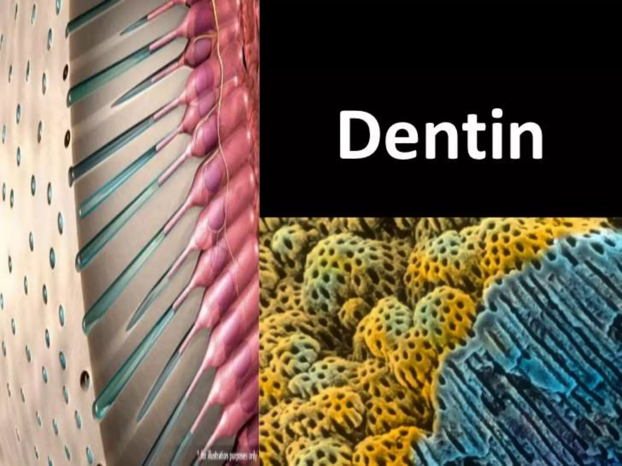
Here are some suggested du'as before and after studying, and during exams:
Before studying:
اللهم أعني على ذكرك وشكرك وحسن عبادتك
O Allah, help me remember You, be grateful to You and worship You in the best way.
اللهم بارك لي في علمي وزدني من فضلك وانفعني بما علمت
O Allah, bless me in my knowledge, increase me in Your bounty and benefit me with what I
This document provides an overview of dentin, including its history, stages of development, physical properties, composition, and age-related changes. Key points include:
- Dentin is the secondary layer of the tooth structure that provides bulk and form. It determines tooth shape and contains dentinal tubules containing odontoblast processes.
- Dentin develops through distinct stages including the lamina, bud, cap, and bell stages. This results in crown formation and root development guided by epithelial cells.
- Dentin is a living tissue composed of collagen, hydroxyapatite crystals, and water. It is harder than bone but softer than enamel. Dentin tubules radiate outward and contain o
Enamel is the hardest and most highly mineralized tissue in the human body. It is composed primarily of hydroxyapatite crystals arranged in prisms or rods called enamel rods. Enamel rods run from the dentinoenamel junction to the outer surface of the enamel in a wavy pattern. The microscopic structure of enamel, including enamel rods, interrod enamel, rod sheaths, cross-striations, Hunter-Schreger bands and enamel spindles provide strength and resilience to the enamel. Ameloblasts are specialized epithelial cells responsible for secreting and mineralizing the enamel matrix in a process called amelogenesis which occurs in several stages over the life of the tooth.
Dentin is the mineralized hard tissue that forms the bulk of the tooth beneath enamel and cementum. It has two main properties that distinguish it from enamel: it is sensitive and forms throughout life at the expense of the dental pulp. Dentinogenesis, or dentin formation, begins when the tooth germ reaches the bell stage. Odontoblasts differentiate from ectomesenchymal cells of the dental papilla and secrete dentin matrix, which then undergoes mineralization to form the bulk of dentin, including mantle dentin and circumpulpal dentin. This process of matrix formation and mineralization by odontoblasts is ongoing throughout life.
The document discusses the peridontium and its components, which include the gingiva, periodontal ligament, cementum, and alveolar bone. It focuses on cementum, describing it as a hard connective tissue that covers tooth roots and provides attachment for collagen fibers. Cementum begins forming at the cementoenamel junction and continues to the root apex. It contains cementoblasts and cementocytes that aid in its formation and structure. Cementum comes in cellular and acellular varieties and demonstrates incremental lines from its continuous deposition over time.
Dentin forms the bulk of the tooth and contains dentinal tubules that run from the outer dentin to the pulp cavity. Dentinal tubules are hollow tubes that carry odontoblastic processes and nerves. They decrease in diameter and become more spaced out from the pulp cavity to the outer dentin. There are different types of dentin including primary dentin which surrounds the pulp and secondary dentin which is laid down after root formation. Tertiary dentin is formed in response to injury or trauma and can be reactionary or reparative depending on the stimulus. The dentin surrounding each tubule, called peritubular dentin, is more mineralized than the intertubular dentin
Enamel is the hardest tissue in the body that covers the tooth crown. It is acellular and highly mineralized, composed mostly of inorganic calcium phosphate in the form of hydroxyapatite crystals. Enamel has a density that decreases from the surface to the dentin junction. It contains enamel rods that weave a wavy path through the enamel and are surrounded by interrod cementing substance. The microscopic structure of enamel includes rods, rod sheaths, and interrod material that give it hardness and strength.
This document provides an overview of the structure of dentin. It discusses the formation of dentin through odontoblast activity and mineralization. It describes the composition and structural components of dentin, including dentinal tubules, peritubular dentin, intertubular dentin, and different types of dentin such as primary, secondary, and tertiary dentin. The document also covers topics like patterns of mineralization, vascular and nerve supply of dentin, physical and chemical properties, and age-related changes in dentin structure.
Cementum is a calcified tissue covering the roots of teeth. It is less hard than dentin and yellowish in color. Cementum is composed of collagen fibers, cells, and inorganic components like hydroxyapatite. It varies in thickness along the root and can be classified as acellular or cellular depending on the presence of cells. Cementum provides attachment of periodontal ligament fibers to the tooth and aids in repair after resorption. Changes in cementum can occur with age, such as increased thickness, or due to conditions like hypercementosis or resorption.
Enamel is the hardest tissue in the body and is derived from the inner enamel epithelium. It is composed mainly of hydroxyapatite crystals and proteins. Enamel is thickest at the incisal edges and cusp tips and thins cervically. Healthy thick enamel appears bluish white while thin enamel near the dentin looks more yellow. Enamel prisms (rods) vary in number between teeth and their twisted structure in cuspal areas adds strength. The rods are surrounded by interprismatic substance and have transverse striations. Enamel is selectively permeable but not impermeable to ions.
Dentin is the hard, bony tissue found in teeth beneath the enamel. It contains dentinal tubules that house odontoblastic processes and nerve fibers. Dentin is classified as primary, secondary, or tertiary depending on the time of its development. Primary dentin forms before and during eruption, secondary forms after root formation is complete, and tertiary dentin is produced in reaction to stimuli like attrition or caries. Tertiary dentin can be reactionary or reparative.
Indian Dental Academy: will be one of the most relevant and exciting training center with best faculty and flexible training programs for dental professionals who wish to advance in their dental practice,Offers certified courses in Dental implants,Orthodontics,Endodontics,Cosmetic Dentistry, Prosthetic Dentistry, Periodontics and General Dentistry.
Dentinogenesis is the formation of dentin by odontoblast cells that differentiate from dental papilla cells. Odontoblasts first form an uncalcified predentin matrix that then undergoes mineralization. There are two types of primary dentin formed - mantle dentin near the enamel and circumpulpal dentin forming the bulk of the tooth. Dentin has a microscopic structure consisting of dentinal tubules containing odontoblast processes, surrounded by highly mineralized peritubular dentin and less mineralized intertubular dentin.
Dentin is the tissue that makes up the bulk of the tooth beneath the enamel. It is less mineralized than enamel but more mineralized than bone. Dentin is made up of hydroxyapatite crystals, collagen, and water. It contains microscopic channels called dentinal tubules that originate from the dental pulp and extend outward. With age, secondary dentin deposition and sclerotic dentin formation cause the dentinal tubules to decrease in diameter or become blocked. Dentin provides structural support and shape to the tooth and transmits sensory input from the pulp via the dentinal tubules.
The pulp is a soft connective tissue located within the tooth. It has several unique features, including being surrounded by rigid dentin walls and susceptible to changes in pressure. The pulp contains odontoblasts, fibroblasts, undifferentiated cells, and defense cells. It is highly vascularized and innervated. During development, dental papilla forms the pulp through proliferation and differentiation of cells. The pulp cavity is divided into coronal and radicular regions. Nerves and blood vessels enter through the apical foramen, supplying the pulp.
This document summarizes a presentation on the periodontal ligament given by Dr. Abhishek Gakhar. It discusses the structure, development, constituents including cells, fibers and blood/nerve supply of the periodontal ligament. The functions of the periodontal ligament and its clinical correlations are also reviewed. Diagrams illustrate the fiber groups and cells found in the periodontal ligament.
The document discusses the dental pulp, including its development, structure, cells, and features. It notes that the dental pulp develops from the dental papilla during tooth formation. The pulp contains coronial and radicular regions, with the radicular pulp terminating at the apical foramen. The pulp has histological zones including the odontoblastic layer and cell-rich and cell-poor zones. Key cells include odontoblasts, fibroblasts, and defense cells. Odontoblasts are responsible for dentin formation and are arranged in palisades along the pulp periphery.
Cementum is the mineralized tissue covering dental roots. It begins at the cementoenamel junction and continues to the root apex. Cementum provides attachment for collagen fibers (Sharpey's fibers) that bind the tooth to surrounding structures. Cementum develops in two stages: the prefunctional stage involving matrix formation and mineralization, and the functional stage where cementum deposition continues throughout life in response to tooth movement and wear. Cementum comes in various forms classified by development, cellularity, and fiber origin and includes acellular, cellular, intrinsic and extrinsic fiber cementum.
Dentin is the hard, bony tissue found underneath the enamel of teeth. It is formed by odontoblast cells in a process called dentinogenesis, which occurs in two phases - organic matrix formation and mineralization. There are different types of dentin based on the time of formation (primary, secondary, tertiary) and relation to dentinal tubules (peritubular, intertubular, intratubular). Dentin contains dentinal tubules that contain odontoblast processes and nerves, and transmit sensations. Theories of dentin sensitivity include the direct nerve stimulation theory, transduction theory, and hydrodynamic theory which suggests that fluid pressure changes in dentinal tubules stimulate nerves to cause
The document summarizes key aspects of enamel structure and formation. It describes that enamel is composed of elongated enamel rods made of hydroxyapatite crystals. Enamel formation (amelogenesis) involves an initial secretory stage where the enamel organic matrix is deposited, followed by a maturation stage where the matrix mineralizes. During the secretory stage, ameloblasts form finger-like projections called Tomes' processes that guide enamel rod formation.
The document discusses tooth development from the intrauterine life stage through the bell stage. It describes the key stages and structures involved, including the dental lamina, bud stage, cap stage, and early bell stage. During these stages, the enamel organ, dental papilla, and dental sac develop and differentiate. Structures like the enamel knot, enamel cord, and cervical loop form transiently to guide tooth morphogenesis. By the early bell stage, the inner dental epithelium induces the underlying mesenchyme to form odontoblasts, marking the beginning of dentin formation.
Dentine is the tissue that makes up the bulk of the tooth beneath enamel. It has both inorganic and organic components. The inorganic portion is made up of hydroxyapatite crystals that are smaller and more carbonate-rich than enamel. Collagen fibrils make up the organic matrix. Dentine also contains non-collagenous proteins and lipids that play roles in mineralization. Dentinal tubules radiate outward from the pulp cavity and contain odontoblastic processes, nerves, and dentinal fluid. With age and in response to stimuli, peritubular dentine deposits form, narrowing the tubules. Secondary and tertiary dentines are laid down over time. Regional variations in
The document provides an overview of the pulp-dentin complex, including dentin and pulp. It discusses the physical and chemical properties of dentin, its structure including dentinal tubules and types of dentin. Dentinogenesis and age-related changes are also covered. The morphology, development, zones and cell types in pulp are summarized. The document establishes that dentin and pulp are embryologically, histologically and functionally the same tissue and should be considered as a complex.
This document summarizes the anatomy and histology of dental pulp. It describes how dental pulp develops from ectomesenchymal cells and resides within the tooth. The pulp contains four zones - the odontoblastic zone nearest the dentin, the cell-free zone of Weil, the cell-rich zone, and the pulp core containing nerves and blood vessels. Principal pulp cells are odontoblasts, fibroblasts, and undifferentiated mesenchymal cells. Blood vessels and nerves enter the pulp through the apex to provide nutrients and sensory functions. Calcified pulp stones may form within the pulp chamber or root canals.
This document provides an overview of dentin, including:
- Its history, development, physical and chemical properties, structure, types, and innervation
- Dentinogenesis is the process by which dentin is formed through the secretion and mineralization of an organic matrix by odontoblasts.
- Dentin's main components are hydroxyapatite crystals, collagen fibers, non-collagenous proteins, and water. Its tubular structure and composition provide mechanical strength and sensitivity.
- Different types of dentin include primary, secondary, and tertiary dentin, which vary in their location, thickness, mineralization, and quality.
Dentin is the mineralized tissue beneath enamel and cementum in teeth. It consists of 70% inorganic hydroxyapatite and 30% organic collagen and proteins. Dentin contains microscopic channels called dentinal tubules that extend from the pulp cavity to the outer surface. The tubules have primary curved shapes near the crown and take on spiral secondary curves in the root. Dentin comes in different types based on its structure, age, and location in the tooth. Primary dentin makes up the bulk of the tooth whereas secondary dentin forms a narrow band near the pulp later in life. Tertiary or reparative dentin can form in response to stimuli like cavity preparation or trauma.
middle layer of tooth the dentin which has yellowish in colorRenu710209
dentin is the resilient structure of tooth which gives yellowish color and protect the underlying dentalpulp and innervated structures from external stimuli
Indian Dental Academy: will be one of the most relevant and exciting training center with best faculty and flexible training programs for dental professionals who wish to advance in their dental practice,Offers certified courses in Dental implants,Orthodontics,Endodontics,Cosmetic Dentistry, Prosthetic Dentistry, Periodontics and General Dentistry.
Dentin is the mineralized connective tissue that makes up the bulk of teeth. It surrounds the dental pulp. Dentin is formed by odontoblasts, cells originating from the dental papilla that differentiate during tooth development. As odontoblasts secrete collagen and other proteins, they become elongated and form dentinal tubules that extend from the pulp cavity to the outer surface of the tooth. Dentin is composed primarily of hydroxyapatite crystals embedded within an organic matrix. The dentin-pulp complex functions together to detect stimuli and initiate responses like additional dentin formation.
Dentin /certified fixed orthodontic courses by Indian dental academy Indian dental academy
Welcome to Indian Dental Academy
The Indian Dental Academy is the Leader in continuing dental education , training dentists in all aspects of dentistry and offering a wide range of dental certified courses in different formats.
Indian dental academy has a unique training program & curriculum that provides students with exceptional clinical skills and enabling them to return to their office with high level confidence and start treating patients
State of the art comprehensive training-Faculty of world wide repute &Very affordable.
Dentin is the hard connective tissue that forms the bulk of the tooth. It consists of tubules throughout its thickness and determines the shape of the tooth crown. Dentin is formed by odontoblasts that differentiate from dental papilla cells and produce an organic matrix that becomes mineralized. There are three types of dentin - primary, secondary, and tertiary. Primary dentin forms most of the tooth, secondary dentin is deposited after root formation, and tertiary dentin is reparative dentin deposited in response to stimuli. Dentin has a tubular structure and contains both collagen and hydroxyapatite crystals.
This document provides an overview of dentin, including:
- Its composition, formation process, and physical properties.
- The roles of odontoblasts and other components in dentinogenesis.
- The different types and structures of dentin, such as peritubular and intertubular dentin.
- Features like dentinal tubules, Von Ebner's lines, and the dentinoenamel junction.
- Its clinical significance, including use of the cementodentinal junction as a reference point in root canals.
- Potential developmental irregularities below the enamel-dentin junction that could predispose to caries.
Dentin is the mineralized hard tissue that forms the bulk of the tooth beneath enamel and cementum. It is sensitive and continues to form throughout life at the expense of the pulp. Dentinogenesis begins when the dental papilla differentiates into odontoblasts. There are three stages of odontoblast development: differentiation, secretory formation of predentin and dentin, and a resting stage. Dentin is composed of hydroxyapatite crystals embedded in a collagen matrix. It has tubules that house odontoblast processes and dentinal fluid. The structure and composition of dentin provides strength and protection for the pulp.
The document summarizes the histogenesis of tooth tissues including dentin, cementum, pulp, and periodontal ligament. It describes how:
1) Odontoblasts differentiate from dental papilla cells and secrete predentin which mineralizes to form dentin around the pulp.
2) Cementoblasts differentiate from dental follicle cells and secrete cementum on the root surface.
3) Periodontal ligament fibers are produced that attach the cementum to alveolar bone, anchoring the tooth.
4) The dental papilla cells develop into the pulp tissue enclosed within the formed dentin.
Indian Dental Academy: will be one of the most relevant and exciting training center with best faculty and flexible training programs for dental professionals who wish to advance in their dental practice,Offers certified courses in Dental implants,Orthodontics,Endodontics,Cosmetic Dentistry, Prosthetic Dentistry, Periodontics and General Dentistry.
This document discusses dentinogenesis, the formation of dentin by odontoblasts. It describes the different types of dentin formed at various stages - mantle dentin, primary dentin, secondary dentin, and tertiary dentin. It also discusses the regional distribution and structure of different zones of dentin, including mantle dentin, circumpulpal dentin, secondary dentin, tertiary dentin, predentin, interglobular dentin, and the granular layer of Tomes. Additionally, it compares intratubular and intertubular dentin, and provides notes on sclerotic dentin, dead tracts, incremental lines of Von Ebner and Andersen, lines of primary
The document discusses the properties and development of dentin. It begins by introducing dentin and its role in tooth structure. Then it covers the physical and chemical properties of dentin, including its composition, hardness, thickness and density. The stages of dentin development and mineralization are described. Histologically, the key features of dentin are dentinal tubules, peritubular dentin, intertubular dentin and predentin. Structural lines like the dentinoenamel junction and Tome's granular layer are also outlined. Finally, the document notes different types of dentin like mantle dentin.
An overview on the Pulp Dentin Complex .pptxperiovista
The document discusses the pulp-dentin complex. It describes dentin as the second layer of the tooth that provides structure and determines tooth shape. Dentin is a living tissue containing odontoblast processes in tubules. Dentin forms through secretion of an organic matrix followed by mineralization. Different types of dentin form throughout life. The pulp contains blood vessels, nerves and odontoblasts that form dentin. Dentin-pulp complex has sensory and protective functions.
The periodontal ligament (PDL) is a soft connective tissue that surrounds tooth roots and attaches them to the alveolar bone in the jaw. It ranges from 0.15-0.38mm in width and is narrowest at the mid-root level. The PDL contains principal collagen fibers, blood vessels, nerves and cells that allow it to absorb forces and remodel throughout life. Diseases can widen the PDL space and disrupt its fibers. The document discusses the development, structure, functions and clinical implications of the PDL.
This document provides an overview of dentin, including its composition, formation process (dentinogenesis), physical properties, histology, types, innervation, and age-related changes. It discusses the differences between primary and permanent dentin, infected vs affected dentin, the smear layer, and dentin bonding systems. The document concludes with a brief discussion of the role of dentin in forensic odontology. Key points include that dentin is a mineralized tissue containing dentinal tubules that contain odontoblast processes, and its formation involves both collagen matrix formation and subsequent mineralization.
4.DENTIN.ppt dental histology 1st year BdsAmulyaSnr
Dentin forms the bulk of the tooth and is the first dental hard tissue to form. It is yellow in color and elastic in nature. Dentin is composed primarily of hydroxyapatite crystals, type 1 collagen, and other organic and inorganic components. Dentin formation begins with the differentiation of odontoblasts from dental papilla cells. Odontoblasts secrete an organic matrix called predentin and initiate its mineralization. Dentin can be divided into primary, secondary, and tertiary types based on the stage of tooth development in which they form. Primary dentin includes mantle and circumpulpal dentin and makes up the bulk of dentin. Secondary dentin forms more slowly and lays down within the pulp
Garima Singh presented on the topic of dentin. Key points included:
- Dentin is the tissue found underneath enamel and makes up the bulk of teeth. It contains dentinal tubules that contain odontoblast processes and connect the pulp chamber.
- Dentin is made up of 70% inorganic material (mainly hydroxyapatite), 20% organic material (mainly type I collagen), and 10% water. It undergoes dentinogenesis through collagen matrix formation and mineralization.
- There are different types of dentin, including primary, secondary, and tertiary dentin which are formed at different stages. Characteristics like tubule orientation and mineralization differ between primary and permanent dentin
Dentin with emphasis on applied physiology and pathology Nadeem Aashiq
This document provides an overview of the structure and properties of dentin. It discusses the composition, physical properties, and microscopic structure of dentin. Key points include that dentin is composed of organic and inorganic materials, including collagen, water and hydroxyapatite crystals. It describes the arrangement of dentinal tubules and peritubular dentin. Primary, secondary and tertiary dentin formation is explained. The roles of odontoblasts and dentinal fluid are also summarized. The document considers age-related changes in dentin like dead tracts and sclerosis.
The periodontal ligament connects the tooth to the alveolar bone and is made of specialized connective tissue. It develops as cells from the dental follicle migrate into the newly formed root cementum. The principal fibers of the ligament develop with tooth eruption, first entering the alveolar bone and later organizing into bundles. Microscopically, the ligament contains fibroblasts, progenitor cells, and extracellular fibers and ground substance. The fibroblasts are responsible for collagen synthesis and turnover and have a higher rate of protein production compared to gingiva or bone fibroblasts.
Does Over-Masturbation Contribute to Chronic Prostatitis.pptxwalterHu5
In some case, your chronic prostatitis may be related to over-masturbation. Generally, natural medicine Diuretic and Anti-inflammatory Pill can help mee get a cure.
Histololgy of Female Reproductive System.pptxAyeshaZaid1
Dive into an in-depth exploration of the histological structure of female reproductive system with this comprehensive lecture. Presented by Dr. Ayesha Irfan, Assistant Professor of Anatomy, this presentation covers the Gross anatomy and functional histology of the female reproductive organs. Ideal for students, educators, and anyone interested in medical science, this lecture provides clear explanations, detailed diagrams, and valuable insights into female reproductive system. Enhance your knowledge and understanding of this essential aspect of human biology.
Basavarajeeyam is a Sreshta Sangraha grantha (Compiled book ), written by Neelkanta kotturu Basavaraja Virachita. It contains 25 Prakaranas, First 24 Chapters related to Rogas& 25th to Rasadravyas.
Integrating Ayurveda into Parkinson’s Management: A Holistic ApproachAyurveda ForAll
Explore the benefits of combining Ayurveda with conventional Parkinson's treatments. Learn how a holistic approach can manage symptoms, enhance well-being, and balance body energies. Discover the steps to safely integrate Ayurvedic practices into your Parkinson’s care plan, including expert guidance on diet, herbal remedies, and lifestyle modifications.
Here is the updated list of Top Best Ayurvedic medicine for Gas and Indigestion and those are Gas-O-Go Syp for Dyspepsia | Lavizyme Syrup for Acidity | Yumzyme Hepatoprotective Capsules etc
Muktapishti is a traditional Ayurvedic preparation made from Shoditha Mukta (Purified Pearl), is believed to help regulate thyroid function and reduce symptoms of hyperthyroidism due to its cooling and balancing properties. Clinical evidence on its efficacy remains limited, necessitating further research to validate its therapeutic benefits.
TEST BANK For An Introduction to Brain and Behavior, 7th Edition by Bryan Kol...rightmanforbloodline
TEST BANK For An Introduction to Brain and Behavior, 7th Edition by Bryan Kolb, Ian Q. Whishaw, Verified Chapters 1 - 16, Complete Newest Versio
TEST BANK For An Introduction to Brain and Behavior, 7th Edition by Bryan Kolb, Ian Q. Whishaw, Verified Chapters 1 - 16, Complete Newest Version
share - Lions, tigers, AI and health misinformation, oh my!.pptxTina Purnat
• Pitfalls and pivots needed to use AI effectively in public health
• Evidence-based strategies to address health misinformation effectively
• Building trust with communities online and offline
• Equipping health professionals to address questions, concerns and health misinformation
• Assessing risk and mitigating harm from adverse health narratives in communities, health workforce and health system
TEST BANK For Community Health Nursing A Canadian Perspective, 5th Edition by...Donc Test
TEST BANK For Community Health Nursing A Canadian Perspective, 5th Edition by Stamler, Verified Chapters 1 - 33, Complete Newest Version Community Health Nursing A Canadian Perspective, 5th Edition by Stamler, Verified Chapters 1 - 33, Complete Newest Version Community Health Nursing A Canadian Perspective, 5th Edition by Stamler Community Health Nursing A Canadian Perspective, 5th Edition TEST BANK by Stamler Test Bank For Community Health Nursing A Canadian Perspective, 5th Edition Pdf Chapters Download Test Bank For Community Health Nursing A Canadian Perspective, 5th Edition Pdf Download Stuvia Test Bank For Community Health Nursing A Canadian Perspective, 5th Edition Study Guide Test Bank For Community Health Nursing A Canadian Perspective, 5th Edition Ebook Download Stuvia Test Bank For Community Health Nursing A Canadian Perspective, 5th Edition Questions and Answers Quizlet Test Bank For Community Health Nursing A Canadian Perspective, 5th Edition Studocu Test Bank For Community Health Nursing A Canadian Perspective, 5th Edition Quizlet Test Bank For Community Health Nursing A Canadian Perspective, 5th Edition Stuvia Community Health Nursing A Canadian Perspective, 5th Edition Pdf Chapters Download Community Health Nursing A Canadian Perspective, 5th Edition Pdf Download Course Hero Community Health Nursing A Canadian Perspective, 5th Edition Answers Quizlet Community Health Nursing A Canadian Perspective, 5th Edition Ebook Download Course hero Community Health Nursing A Canadian Perspective, 5th Edition Questions and Answers Community Health Nursing A Canadian Perspective, 5th Edition Studocu Community Health Nursing A Canadian Perspective, 5th Edition Quizlet Community Health Nursing A Canadian Perspective, 5th Edition Stuvia Community Health Nursing A Canadian Perspective, 5th Edition Test Bank Pdf Chapters Download Community Health Nursing A Canadian Perspective, 5th Edition Test Bank Pdf Download Stuvia Community Health Nursing A Canadian Perspective, 5th Edition Test Bank Study Guide Questions and Answers Community Health Nursing A Canadian Perspective, 5th Edition Test Bank Ebook Download Stuvia Community Health Nursing A Canadian Perspective, 5th Edition Test Bank Questions Quizlet Community Health Nursing A Canadian Perspective, 5th Edition Test Bank Studocu Community Health Nursing A Canadian Perspective, 5th Edition Test Bank Quizlet Community Health Nursing A Canadian Perspective, 5th Edition Test Bank Stuvia
Top 10 Best Ayurvedic Kidney Stone Syrups in India
Dentin
1.
2. INTRODUCTION
DENTINO-PULPAL COMPLEX
HISTORY
DENTINOGENESIS
COMPOSITION
PROPERTIES OF DENTIN
HISTOLOGY OF DENTIN
FUNCTION OF DENTIN
CLINICAL SIGNIFICANCE
AGE AND FUNCTIONAL CHANGES
DEVELOPMENTAL ANOMALIES
CONCLUSION
REFERENCES
3. INTRODUCTION
Most Voluminous Mineralized
Connective Tissue Of Tooth
Hard Portion Of Dentino-Pulpal
Complex
Dentin = Vital Tissue with Living
Protoplasm
5. The dental pulp and dentin functions as a unit.
Odontoblasts are located in the periphery of the
pulp tissue with extensions into the inner part of
the dentin.
Dentin would not exist unless produced by
odontoblasts and dental pulp is dependant
on the dentin for protection(alongwith enamel).
Likewise, integrated dynamics of the pulp-
dentin complex imply that impacts on the dentin
may affect the pulpal component and that
disturbances in the pulpal will in turn affect the
quantity and quality of dentin produced.
6. ODONTOBLASTS
The odontoblasts line the entire pulpal aspect of the
predentin.
The odontoblasts are tall columnar cells or
sometimes somewhat cuboidal in shape (especially
in the root).
The nucleus is oval, has one or two nucleoli. The
cytoplasm of the cell body contains numerous
organelles rough surfaced endoplasmic reticulum
(arranged in longitudinally running cisternae) a well
developed Golgi apparatus, Mitochondria, Vesicles.
Vacuoles, Electron bodies, Microtubules and fibrils.
The odontoblasts cells bodies are approximately 7
micrometre in diameter & 40 micrometre in length.
7.
8. ODONTOBLASTIC
PROCESS
The odontoblast processes are the
cytoplasmic extensions of the
odontoblasts. The odontoblasts cells reside
in the peripheral pulp at the pulp-predentin
border and their processes extend in to the
dentinal tubules .
The processes are largest in diameter near
the pulp (3 to 4 mm) and taper into the
dentin. The odontoblast processes divide
near the dentino enamel junction and may
indeed extend in to enamel in the “enamel
spindles”
10. The biological structure of dentin has been
investigated since time of:
1) Anton Van Leeuwenhock : Explained the
tubular structure of dentin.
2) Von Purkinje and Retrices : Explained abt.
dentinal tubules.
3) John Hunter : Specified it as hard tissue
4) Cuvien : coined the term IVORY for dentin
11. Numerous structural elements of dentin carry
the name of well-known histologists of 19th
century:
1) Owen: Contour lines of Owen (1840)
2) Von Ebner: Ebner’s growth lines (1891)
3) Neumann: Neumann’s sheath (1863)
4) John Tomes: Tomes granular layer (1838)
Tomes process (1856)
5) Von Korff: Korff’s fibers (1906)
13. Dentin is formed by cells called odontoblasts
that differentiates from ectomesenchymal
cells of dental papilla following an organizing
influence that emanates from inner dental
epithelium.
Dental papilla is the formative organ of dentin
and eventually becomes the pulp of the teeth,
a change in terminology generally associated
with the moment dentin formation begins.
14.
15. Dentinogenesis is composed of three stages
i.e.
1) CYTODIFFERENTIATION
2) MATRIX FORMATION
3) MINERALIZATION
16. CYTODIFFERENTIATION
* This takes place during the BELL STAGE.
* In this stage, the cells from the papillae sac
differentiates to produce odontoblasts under
the influence of the Inner dental epithelium
starting at the cuspal or incisal region.
* The cells become cylindrical in shape and
are sometimes referred as Preodontoblasts.
* The no. of organelles markedly increases
with the cell becoming tall and columnar
located at end farthest from basement
membrane of Inner dental epithelia. These cells
are known as Odontoblast.
17. * Concomitant with the formation of predentin
at cuspal and incisal, continued differentiation
of new odontoblasts takes place further apic-
-ally in dental papilla.
* After the crown is fully formed, the rate of
dentin slows down markedly(2-3yrs post-
-natal) until root formation starts i.e. similar
to cytodifferentiation in crown.
18. MATRIX FORMATION
* The odontoblasts exhibits all the characterstics
of matrix-producing cells i.e. an abundance
of RER, a well developed Golgi apparatus,
mitochondria and secretory granules.
* Pro-collagen is synthesized by RER, transferr-
-ed to Golgi apparatus and finally, secreted by
secretory granules.
* The collagen, glycoprotein and proteoglycan
complexes constitutes the organic matrix of
dentin prior to mineralization, two layer in pre-
-dentin may be differentiated.
19. * The fibers involved in the formation of the
initial predentin are argyrophilic and k/a
VON KROFF’S FIBERS.
* VON KROFF’S FIBERS are the first fibers
that signifies the first formed dentin. Due
to the argyrophilic rxn, it was long believ-
-ed that bundles of collagen formed
among the odontoblasts.
* Recently EM studies revealed that the
staining is of ground substance among the
cells and not collagen.
20. * As the matrix formation continues, the
odontoblastic process lenghtens, as does
the dentinal tubule.
* Initially, 4µ of daily increments are added.
This continues until the crown is formed
and the teeth move into occlusion. After
this time a daily incremental deposit of 1µ
takes place.
21. MINERALIZATION
* Basically, it occurs by globular calcification.
* Earliest crystal deposition takes place in the
form of very fine plates of hydroxyapatite on
the surface of collagen fibrils in the ground
substance.
* The crystals are laid down within the fibrils
themselves with the crystals that are
associated with collagen being in orderly
manner( long axis parallel to fibrils long axis)
and in rows conforming to 64nm striation
pattern within the globular islands of mineraliz-
-ation .
22. * The crystal deposition appears to take place
readily from common centers in so-called
Spherulite form.
* The peritubular region becomes highly minera-
-lization at a very early stage. The ultimate
crystal size remains very small about 3mm
in thickness and 100nm in length
* The hydroxyapatite crystal of dentin resembles
bone and cementum. They are 300 times
smaller than enamel crystals.
24. COMPOSITION PERCENTAGE CONTENT
INORGANIC 70%(by weight)
45%(by volume)
CALCIUM HYDROXYAPATITE
ORGANIC 20%(by weight)
33%(by volume)
1)TYPE I COLLAGEN
(minor type III and IV)
2) MINOR FRACTION
a) Lipids
b) Non-collagenous matrix
protein
WATER 10%(by weight)
22%(by volume)
26. COMPOSITION(contd.)
NON COLLAGENOUS MATRIX PROTEIN
• Regulates Mineral Deposition and
can act as Promoter or Inhibitor.
• Pack the space between Collagen
fibrils and accumulates along the
periphery of dentinal tubules.
(Peritubular Dentin)
27. COMPOSITION(contd.)
IMP. NON COLLAGENOUS MATRIX
PROTEINS
1) Dentin Sialophosphoprotein
2) Dentin Matrix Protein 1
3) Osteonectin(SPARC)
4) Osteocalcin
5) Osteopontin
6) Bone Sialoprotein
7) Matrix Extracellular Phosphoglycoprotein
29. S.No. PROPERTY EXPLAINATION SIGNIFICANCE
1 COLOR DARKER THAN ENAMEL
(Light yellow in young
adult and Dark yellow
in elderly)
EBURNATED
DENTIN
2 THICKNESS MAXIMAL
1) Cuspal height
2) Incisal edge
MINIMAL
Cervical areas
1)Thickness
increases with age.
2) 3-3.5 mm in
Coronal aspect.
3 HARDNESS 1/5th of Enamel
(High Compressive
strength and low Tensile
strength)
MOE= 1670000 Psi
(more flexibility hence
shows Cushion
Effect)
33. The dentinal tubules follow a Gentle ‘S’
shaped curve in crown and is straighter in
incisal edges, cusps and root areas.
Its perpendicular to CEJ and DEJ.
They have lateral branches throughout dentin
k/a CANALICULI or MICROTUBULES.
No. and Diameter of dentinal tubules
NUMBER DIAMETER(in µ)
PULP 45000-65000 2-3
DEJ 15000-20000 0.5-0.9
35. First formed dentin i.e. not mineralized.
10-30 µm zone of unmineralized zone
between Odontoblasts and Mineralized
dentin.
This layer of dentin lies very close pulp
tissue i.e. just next to CELL BODIES
OF ODONTOBLASTS .
37. PERITUBULAR DENTIN
• Dentin lining the Dentinal Tubules
• More mineralized than Intertubular
dentin.
INTERTUBULAR DENTIN
• Dentin i.e. present between the tubules
and is less mineralized compared to
Peritubular dentin.
• It determines ELASTICITY of dental
matrix
39. PRIMARY
DENTINE
SECONDARY
DENTINE
TERTIARY
DENTINE
DEFINITION Dentine formed
before root
completion
Formed after root
completion
Formed in response
to external stimuli
TYPE OF CELLS 1º Odontoblasts 1º Odontoblasts 2º Odontoblasts
or
undiff. Mesenchymal
cells of pulp
LOCATION All areas of dentine Not uniform
(roof and floor of
pulp )
Localized in areas of
ext. stimuli
ORIENTATION OF
TUBULES
Regular Irregular Atubular
RATE OF
FORMATION
Rapid Slow Rapid
(1.5-3.5 µm/day)
PERMEABILITY More Less Least
41. CAUSE
1) Aging patients
2) Mild irritation(Eg. Slowly advancing
caries)
HISTOLOGICAL CHANGES
Increased deposition of calcified
materials from enamel to pulp leading
to widening of PERITUBULAR DENTIN.
CLINICAL SIGNIFICANCE
Area becomes HARDER,DENSER,LESS
SENSITIVE and MORE PROTECTIVE
against irritation.
42. TYPES OF SCLEROTIC DENTIN
1)PHYSIOLOGICAL SCLEROTIC DENTIN
Sclerotic dentin due to aging.
2)REACTIVE SCLEROTIC DENTIN
Sclerotic dentin due to irritants.
3)EBURNATED DENTIN
Type of Reactive Sclerotic dentin
i.e. formed due to destruction by
slow carious process or mild chronic
irritation leading to HARD ,
DARKENED CLEANABLE on outward
portion of Reactive dentin.
44. Severe Stimulation to Dentin leads to
Destruction and Disintegration of Odontoblasts
and Odontoblastic Process.
The Dentin Tubules are empty and filled with
air.(often surrounded by Sclerotic Dentin)
Appears Black in Ground Sections.
Commonly seen in areas of narrow Pulpal
Horns due to Odontoblastic crowding.
48. Also known as Incremental lines of Von
Ebner.
Growth lines that is seen in ground section
of dentin parallel to the dentinal tubules.
These lines represents the rest phase in
dentin deposition with dentin deposition
rate of 6 µ/day in crown to 3.5 µ/day in
roots
49. 10) OSTEODENTIN
Seen in tertiary dentin due to entrapment of
odontoblast cells in the dentin. Since it
appears like bones so k/a OSTEODENTIN.
53. 1) HYPERSENSITIVITY
Dentinal hypersensitivity is the term used to
describe clinical condition of an exaggerated
response to an exogenous stimuli.
Dental hypersensitivity is defined as “sharp,
short pain arising from exposed dentin in
response to stimuli typically thermal, chemical,
tactile or osmotic and which cannot be ascribed
to any other form of dental defect or
pathology.”(Holland et al,1997)
54. Theories of Dentinal Hypersensitivity are
1) Transducer Theory
* The theory assumed that odontoblast
extends to the periphery and its
membrane comes in close apposition
to the nerve endings in the pulp and so
transmit excitation.
* Thomas,1984 indicated that process of
odontoblasts are restricted to the inner
third of the dentinal tubules.
55. 2) Neural Theory
* The neural theory attributes to activation
of the nerve endings in the tubules which
conducts the stimuli along the parent 1º
afferent nerve fibers in the pulp into the
dental nerve branch.
* According to this theory, nerve is located
throughout the thickness of the dentin, but
this was not found so in ground sections.
56. 3) Hydrodynamic Theory
* Put forth by Brannstrom,1962.
* According to this theory, hypersensitivity
is due to displacement of the fluids in the
dentinal tubules.
* Mathews et al,1994 noted that stimuli such
as cold causes fluids to flow away from the
pulp, produces more rapid and greater pulp
nerve responses than those such as heat,
which causes inward flow of fluid.
57.
58. * When exposed dentin is dehydrated using
air blasts, it causes outward fluid movt. and
stimulates the mechanoreceptor to cause
pain.
* When overexposed to such stimuli, dentinal
plugs are formed that protect the pulp from
further such stimuli.
59. Etiology of hypersensitivity
* Main etiology is exposure of the dentinal tubules
due to either of the two causes i.e. loss of
covering enamel or loss of gingival attachment.
* Loss of gingival attachment is the most common
cause of dentinal hypersensitivity.
Various factors causing
gingival attachment loss are as follows:
1) Inadequate attached gingiva
2) Improper brushing technique
3) Periodontal surgery
60. 4) Overzealous tooth cleaning
5) Oral habits
6) Recession secondary to diseases such
as NUG, Periodontitis, Herpetic gingivo-
-stomatitis
7) Crown preparation
* Once the dentinal tubules are exposed there are
process to maintain its patency i.e.
1) Poor plaque control
2) Enamel wear
3) Improper oral hygiene technique
4) Cervical erosion
5) Exposure to acids
61. Management of Hypersensitivity
1) Desensitizing by occlusion of dentinal tubules
a) formation of smear layer by burnishing with
orangewood sticks.
b) Use of topical agents such as
i) Calcium hydroxide
ii) Calcium phosphate paste
iii) Silver nitrate
iv) Strontium chloride
v) Flouride
vi) Potassium oxalate
vii) Varnish
viii) Ionophoresis
62. c) Placement of restoration
i) GIC
ii) Composite cements
d) Uses of lasers( carbon dioxide, Nd:YAG,
Er:YAG, He:Ne)
2) Desensitization by blocking pulpal sensory
nerves
E.g.. Potassium nitrate in pastes in which
potassium ions passes via tubules and
desensitizes the nerves.
63. 2) PULP PROTECTION
The cells of exposed dentin should be
insulated against bacterial toxins, strong
drugs and undue operative procedures.
When 1mm of dentin is exposed, 30000
living cells are exposed. Hence, its
advisable to provide thermal and chemical
protection.
The protocol is defined by the concept of
REMAINING DENTIN THICKNESS(RDT).
64. RDT > 2mm
(SHALLOW)
RDT (0.5-2mm)
MODERATE
RDT < 0.5mm
DEEP
SILVER
AMALGAM
VARNISH BASE OF ZnPolyF
and GIC
Ca HYDROXIDE
WITH BASE
GLASS IONOMER
CEMENT
NOT REQUIRED NOT REQUIRED CALCIUM
HYDROXIDE
LINER
COMPOSITE DBA DBA CALCIUM
HYDOXIDE + GIC
+ DBA
CAST GOLD NOT REQUIRED BASE CALCIUM
HYDROXIDE +
BASE
65. 3) DENTINAL CARIES
The rapid penetration and spread of
caries is attributed to its low Calcium
content and dentinal tubule system.
The enamel may be undermined at DEJ
but due to dentinal tubules, a passage is
formed for bacteria.
The electron microscopy of carious
dentin shows 5 distinct histological
zones.
66.
67. ZONE 1 : NORMAL DENTIN
Deepest area is normal dentin which has
tubules with odontoblastic process that are
smooth and no crystals in the lumens.
The inter tubular dentin has normal cross
banded collagen and normal dense apatite
crystals.
No bacteria in the tubules.
Stimulation of dentin (e.g. :by osmotic
gradient, a bur, a dragging instrument or air
blow) produces a sharp pain.
68. ZONE 2 : SUBTRANSPARENT
DENTIN
Zone of demineralization of the intertubular
dentin and initial formation of very fine crystals in
the tubule lumen at the advancing front.
Odontoblastic process damage is evident.
No bacteria are found in this zone.
Stimulation of dentin produces pain.
Dentin is capable of remineralization
69. ZONE 3 : TRANSPARENT DENTIN
This dentin is softer than normal dentin and
shows further loss of mineral from the inter
tubular dentin and many large crystals in the
lumen of the tubules.
Stimulation produces pain.
Intact collagen can serve as a template for
the remineralization of the inter tubular
dentin and thus this region is capable of self
repair , provided the pulp remains vital.
70. ZONE 4: TURBID DENTIN
Zone of bacterial invasion and is marked by
widening and distortion of the dentinal
tubules which are filled with bacteria.
There is very little mineral present and the
collagen is irreversibly denatured.
Dentin in this zone will not self repair .This
zone cannot be remineralized and must be
removed before restoration.
71. ZONE 5 : INFECTED DENTIN
Outer most decomposed dentin that is
teeming with bacteria.
No recognizable structure to the dentin
and collagen ,and minerals are absent.
Removal of infected dentin is essential to
sound, successful restorative procedure
as well as prevention of spreading of
infections.
72. It is also important to understand the concept
of Affected and Effected dentin
AFFECTED DENTIN EFFECTED DENTIN
1. SOFT DEMINERALIZED
DENTIN INVADED BY
BACTERIA.
DENTIN IS DEMINERALIZED
BUT NOT INVADED BY
BACTERIA.
2. SOFT LEATHERY SURFACE
WHICH IS EASILY REMOVED.
SOFT SURFACE BUT NOT
EASILY REMOVED.
3. IRREVERSIBLE
DEGRADATION OF DENTIN.
UNINTERRUPTED COLLAGEN
CROSS LINKAGE.
4. CANT BE REMINERALIZED. CAN BE REMINERALIZED.
5. CARIES STAINING DRUGS
STAINS IT.
CARIES DRUGS CANT STAIN IT.
73. 4) CAVITY FLOOR
Cavity’s pulpal floor must lie in dentin.
Due to low Modulus of Elasticity of
Dentin(compared to Enamel) it acts as a
resilient base below the restoration and
so are able to absorb the masticatory
forces without fracture.
74. 5) OPERATIVE
PROCEDURES
Undue trauma from operative procedures can
damage the pulp.
PRESSURE Air driven cutting instrument can
dislodge the odontoblast and aspiration within
the dentinal tubule which could be an important
factor of inflamed pulp.
HEAT PRODUCTION due to rotation of the
rotary instruments is important damaging
factor. If temperature is elevated by 11ºF,
destructive rxn. will occur even in a normal and
vital periodontal organ.
75. SPEED : If speed is b/w 3000-30,000 rpm
without coolant, then, pulpal damage will
occur.
Dull and worn out instrument leads to
increased vibration and reduced cutting
efficiency leading to rise in heat.
VIBRATION : Vibration leads to damage to
pulp and higher vibration leads to
development of cracks in dentin and
enamel.
76. 6) SMEAR LAYER
Presence of smear layer was first demonstrated by
BOYDE et al,1963.
Smear layer is the debris smeared on the surface of
enamel and dentin by cutting of tooth.
Cameron(1983) and Mader(1984) examined smear
layer under EM and concluded that it’s amorphous,
irregular and granular in appearance and consist of
2 layers i.e.
1) Superficial and loosely attached to underlying
dentin.
2) Deeper layer consisting of dental plugs in
dentinal tubules
77. Smear layer is composed of both Inorganic and
Organic component .
1) The organic portion is comprised of
microbes, heat coagulated proteins, necrotic pulp
tissue, odontoblastic process, saliva and blood
cells.
2)The inorganic component of smear layer is
comprised of the tooth debris.
Significance of Smear layer
1) Interferes with bonding of GIC and
composites.
2) Bacteria laden mass whose toxic product
may migrate to pulp.
78. Imp. Agent responsible for removal of smear
layer are:
1) 6% Citric acid for 60s
2) Polyacrylic acid for 5s
3) EDTA
4) Maleic acid
79. 7) ETCHING OF DENTIN
In 1955, S. Buonocore developed the technique of acid
etching for bonding of restoration to enamel.
But etching of tooth was done far before it though
unknowingly.( Soderholm JKM. Dental Adhesives how it
started and Later evolved. J Adhes Dent 2007;9:227-230)
1) In Pre-Columbian era, the Mayans used to cement their
inlays to the tooth surface by acidic cements .
2) Also dentists have been using several acidic cements for
centuries in which acid was found in the cement even after
setting which etches the dentin surface to produce rougher
surface for good adhesion.
Hence, its difficult to decide who discovered ACID ETCHING:
the Mayans or Buonocore.
80. But at that time, it was confined to enamel
and dentin was not etched.
In 1979, Fusayama used 37%
orthophosphoric acid and showed Dentin
etching doesn’t damage the pulp
In 1982, Nakabayashi discovered formation
of HYBRID LAYER OF RESIN INFILTRATED
DENTIN.
Around 1990’s, ETCHING OF DENTIN
became a common practice.
81. Significance of acid etching
1) Removal of smear layer and plugs
2) Opening dentinal tubules and deminer-
-alize Peri-and intertubular dentin, thus
increasing dentin permeability( Pashley
DIL, Carvahlo RM. Dentin permeability
and dentin adhesion. J Dent 1997;25:
355-72)
82. Depth range of decalcifications by acid etching
varies from 2-4µ and depends on the following
factors:
1) Etchant pH
2) Etchant type
3) Etchant concentration
4) Etchant viscosity
5) Application time
Perdigao J, Lambreehts P, Van Meerbeek B,
Tome AR, Vanherle G, Lopes AB. Morphological
field emission-SEM study of the effects of six
phosphoric acid etching agents on human dentin.
Dent Mater 1996;12:262-71
83. Even though, acid etching promotes profound
changes in chemical composition and
physical properties of the dental matrix, it is
widely used in restorative dentistry.
Frankenberger R, Lobbaner U, Roggendorf MJ,
Naumann M, Taschner M. Selective enamel
etching reconsidered : better than etch and
rinse and self etch? J Adhes Dent 2008; 10:
339-334
84. 8) BONDING AGENT
Bonding agents are an integral part of
restorative dentistry.
The enamel bonding agent failed when
employed for dentin. This can be attributed to
following causes:
1) Complex surface of the dentin with lesser
calcium content.
2) Remaining water of about 10% that oozes
85. out of dentinal tubules exposed due to
tooth preparation contaminates the
field.
3) Presence of smear layer.
4) Controversy regarding etching of
dentinal surface.
5) Biological sensitivity of dentin.
6) Low organic material surface energy.
Hence an effort was done to develop that
shows bonding to dentin
86. GENERATION IMPORTANT FEATURES EXAMPLE
1ST
GENERATION
Very weak bond
2ND
GENERATION
Weak bond(1-10MPa) with main
component HEMA or Bis-GMA. No
consideration given to shrinkage or
smear layer.
Scotchbond, Bondlite
3RD
GENERATION
Modification or removal of smear layer
for improvement in bond strength.
Scotchbond II, Gluma,
Prisma, Universal Bond
2/3
4TH
GENERATION
Adoption of total etch tech. for
complete removal of smear layer.
Three step bonding.
Scotchbond multipurpose,
All bond 2, Optibond FL
5TH
GENERATION
Same principle as 4th generation with
self etching primer or self priming
adhesive.
Self etching primer(Clearfil
SE bond)
Self priming
adhesives(Prime n Bond
NT)
6TH
GENERATION
Self etching adhesives, two bottle
systems mixed before application
Prompt L-Pop, Xeno IV
7TH
GENERATION
Single bottle adhesives I-Bond ,G-Bond, Xeno V
87. 9) DENTINO-ENAMEL
JUNCTION The DEJ is a scalloped junction between
enamel and dentin.
SCALLOPED NATURE OF DEJ
1) If DEJ is flat, tensile strength act at DEJ over
continuous areas would push the two areas apart ,
thus leading to delamination.
2) In scalloped DEJ, the net compression towards
the DEJ was consistently higher than net tension
away from it. As a consequence, at DEJ, enamel and
dentin are pushed towards one another during
mastication .
( Shimizu D, Macho GA. Functional significance of
microstructural detail of primate dentino-enamel
junction: a possible example of exaption. J Hum Evol
2007;52(1):103-11)
88. 10) DENTINO-CEMENTAL
JUNCTION
The dentino-cemental junction is of special
concern as it forms the apical constriction.
Apical constriction is the narrowest diameter
upto which Biomechanical preparation and
Obturation is done.
Its usually 0.5-1.0 mm short of apical foramen.
89. 11) IPC/DPC
Indirect Pulp Capping
Reparative dentin can be stimulated by
cavity lining material since dentin is
produced throughout life.
* Deep penetrating lesions is treated by
partial removal of effected dentin and
leaving the affected dentin intact. After
this, we leave a calcium hydroxide liner.
Then dressing is done for minimum three
weeks.
* The liner stimulates the odontoblasts to form
new dentin along the pulpal surface.
90. Direct Pulp Capping
Direct pulp capping can be done to stimulate
reparative dentin young, non inflamed pulps
to secrete reparative dentin.
93. VITALITY OF DENTIN
Since the odontoblast and its process are an integral
part of the dentin. There is no doubt that dentin is a
vital tissue.
Again, if vitality is understood to be the capacity of the
tissue to react to physiologic and pathologic stimuli
dentin must be considered a vital tissue.
Dentin is laid down through out life, although after the
teeth have erupted and have been functioning for a
short time, Dentinogenesis slows, and further dentin
formation is at a much slower rate.
As peritubular diameter increases tubule caliber is
diminished. Pathologic effects of dental caries,
abrasion, attrition etc cause changes in dentin. With
age, tubule closure progress pulp ward and may affect
tubules of greater diameter too)
94. DEAD TRACTS
When a ground section of tooth is observed by
transmitted light, sclerotic dentin appears light, but other
parts of the dentin frequently appear as dark bands.
These areas are called “dead tracts” and represents
groups of empty air filled dentinal tubules.
They appear black in transmitted and white in reflected
light.
These dead tracts mainly appears due to loss of
odontoblastic processes as a result of caries, attrition,
abrasion, cavity preparation or erosion
Their degeneration is often observed in the area of
narrow pulpal horns because of crowding of odontoblasts.
Dentin areas characterized by degenerated odontoblasts
processes give rise to dead tracts. These areas
decreased sensitivity and appear to a greater extent in
older teeth
95. SCLEROTIC OR TRANSPARENT DENTIN
In cases of caries, attrition, abrasion, erosion or cavity
preparation, sufficient stimuli are generated to cause
collagen fibers and apatite crystals to begin appearing in
the dentinal tubules, especially in older individuals.
In such cases blocking of the tubules is considered a
defensive reaction of dentin. The calcified tubular space
assumes a different refractive index becoming
transparent. This calcified predentin and process of
tubule is called sclerotic or transparent dentin.
It is filled with a fine meshwork of crystals and the mineral
density is greater in this area.
It appears transparent or light in transmitted light and dark
in reflected light. It is more frequently found in root and
slowly progressive caries as well as cervical areas of
older teeth with cervical cementum exposure and may
prolong vitality
98. DENTINOGENSIS
IMPERFECTA
# WHAT IS IT?
Dentinogenesis imperfecta is an
AUTOSOMAL DOMINANT trait seen
due to mutation in DSPP.
Characteristic feature of the condition is
OBLITEARTION OF DENTINAL PULP
99. # CLASSIFICATION
1) Dentinogenesis Imperfecta Type I
Dentinogenesis imperfecta with
osteogenesis imperfecta.
Error in Collagen type I synthesis
2) Dentinogenesis Imperfecta Type II
Not associated with osteogenesis
imperfecta
Opalescent Teeth
3) Dentinogenesis Imperfecta Type III
Associated with Opalescent teeth but
with quite varied clinical manifestation.
Also k/a BRANDYWINE TYPE
100. # CLINICAL FEATURE
Mostly they share a lot of common clinical features i.e.
Also k/a Tulip or Bell shaped teeth(excessive cervical
constriction)
S.N
o.
PROPER
TIES
EXPLANATION
1 Color Yellowish brown to bluish grey
(due to abnormal dentin)
2 Enamel Though quite normal but fractures
quite easily(abnormal dentin and lack
of scalloping at DEJ)
3 Roots Shortened and Blunt
4 Susceptibilities
to caries
Slight resistant due to loss of contact
101. Type I is different from Type II as it is
associated with Osteogenesis
Imperfecta.
Type III D.I presents with some extra
features compared to Type I and II i.e.
1) Multiple pulp exposures
2) Periapical radiolucency
3) Variant radiological feature.
102. # RADIOLOGICAL FEATURE
1) Type I and II D.I.
a) Opaque pulpal chamber and canal
b) Bell shaped or Tulip shaped teeth
c) Short and blunt roots
2) Type III D.I.
Extremely large pulpal chambers and
canals with very thin dentin, so k/a
SHELL TEETH
103. # TREATMENT
No specific treatment
Use of full crowns for aesthetics
104. REGIONAL
ODONTODYSPLASIA
# WHAT IS IT?
Anomaly of teeth that involves all the hard
tissue i.e. enamel, dentin and cementum.
Unknown etiology with the most popular
being local vascular defect.
105. # CLINICAL FEATURE
SITE
Maxillary teeth are more commonly
affected.
Central > Lateral > Cuspid
Incisors Incisors
SYMPTOM
a) Delayed or no eruption is observed
b) Features of teeth
i) Short root
ii) Large pupal chambers and canals with
open apical foramina
iii) Increase fractures due to poor mineral-
-ization.
iv) Irregular shape with mottled appearance
106. # RADIOLOGICAL FEATURES
Marked reduction of radio-opacity of
enamel and dentin with increase in
pulpal volume, so k/a GHOST TEETH
# TREATMENT
Extraction and replacement with
prosthesis
107. DENTIN DYSPLASIA
(Rushton,1939)
# INTRODUCTION
Autosomal dominant disorder which
manifests with normal enamel, atypical
dentin and abnormal pulp morphology.
Witkop classified it into two type i.e.
a) Radicular Dentin Dysplasia
(Type I dentin dysplasia)
b) Coronal Dentin Dysplasia
( Type II dentin dysplasia)
108. # CLINICAL AND RADIOLOGICAL FEATURE
FEATURE RADICULAR DENTIN
DYSPLASIA
CORONAL DENTIN
DYSPLASIA
CLINICAL a) Both types of dentition are
involved.
b) Look normal in
appearance though have
AMBER color appearance.
c) Short roots.
d) Delayed eruptions.
a) Both type of dentition are
involved.
b) 1º dentition shows
yellowish-brown to bluish-
grey color.
c) 2º dentition shows normal
picture.
RADIOLOGICAL a) Obliterated pulp chamber
b) Periapical radiolucency
c) Short, conical and blunt
roots
d) Crescent shaped pulpal
remnant in some 2º
dentition.
a) 1º dentition
Obliterated pulp chamber.
b) 2º dentition
Abnormally large pulp
chamber with pulp stone.
112. The Dentino-Pulpal complex is a very
important partnership. What happens to
one, affects the other.
Hence, it is quite important to
understand the dentin in order to attain
success in the field of conservative
dentistry and endodontics.
113. IMPORTANT FACTS
XENO V is an important 7th generation Dentin
Bonding agent.
Important liners seen under the composite
resins in deep carious lesion(RDT<0.5mm) are
Light Cure GIC followed by Type I GIC due to
their short setting time.
Affected dentin comprises of the INFECTED
ZONE(Zone 5) and the TURBID ZONE(Zone
6).
114. Effected dentin comprises of SUBTRANSPARENT
DENTIN (Zone 2) and TRANSPARENT DENTIN
(Zone 3).
Apical gauging is a very important procedure as it
gives a good approximation of canal diameter in
the most important apical 3-5mm of the root canal.
For e.g. if apical diameter of an root canal is
0.4mm then its not advisable to instrument this
canal to less than 40 K-File. Hence, it acts as a
guide.