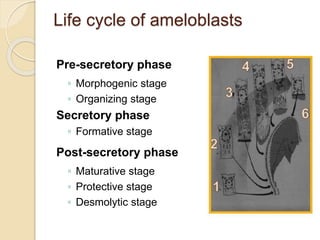
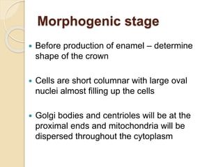
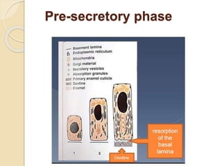
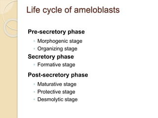
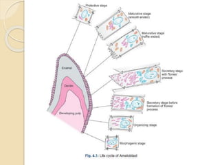
Amelogenesis
Dr. Arun Mohan
Dept. Of Oral Pathology
Royal Dental College
Life cycle of ameloblasts
Pre-secretory phase
◦ Morphogenic stage
◦ Organizing stage
Secretory phase
◦ Formative stage
Post-secretory phase
◦ Maturative stage
◦ Protective stage
◦ Desmolytic stage
Morphogenic stage
 Before production of enamel – determine
shape of the crown
 Cells are short columnar with large oval
nuclei almost filling up the cells
 Golgi bodies and centrioles will be at the
proximal ends and mitochondria will be
dispersed throughout the cytoplasm
 Proximal and distal terminal bars will develop
- intercellular attachments
- formed by thickening of cell membranes,
caused due to condensation of underlying
cytoplasm
 Cells are separated from d.papilla by a delicate
basal lamina
 Adjacent d.papilla is a light, cell-free zone
Organizing stage/Differentiation
stage
 Differentiates into ameloblasts
- becomes tall columnar (40µ in length)
- reversal of polarity (nucleus shifts to proximal
end)
- golgi apparatus and centrioles shift to distal
part
- mitochondria concentrate in the proximal
cytoplasm
 In this stage ameloblasts exert an organizing
influence on adjacent dental papilla cells – and
 Towards the end of this stage odontoblasts
secrete dentin
 Deposition of dentin helps ameloblasts attain
secretory function – reciprocal induction
 Source of nutrition changes to dental sac –
more capillaries in dental sac and collapse of
stellate reticulum
 Basal lamina disintegrates
Pre-secretory phase
Life cycle of ameloblasts
Pre-secretory phase
◦ Morphogenic stage
◦ Organizing stage
Secretory phase
◦ Formative stage
Post-secretory phase
◦ Maturative stage
◦ Protective stage
◦ Desmolytic stage
Formative stage
Deposition of enamel matrix (proteins)
and partial mineralization
 In this stage amelobalsts are fully differentiated
and and are structurally suited for secretion
- cells have abundant mitochondria, RERs,
free ribosomes, well developed golgi bodies
- basal cytoplasm has abundant secretory
granules packed with enamel proteins
- abundant microtubules are present which
helps in movement of secretory granules to
basal plasma membrane
 Secretory granules filled with
synthesised enamel proteins – move
towards basal plasma membrane and
release the proteins by exocytosis
 Proximal and distal terminal bars –
provides attachment between cells
 Distal terminal bars prevents entry of
Calcium ions and other molecules
from ECF
 In the initial stage of secretory stage, basal
region is flat.
 After initial deposition of enamel (aprismatic
enamel), a conical cytoplasmic process
develops at the base
– Tome’s process
 Cytoplasm of Tome’s process contain secretory
granules, microtubules and few mitochondria
 Tome’s process is responsible for
enamel rods and interrod enamel
 It is lost before the last phase of
secretion
- just before deposition of surface
aprismatic enamel
Life cycle of ameloblasts
Pre-secretory phase
◦ Morphogenic stage
◦ Organizing stage
Secretory phase
◦ Formative stage
Post-secretory phase
◦ Maturative stage
◦ Protective stage
◦ Desmolytic stage
Maturative stage
 Enamel maturation (full mineralization), occurs in
incisal and occlusal areas first and then extends to
cervical region.
 During maturative stage ameloblasts- reduce in
height, volume and organelle content
 They have dual functions
- Ruffle bordered : microvilli in their distal
extrimities
- mineralization
- Smooth bordered : reabsorbs protein and water
 Stratum intermedium lose their
cuboidal shape and becomes spindle
shaped
Protective stage
 Basal plasma membrane loses ruffled ended
borders.
 Secrete a protein similar to basal lamina onto
the surface of newly formed enamel –
Nasmyth’s membrane
 Ameloblasts develop hemidesmosomal
attachments to dental lamina
 After deposition of full thickness of epithelium,
ameloblasts cease to exist as a well- defined
layer
 It becomes indistinguishable from OEE and
stratum intermedium – form a 2-3 layered
stratified epithelial covering of enamel – Reduce
Enamel Epithelium
 REE protects mature enamel from coming into
contact with CT ( - thus prevents resorption of
enamel and deposition of cementum over
enamel), till the tooth erupts
Desmolytic stage
 REE induces atrophy of CT separating it
from oral epithelium - facilitating eruption of
tooth
 REE secretes enzymes like collagenases
 REE proliferates and fuses with the oral
epithelum to form a solid plug of epithelial
cells – its central cells degenerate and to
from a canal through which bloodless
eruption of tooth takes palce
Amelogenesis
 Enamel matrix deposition
 Development of Tome’s process
 Mineralization
Enamel matrix deposition
 The secretory ameloblasts which are
structurally suited for synthesis and secretion
of enamel proteins, start secretory function
after a layer of dentin is deposited
 After a layer of dentin is deposited,
ameloblasts synthesize and secrete enamel
through the distal part.
 The secretory granules packed with enamel
proteins fuses with the basal plasma
membrane and release the matrix protein
against the newly formed dentin by a process
called exocytosis.
 Begins in cusp tips and progresses outward
and cervically
 With progress of matrix deposition –
 In the early stages the matrix contains 20-
30% proteins – amelogenin and non-
amelogenin (enamelin, tuftelin,
ameloblastin)
 The proportion of proteins gradually
decreases during mineralization
 Tuftelin– hypothesized to induce nucleation
 Amelogenins found in intercrystalline
spaces – hypothesized to control crystal
growth
Development of Tome’s
process
 After deposition of about
30µ thickness of matrix is
deposited – ameloblasts
develop a conical process
at the base – Tome’s
process
 It extends beyond the
distal terminal bars
 Matrix deposition and
mineralization takes place
only through this area – in
 The distal terminal bars have fine radiating
actin filaments extending into cytoplasm –
forming a septa which partially separates
Tome’s process from rest
 Mostly contains secretory granules,
microfilaments, mitochondria and some
Golgi bodies
 Secretions from proximal part of
secretory end, occur early and form
interrod enamel
 Secretions from distal part (Tome’s
process) secrete rod enamel
(perpendicular to interrod enamel) into
the pit like outline formed by interrod
enamel
 Head of one enamel rod is formed by
one ameloblast and tail is formed by
three surrounding amelobalsts
 Thus each enamel rod is formed by 4
ameloblasts and each ameloblast
contributes to 4 enamel rods
Mineralization
 Ameloblasts are involved in both secretion
of enamel matrix and its mineralization
 Ca2+ ions circulation ECF
actively transported to cell
attached to Calcium binding proteins
transported to distal cytoplasm
Ca2+ ions are extruded through secretory
end
by active process
 2 stages :
- Immediate partial mineralization
- Maturation
Immediate partial
mineralization
 In formative stage, immediately after matrix is laid
down
 25-30% of eventual total mineral content
 Mineralization begins in DEJ – rich in Tuftelin
- crystals perpendicular to DEJ
 After nucleation ameloblasts secrete matrix rich in
amelogenin – regulate crystal size
 First forms octacalcium – unstable – immediately
converts to HA
Maturation
 Gradual completion of
mineralization
 Each rod matures
from DEJ to surface
and the sequence of
maturing rods is from
cusps/incisal edges
towards cervical line
 Thus, occlusal
regions mature ahead
of cervical regions
 Growth of crystals formed in primary
mineralization stage – regulated by enamel
proteins, mainly amelogenin
( proteolytic enzymes cleave amelogenin
protein and thus helps in growth of individual
crystals)
 During maturation, crystal thickness increases
from 1.5µ to 25µ
 Rate of deposition - 4µ/day
Ameloblast modulation
 As maturation progresses, proteins and
water needs to be reabsorbed and broken
down by ameloblasts – to create space for
growing crystals
 Leads to modulation – alternating cycles of
ruffle-ended and smooth-ended
ameloblasts
 Ruffle ended ameloblasts have microvilli
projections in basal membrane, leaky
proximal and tight distal terminal bars –
contains lysosomes, and vesicles containing
matrix proteins
 Smooth ended ameloblasts have smooth
basal membrane, tight proximal and leaky
distal terminal bars
 Ruffle-ended cells are believed to actively secerte
Ca ions and release various proteases that break
down enamel proteins
 Broken down matrix remnants are reabsorbed or
leaks through the space in b/w smooth-ended
ameloblasts
 Thus about 90 % of enamel proteins
secreted are reabsorbed – process
unique to enamel formation
 Finally forms enamel with 96% mineral
content

Amelogenesis Slideshare.pptx

  • 1.
    Amelogenesis Dr. Arun Mohan Dept.Of Oral Pathology Royal Dental College
  • 2.
    Life cycle ofameloblasts Pre-secretory phase ◦ Morphogenic stage ◦ Organizing stage Secretory phase ◦ Formative stage Post-secretory phase ◦ Maturative stage ◦ Protective stage ◦ Desmolytic stage
  • 3.
    Morphogenic stage  Beforeproduction of enamel – determine shape of the crown  Cells are short columnar with large oval nuclei almost filling up the cells  Golgi bodies and centrioles will be at the proximal ends and mitochondria will be dispersed throughout the cytoplasm
  • 4.
     Proximal anddistal terminal bars will develop - intercellular attachments - formed by thickening of cell membranes, caused due to condensation of underlying cytoplasm  Cells are separated from d.papilla by a delicate basal lamina  Adjacent d.papilla is a light, cell-free zone
  • 5.
    Organizing stage/Differentiation stage  Differentiatesinto ameloblasts - becomes tall columnar (40µ in length) - reversal of polarity (nucleus shifts to proximal end) - golgi apparatus and centrioles shift to distal part - mitochondria concentrate in the proximal cytoplasm  In this stage ameloblasts exert an organizing influence on adjacent dental papilla cells – and
  • 6.
     Towards theend of this stage odontoblasts secrete dentin  Deposition of dentin helps ameloblasts attain secretory function – reciprocal induction  Source of nutrition changes to dental sac – more capillaries in dental sac and collapse of stellate reticulum  Basal lamina disintegrates
  • 7.
  • 8.
    Life cycle ofameloblasts Pre-secretory phase ◦ Morphogenic stage ◦ Organizing stage Secretory phase ◦ Formative stage Post-secretory phase ◦ Maturative stage ◦ Protective stage ◦ Desmolytic stage
  • 9.
    Formative stage Deposition ofenamel matrix (proteins) and partial mineralization
  • 10.
     In thisstage amelobalsts are fully differentiated and and are structurally suited for secretion - cells have abundant mitochondria, RERs, free ribosomes, well developed golgi bodies - basal cytoplasm has abundant secretory granules packed with enamel proteins - abundant microtubules are present which helps in movement of secretory granules to basal plasma membrane
  • 11.
     Secretory granulesfilled with synthesised enamel proteins – move towards basal plasma membrane and release the proteins by exocytosis
  • 12.
     Proximal anddistal terminal bars – provides attachment between cells  Distal terminal bars prevents entry of Calcium ions and other molecules from ECF
  • 13.
     In theinitial stage of secretory stage, basal region is flat.  After initial deposition of enamel (aprismatic enamel), a conical cytoplasmic process develops at the base – Tome’s process  Cytoplasm of Tome’s process contain secretory granules, microtubules and few mitochondria
  • 15.
     Tome’s processis responsible for enamel rods and interrod enamel  It is lost before the last phase of secretion - just before deposition of surface aprismatic enamel
  • 16.
    Life cycle ofameloblasts Pre-secretory phase ◦ Morphogenic stage ◦ Organizing stage Secretory phase ◦ Formative stage Post-secretory phase ◦ Maturative stage ◦ Protective stage ◦ Desmolytic stage
  • 17.
    Maturative stage  Enamelmaturation (full mineralization), occurs in incisal and occlusal areas first and then extends to cervical region.  During maturative stage ameloblasts- reduce in height, volume and organelle content  They have dual functions - Ruffle bordered : microvilli in their distal extrimities - mineralization - Smooth bordered : reabsorbs protein and water
  • 18.
     Stratum intermediumlose their cuboidal shape and becomes spindle shaped
  • 19.
    Protective stage  Basalplasma membrane loses ruffled ended borders.  Secrete a protein similar to basal lamina onto the surface of newly formed enamel – Nasmyth’s membrane  Ameloblasts develop hemidesmosomal attachments to dental lamina
  • 20.
     After depositionof full thickness of epithelium, ameloblasts cease to exist as a well- defined layer  It becomes indistinguishable from OEE and stratum intermedium – form a 2-3 layered stratified epithelial covering of enamel – Reduce Enamel Epithelium  REE protects mature enamel from coming into contact with CT ( - thus prevents resorption of enamel and deposition of cementum over enamel), till the tooth erupts
  • 22.
    Desmolytic stage  REEinduces atrophy of CT separating it from oral epithelium - facilitating eruption of tooth  REE secretes enzymes like collagenases  REE proliferates and fuses with the oral epithelum to form a solid plug of epithelial cells – its central cells degenerate and to from a canal through which bloodless eruption of tooth takes palce
  • 25.
    Amelogenesis  Enamel matrixdeposition  Development of Tome’s process  Mineralization
  • 26.
    Enamel matrix deposition The secretory ameloblasts which are structurally suited for synthesis and secretion of enamel proteins, start secretory function after a layer of dentin is deposited
  • 28.
     After alayer of dentin is deposited, ameloblasts synthesize and secrete enamel through the distal part.  The secretory granules packed with enamel proteins fuses with the basal plasma membrane and release the matrix protein against the newly formed dentin by a process called exocytosis.  Begins in cusp tips and progresses outward and cervically  With progress of matrix deposition –
  • 30.
     In theearly stages the matrix contains 20- 30% proteins – amelogenin and non- amelogenin (enamelin, tuftelin, ameloblastin)  The proportion of proteins gradually decreases during mineralization  Tuftelin– hypothesized to induce nucleation  Amelogenins found in intercrystalline spaces – hypothesized to control crystal growth
  • 31.
    Development of Tome’s process After deposition of about 30µ thickness of matrix is deposited – ameloblasts develop a conical process at the base – Tome’s process  It extends beyond the distal terminal bars  Matrix deposition and mineralization takes place only through this area – in
  • 32.
     The distalterminal bars have fine radiating actin filaments extending into cytoplasm – forming a septa which partially separates Tome’s process from rest  Mostly contains secretory granules, microfilaments, mitochondria and some Golgi bodies
  • 33.
     Secretions fromproximal part of secretory end, occur early and form interrod enamel  Secretions from distal part (Tome’s process) secrete rod enamel (perpendicular to interrod enamel) into the pit like outline formed by interrod enamel
  • 37.
     Head ofone enamel rod is formed by one ameloblast and tail is formed by three surrounding amelobalsts  Thus each enamel rod is formed by 4 ameloblasts and each ameloblast contributes to 4 enamel rods
  • 40.
    Mineralization  Ameloblasts areinvolved in both secretion of enamel matrix and its mineralization  Ca2+ ions circulation ECF actively transported to cell attached to Calcium binding proteins transported to distal cytoplasm Ca2+ ions are extruded through secretory end by active process
  • 41.
     2 stages: - Immediate partial mineralization - Maturation
  • 42.
    Immediate partial mineralization  Informative stage, immediately after matrix is laid down  25-30% of eventual total mineral content  Mineralization begins in DEJ – rich in Tuftelin - crystals perpendicular to DEJ  After nucleation ameloblasts secrete matrix rich in amelogenin – regulate crystal size  First forms octacalcium – unstable – immediately converts to HA
  • 43.
    Maturation  Gradual completionof mineralization  Each rod matures from DEJ to surface and the sequence of maturing rods is from cusps/incisal edges towards cervical line  Thus, occlusal regions mature ahead of cervical regions
  • 44.
     Growth ofcrystals formed in primary mineralization stage – regulated by enamel proteins, mainly amelogenin ( proteolytic enzymes cleave amelogenin protein and thus helps in growth of individual crystals)  During maturation, crystal thickness increases from 1.5µ to 25µ  Rate of deposition - 4µ/day
  • 45.
    Ameloblast modulation  Asmaturation progresses, proteins and water needs to be reabsorbed and broken down by ameloblasts – to create space for growing crystals  Leads to modulation – alternating cycles of ruffle-ended and smooth-ended ameloblasts
  • 46.
     Ruffle endedameloblasts have microvilli projections in basal membrane, leaky proximal and tight distal terminal bars – contains lysosomes, and vesicles containing matrix proteins  Smooth ended ameloblasts have smooth basal membrane, tight proximal and leaky distal terminal bars
  • 47.
     Ruffle-ended cellsare believed to actively secerte Ca ions and release various proteases that break down enamel proteins  Broken down matrix remnants are reabsorbed or leaks through the space in b/w smooth-ended ameloblasts
  • 48.
     Thus about90 % of enamel proteins secreted are reabsorbed – process unique to enamel formation  Finally forms enamel with 96% mineral content