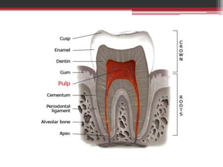
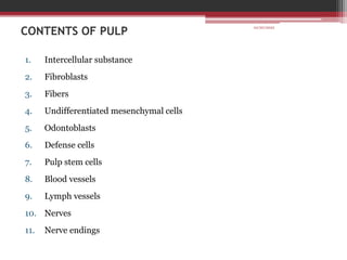
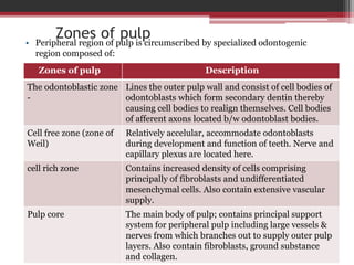
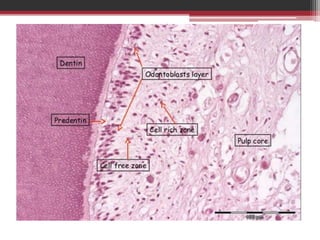
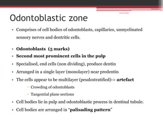
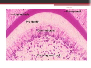
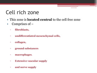
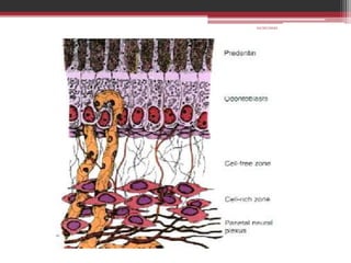
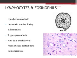
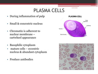
The document provides information on the structure and functions of the dental pulp. It begins with definitions and general anatomy, describing the pulp as a soft connective tissue enclosed within dentin. It then discusses the zones and structural features of the pulp in more detail. This includes the odontoblastic zone containing odontoblasts and nerve endings, the cell-free zone with capillaries and nerves, and the cell-rich zone with fibroblasts and blood vessels. Key cell types like odontoblasts, fibroblasts, and immune cells are also described. The functions of the pulp in dentin formation, nutrition, and defense are highlighted.
PHYSICAL PROPERTIES
CHEMICAL PROPERTIES
STRUCTURE OF ENAMEL
DEVELOPMENT OF ENAMEL
EPITHELIAL ENAMEL ORGAN
AMELOGENESIS
LIFE CYCLE OF AMELOBLASTS
AGE CHANGES IN ENAMEL
DEFECTS OF AMELOGENESIS
CLINICAL IMPLICATIONS
I prepared this presentation during the first year of my MDS. This will give you a basic idea and necessary information about the pulp of the teeth and its histology. Hope you guys find it useful.
Cementum also commonly known as root cementum , is a highly mineralized tissue covering the entire root surface.
Cementum is also often referred to as a bone-like tissue. Cementum contains two types of fibers, mainly extrinsic (Sharpey's) fibers and intrinsic fibers. Fibroblasts and cementoblasts are the fiber secreting cells.
An odontoblast is a biological cell of neural crest origin whose main function is formation of dentin.
This slide gives a detailed explanation of the same.
https://userupload.net/3ppacneii1wj
Toxicologic Pathology (Second Edition), 2010
INTRODUCTION
The oral mucosa is, in many ways, similar to the skin in its architecture, function, and reaction patterns. This section only emphasizes those characteristics of the oral mucosa that influence or result in a distinct group of pathologic entities.
Because of its location at the entrance of the digestive and respiratory tracts and its proximity to the teeth, the oral mucosa is subjected to numerous natural and man-made xenobiotics. The peculiar architecture and absorption characteristics of the oral mucosa, especially in areas of extreme thinness, coupled with the rich microorganism flora of the mouth, makes the oral mucosa a peculiar site deserving separate discussion.
Dental Pulp: development, innervation, vascular functions, pathways of pain, sensitivity and sensibility tests, pulpal diagnosis as applied to pediatric dentistry.
seminar on gingiva
contents:
Introduction
Definition
Development of gingiva
Macroscopic anatomy
Microscopic anatomy
Blood supply
Lymphatic drainage
Nerve supply
Correlation of clinical and microscopic features
Repair/healing of gingiva
Age changes
Gingival diseases
Clinical considerations
Conclusion
References
PDL is complex vascular structure which surround tooth root and connect it to the alveolar bone. In this we discuss development, macroscopic and microscopic features, functions and it’s supply and clinical consideration.
Title: Sense of Taste
Presenter: Dr. Faiza, Assistant Professor of Physiology
Qualifications:
MBBS (Best Graduate, AIMC Lahore)
FCPS Physiology
ICMT, CHPE, DHPE (STMU)
MPH (GC University, Faisalabad)
MBA (Virtual University of Pakistan)
Learning Objectives:
Describe the structure and function of taste buds.
Describe the relationship between the taste threshold and taste index of common substances.
Explain the chemical basis and signal transduction of taste perception for each type of primary taste sensation.
Recognize different abnormalities of taste perception and their causes.
Key Topics:
Significance of Taste Sensation:
Differentiation between pleasant and harmful food
Influence on behavior
Selection of food based on metabolic needs
Receptors of Taste:
Taste buds on the tongue
Influence of sense of smell, texture of food, and pain stimulation (e.g., by pepper)
Primary and Secondary Taste Sensations:
Primary taste sensations: Sweet, Sour, Salty, Bitter, Umami
Chemical basis and signal transduction mechanisms for each taste
Taste Threshold and Index:
Taste threshold values for Sweet (sucrose), Salty (NaCl), Sour (HCl), and Bitter (Quinine)
Taste index relationship: Inversely proportional to taste threshold
Taste Blindness:
Inability to taste certain substances, particularly thiourea compounds
Example: Phenylthiocarbamide
Structure and Function of Taste Buds:
Composition: Epithelial cells, Sustentacular/Supporting cells, Taste cells, Basal cells
Features: Taste pores, Taste hairs/microvilli, and Taste nerve fibers
Location of Taste Buds:
Found in papillae of the tongue (Fungiform, Circumvallate, Foliate)
Also present on the palate, tonsillar pillars, epiglottis, and proximal esophagus
Mechanism of Taste Stimulation:
Interaction of taste substances with receptors on microvilli
Signal transduction pathways for Umami, Sweet, Bitter, Sour, and Salty tastes
Taste Sensitivity and Adaptation:
Decrease in sensitivity with age
Rapid adaptation of taste sensation
Role of Saliva in Taste:
Dissolution of tastants to reach receptors
Washing away the stimulus
Taste Preferences and Aversions:
Mechanisms behind taste preference and aversion
Influence of receptors and neural pathways
Impact of Sensory Nerve Damage:
Degeneration of taste buds if the sensory nerve fiber is cut
Abnormalities of Taste Detection:
Conditions: Ageusia, Hypogeusia, Dysgeusia (parageusia)
Causes: Nerve damage, neurological disorders, infections, poor oral hygiene, adverse drug effects, deficiencies, aging, tobacco use, altered neurotransmitter levels
Neurotransmitters and Taste Threshold:
Effects of serotonin (5-HT) and norepinephrine (NE) on taste sensitivity
Supertasters:
25% of the population with heightened sensitivity to taste, especially bitterness
Increased number of fungiform papillae
Ozempic: Preoperative Management of Patients on GLP-1 Receptor Agonists Saeid Safari
Preoperative Management of Patients on GLP-1 Receptor Agonists like Ozempic and Semiglutide
ASA GUIDELINE
NYSORA Guideline
2 Case Reports of Gastric Ultrasound
These lecture slides, by Dr Sidra Arshad, offer a quick overview of physiological basis of a normal electrocardiogram.
Learning objectives:
1. Define an electrocardiogram (ECG) and electrocardiography
2. Describe how dipoles generated by the heart produce the waveforms of the ECG
3. Describe the components of a normal electrocardiogram of a typical bipolar leads (limb II)
4. Differentiate between intervals and segments
5. Enlist some common indications for obtaining an ECG
Study Resources:
1. Chapter 11, Guyton and Hall Textbook of Medical Physiology, 14th edition
2. Chapter 9, Human Physiology - From Cells to Systems, Lauralee Sherwood, 9th edition
3. Chapter 29, Ganong’s Review of Medical Physiology, 26th edition
4. Electrocardiogram, StatPearls - https://www.ncbi.nlm.nih.gov/books/NBK549803/
5. ECG in Medical Practice by ABM Abdullah, 4th edition
6. ECG Basics, http://www.nataliescasebook.com/tag/e-c-g-basics
Prix Galien International 2024 Forum ProgramLevi Shapiro
June 20, 2024, Prix Galien International and Jerusalem Ethics Forum in ROME. Detailed agenda including panels:
- ADVANCES IN CARDIOLOGY: A NEW PARADIGM IS COMING
- WOMEN’S HEALTH: FERTILITY PRESERVATION
- WHAT’S NEW IN THE TREATMENT OF INFECTIOUS,
ONCOLOGICAL AND INFLAMMATORY SKIN DISEASES?
- ARTIFICIAL INTELLIGENCE AND ETHICS
- GENE THERAPY
- BEYOND BORDERS: GLOBAL INITIATIVES FOR DEMOCRATIZING LIFE SCIENCE TECHNOLOGIES AND PROMOTING ACCESS TO HEALTHCARE
- ETHICAL CHALLENGES IN LIFE SCIENCES
- Prix Galien International Awards Ceremony
Tom Selleck Health: A Comprehensive Look at the Iconic Actor’s Wellness Journeygreendigital
Tom Selleck, an enduring figure in Hollywood. has captivated audiences for decades with his rugged charm, iconic moustache. and memorable roles in television and film. From his breakout role as Thomas Magnum in Magnum P.I. to his current portrayal of Frank Reagan in Blue Bloods. Selleck's career has spanned over 50 years. But beyond his professional achievements. fans have often been curious about Tom Selleck Health. especially as he has aged in the public eye.
Follow us on: Pinterest
Introduction
Many have been interested in Tom Selleck health. not only because of his enduring presence on screen but also because of the challenges. and lifestyle choices he has faced and made over the years. This article delves into the various aspects of Tom Selleck health. exploring his fitness regimen, diet, mental health. and the challenges he has encountered as he ages. We'll look at how he maintains his well-being. the health issues he has faced, and his approach to ageing .
Early Life and Career
Childhood and Athletic Beginnings
Tom Selleck was born on January 29, 1945, in Detroit, Michigan, and grew up in Sherman Oaks, California. From an early age, he was involved in sports, particularly basketball. which played a significant role in his physical development. His athletic pursuits continued into college. where he attended the University of Southern California (USC) on a basketball scholarship. This early involvement in sports laid a strong foundation for his physical health and disciplined lifestyle.
Transition to Acting
Selleck's transition from an athlete to an actor came with its physical demands. His first significant role in "Magnum P.I." required him to perform various stunts and maintain a fit appearance. This role, which he played from 1980 to 1988. necessitated a rigorous fitness routine to meet the show's demands. setting the stage for his long-term commitment to health and wellness.
Fitness Regimen
Workout Routine
Tom Selleck health and fitness regimen has evolved. adapting to his changing roles and age. During his "Magnum, P.I." days. Selleck's workouts were intense and focused on building and maintaining muscle mass. His routine included weightlifting, cardiovascular exercises. and specific training for the stunts he performed on the show.
Selleck adjusted his fitness routine as he aged to suit his body's needs. Today, his workouts focus on maintaining flexibility, strength, and cardiovascular health. He incorporates low-impact exercises such as swimming, walking, and light weightlifting. This balanced approach helps him stay fit without putting undue strain on his joints and muscles.
Importance of Flexibility and Mobility
In recent years, Selleck has emphasized the importance of flexibility and mobility in his fitness regimen. Understanding the natural decline in muscle mass and joint flexibility with age. he includes stretching and yoga in his routine. These practices help prevent injuries, improve posture, and maintain mobilit
Lung Cancer: Artificial Intelligence, Synergetics, Complex System Analysis, S...Oleg Kshivets
RESULTS: Overall life span (LS) was 2252.1±1742.5 days and cumulative 5-year survival (5YS) reached 73.2%, 10 years – 64.8%, 20 years – 42.5%. 513 LCP lived more than 5 years (LS=3124.6±1525.6 days), 148 LCP – more than 10 years (LS=5054.4±1504.1 days).199 LCP died because of LC (LS=562.7±374.5 days). 5YS of LCP after bi/lobectomies was significantly superior in comparison with LCP after pneumonectomies (78.1% vs.63.7%, P=0.00001 by log-rank test). AT significantly improved 5YS (66.3% vs. 34.8%) (P=0.00000 by log-rank test) only for LCP with N1-2. Cox modeling displayed that 5YS of LCP significantly depended on: phase transition (PT) early-invasive LC in terms of synergetics, PT N0—N12, cell ratio factors (ratio between cancer cells- CC and blood cells subpopulations), G1-3, histology, glucose, AT, blood cell circuit, prothrombin index, heparin tolerance, recalcification time (P=0.000-0.038). Neural networks, genetic algorithm selection and bootstrap simulation revealed relationships between 5YS and PT early-invasive LC (rank=1), PT N0—N12 (rank=2), thrombocytes/CC (3), erythrocytes/CC (4), eosinophils/CC (5), healthy cells/CC (6), lymphocytes/CC (7), segmented neutrophils/CC (8), stick neutrophils/CC (9), monocytes/CC (10); leucocytes/CC (11). Correct prediction of 5YS was 100% by neural networks computing (area under ROC curve=1.0; error=0.0).
CONCLUSIONS: 5YS of LCP after radical procedures significantly depended on: 1) PT early-invasive cancer; 2) PT N0--N12; 3) cell ratio factors; 4) blood cell circuit; 5) biochemical factors; 6) hemostasis system; 7) AT; 8) LC characteristics; 9) LC cell dynamics; 10) surgery type: lobectomy/pneumonectomy; 11) anthropometric data. Optimal diagnosis and treatment strategies for LC are: 1) screening and early detection of LC; 2) availability of experienced thoracic surgeons because of complexity of radical procedures; 3) aggressive en block surgery and adequate lymph node dissection for completeness; 4) precise prediction; 5) adjuvant chemoimmunoradiotherapy for LCP with unfavorable prognosis.
Knee anatomy and clinical tests 2024.pdfvimalpl1234
This includes all relevant anatomy and clinical tests compiled from standard textbooks, Campbell,netter etc..It is comprehensive and best suited for orthopaedicians and orthopaedic residents.
Recomendações da OMS sobre cuidados maternos e neonatais para uma experiência pós-natal positiva.
Em consonância com os ODS – Objetivos do Desenvolvimento Sustentável e a Estratégia Global para a Saúde das Mulheres, Crianças e Adolescentes, e aplicando uma abordagem baseada nos direitos humanos, os esforços de cuidados pós-natais devem expandir-se para além da cobertura e da simples sobrevivência, de modo a incluir cuidados de qualidade.
Estas diretrizes visam melhorar a qualidade dos cuidados pós-natais essenciais e de rotina prestados às mulheres e aos recém-nascidos, com o objetivo final de melhorar a saúde e o bem-estar materno e neonatal.
Uma “experiência pós-natal positiva” é um resultado importante para todas as mulheres que dão à luz e para os seus recém-nascidos, estabelecendo as bases para a melhoria da saúde e do bem-estar a curto e longo prazo. Uma experiência pós-natal positiva é definida como aquela em que as mulheres, pessoas que gestam, os recém-nascidos, os casais, os pais, os cuidadores e as famílias recebem informação consistente, garantia e apoio de profissionais de saúde motivados; e onde um sistema de saúde flexível e com recursos reconheça as necessidades das mulheres e dos bebês e respeite o seu contexto cultural.
Estas diretrizes consolidadas apresentam algumas recomendações novas e já bem fundamentadas sobre cuidados pós-natais de rotina para mulheres e neonatos que recebem cuidados no pós-parto em unidades de saúde ou na comunidade, independentemente dos recursos disponíveis.
É fornecido um conjunto abrangente de recomendações para cuidados durante o período puerperal, com ênfase nos cuidados essenciais que todas as mulheres e recém-nascidos devem receber, e com a devida atenção à qualidade dos cuidados; isto é, a entrega e a experiência do cuidado recebido. Estas diretrizes atualizam e ampliam as recomendações da OMS de 2014 sobre cuidados pós-natais da mãe e do recém-nascido e complementam as atuais diretrizes da OMS sobre a gestão de complicações pós-natais.
O estabelecimento da amamentação e o manejo das principais intercorrências é contemplada.
Recomendamos muito.
Vamos discutir essas recomendações no nosso curso de pós-graduação em Aleitamento no Instituto Ciclos.
Esta publicação só está disponível em inglês até o momento.
Prof. Marcus Renato de Carvalho
www.agostodourado.com
NVBDCP.pptx Nation vector borne disease control programSapna Thakur
NVBDCP was launched in 2003-2004 . Vector-Borne Disease: Disease that results from an infection transmitted to humans and other animals by blood-feeding arthropods, such as mosquitoes, ticks, and fleas. Examples of vector-borne diseases include Dengue fever, West Nile Virus, Lyme disease, and malaria.
HOT NEW PRODUCT! BIG SALES FAST SHIPPING NOW FROM CHINA!! EU KU DB BK substit...GL Anaacs
Contact us if you are interested:
Email / Skype : kefaya1771@gmail.com
Threema: PXHY5PDH
New BATCH Ku !!! MUCH IN DEMAND FAST SALE EVERY BATCH HAPPY GOOD EFFECT BIG BATCH !
Contact me on Threema or skype to start big business!!
Hot-sale products:
NEW HOT EUTYLONE WHITE CRYSTAL!!
5cl-adba precursor (semi finished )
5cl-adba raw materials
ADBB precursor (semi finished )
ADBB raw materials
APVP powder
5fadb/4f-adb
Jwh018 / Jwh210
Eutylone crystal
Protonitazene (hydrochloride) CAS: 119276-01-6
Flubrotizolam CAS: 57801-95-3
Metonitazene CAS: 14680-51-4
Payment terms: Western Union,MoneyGram,Bitcoin or USDT.
Deliver Time: Usually 7-15days
Shipping method: FedEx, TNT, DHL,UPS etc.Our deliveries are 100% safe, fast, reliable and discreet.
Samples will be sent for your evaluation!If you are interested in, please contact me, let's talk details.
We specializes in exporting high quality Research chemical, medical intermediate, Pharmaceutical chemicals and so on. Products are exported to USA, Canada, France, Korea, Japan,Russia, Southeast Asia and other countries.
4. INTRODUCTION
Definition:
“A richly vascularized and innervated connective tissue of mesodermal
origin enclosed by dentin with communications to the periodontal
ligament.”
The term ‘ pulp’ is derived from Latin word “pulpa” – animal or plant
tissue which is moist or soft occurring in the form of cohesive mass.
5. GENERAL ANATOMY
• Occupies centre of tooth – pulp
chamber & root canals
• Soft connective tissue
• Crown – coronal pulp
• Root – radicular pulp
• Total volume
• All permanent teeth pulp – 0.38 cc
• MV – single tooth – 0.02 cc
• Molar pulp 3 -4 times larger than
Incisor pulp
6. Coronal pulp
• In young tooth resemble outer
surface of coronal dentin.
• 6 surfaces
• Occlusal or roof
• Mesial
• Distal
• Buccal
• Lingual
• Floor
• Pulp horns – no of cusps
• Pulpal constriction by dentin
deposition esp at floor
7. Radicular pulp
• From cervical region to root apex
• Single to multiple
• Tubular shape due to dentinal wall tapering
• Continuous with periapical connective tissues through apical foramen
• During the root formation the apical end is a wide opening limited by
epithelial diaphragm
• Apical pulp canal becomes smaller with cementum deposition
8.
9. Apical foramen
• Av size – 0.4mm – 0.3 mm
• Location & size of apical foramen alters due to functional influences
• Periapical tissue exerts pressure on apical foramen – resorption
• At same time cementum is laid down in the opposite side – shifting
of foramen
• More lateral position
• Or 2 foramina separated by dentin & or cementum
10. Accessory canals
• Canals from radicular pulp laterally through root dentin to
periodontal tissues
• More in apical 3rd region
• Clinically significant – spread of infection
• Unknown origin
• But could be due to loss of HERS cells in these areas which have to
form dentin OR
• Due to presence of blood vessel, dentin is formed around to develop
the canal
13. INDUCTIVE
Primary role – interact with oral ectoderm to form DL & EO
Also interact with enamel organ to decide the particular tooth type.
FORMATIVE
Pulp organ cells – odontoblast – dentin – protect pulp
NUTRITIVE
Nourishes the dentin through odontoblasts & their processes by
vascular supply
PROTECTIVE
Sensory nerves respond to noxious stimuli with pain. Nerves also
initiate reflexes (sympathetic function) that control circulation of
pulp
10/20/2022
17. Zones of pulp
• Peripheral region of pulp is circumscribed by specialized odontogenic
region composed of:
Zones of pulp Description
The odontoblastic zone
-
Lines the outer pulp wall and consist of cell bodies of
odontoblasts which form secondary dentin thereby
causing cell bodies to realign themselves. Cell bodies
of afferent axons located b/w odontoblast bodies.
Cell free zone (zone of
Weil)
Relatively accelular, accommodate odontoblasts
during development and function of teeth. Nerve and
capillary plexus are located here.
cell rich zone Contains increased density of cells comprising
principally of fibroblasts and undifferentiated
mesenchymal cells. Also contain extensive vascular
supply.
Pulp core The main body of pulp; contains principal support
system for peripheral pulp including large vessels &
nerves from which branches out to supply outer pulp
layers. Also contain fibroblasts, ground substance
and collagen.
19. Odontoblastic zone
• Comprises of cell bodies of odontoblasts, capillaries, unmyelinated
sensory nerves and dentritic cells.
• Odontoblasts (5 marks)
• Second most prominent cells in the pulp
• Specialised, end cells (non dividing), produce dentin
• Arranged in a single layer (monolayer) near predentin
• The cells appear to be multilayer (peudostratified)-> artefact
Crowding of odontoblasts
Tangential plane sections
• Cell bodies lie in pulp and odontoblastic process in dentinal tubule.
• Cell bodies are arranged in “palisading pattern”
10/20/2022
21. • The capillary network passes among odontoblasts .
• The terminal capillaries loops are located among the odontoblasts .
• They have fenestrations in the endothelial cells .
• These pores are involved in the rapid transport of metabolites at the
time of dentinogenesis.
• Unmyelinated sensory nerves terminate among or pass between
odontoblasts to enter into dentinal tubules.
• The nerve terminals contain round or oval enlargements
• They are terminal filaments that contain microvesicles , small dark
granular bodies and mitochondria.
• They are located very close to the odontoblast plasma membrane
• Act as sensory receptors to pain.
• The sensory response in pulp cannot differentiate between heat, touch,
pressure or chemicals.
10/20/2022
22. Cell free zone (zone of weil)
• Relatively acellular zone, located central to odontoblastic zone
• Contains capillary plexus, plexus of Raschkow and ground
substances
• Also contains some fibroblasts, undifferentiated mesenchymal
cells and macrophages
• Fibroblasts produce reticular fibers
• If odontoblasts are destroyed , new ones are formed from
undifferentiated mesenchymal cells
• Macrophages -> phagocytosis
• This layer is inconspicous during rapid dentinogenesis.
• Capillary plexus – nutrition of odontoblasts and cells of this zone and
is clearly visible only in the period of dentinogenesis and inflammation
10/20/2022
23. • Plexus of Raschkow / parietal layer of nerves/
subodontoblastic plexus of Raschkow
• The peripheral axons of nerves forms a network in this zone
• Both myelinated axons , ranging from 2-5 µm in diameter and minute non
myelinated fibres approx 200 – 1600 µm in size make up this layer
• This layer is primarily located in the roof and lateral walls of coronal
pulp and less developed in root canal.
• The parietal layer develops gradually becoming prominent when root
formation is complete , thus plexus is established after tooth eruption.
• From the plexus the fibers extend as a unmyelinated form towards the
odontoblasts where they lose their myelin sheath.
• The fibers terminate as free nerve endings near odontoblasts and extend
between them or further up to dentinal tubules.
• They function transmitting pain stimuli from heat, cold, or pressure.
10/20/2022
24. Cell rich zone
• This zone is located central to the cell free zone
• Comprises of –
▫ fibroblasts,
▫ undifferentiated mesenchymal cells,
▫ collagen,
▫ ground substances
▫ macrophages.
▫ Extensive vascular supply
▫ and nerve supply
10/20/2022
25. Pulp core
• Central zone
• Pulp proper
• Comprises of
▫ Fibroblasts
▫ Collagen
▫ Blood vessels
▫ Nerves
▫ Ground substances.
10/20/2022
27. INTERCELLULAR SUBSTANCES
• Ground substances which are dense & gel like nature varies in
nature from finely granular to fibrillar
1. Glycosaminoglycans –
1. Chondroitin A & B -
2. Hyaluronan – mechanical function and cell migration
2. Proteoglycans –
1. versican, - form bulk of proteoglycan
2. syndecan - act as a adhesion molecule between fibroblast and collagen
3. Glycoproteins –
1. Integrins – cell surface adhesion receptors
2. tenascin -
3. fibronectin - cell adhesion and cell migration
4. laminin – basement membrane of b.v. and and coats odontoblastic
cell membrane.
28. FIBROBLASTS
• Most predominant cell type of the pulp
• Origin – undifferentiated mesenchymal cells & dividing fibroblasts
• Secretion – collagen formation and ground substance
• Location – numerous in cell rich zone ,ie, central part of coronal
pulp
• Structure of an active fibroblast - Typical stellate shape
• extensive processes that link other fibroblasts and odontoblasts by
intercellular junctions – “Arms of stars linking”
• Deeply stained nuclei & lighter cytoplasm
• EM – RER, mtc & others
29. • In young pulp cells divide & are active in protein synthesis
• Older pulp – spindle shape, short process & fewer organelles
• - fibrocytes
• Function :
1. Forms pulp matrix – collagen bundles and ground substances,
even can form calcified tissue, also can replace dead odontoblast –
reparative dentine formation
2. degradation of matrix (dual function)
3. inflammation – secrete colony stimulating factor that attract
inflammatory cells and secrete inflammatory mediators
4. healing – secrete angiogenic factors like FGF2 and VEGF
10/20/2022
32. FIBERS
Mainly collagen fibers
Type I – 60% and type III – 40 %
They have a 64nm cross striations but differ from type III having only
alpha 1 chain and type I – alpha 1 and alpha 2 chains.
Collagen fibrils of 10 – 100 nm diameter grouped into fibres thinly and
and irregularly scattered through out tissue
Near predentin – they are more organised they have a parallel
arrangement
Fibrillin – a large glycoprotei that form non collagenous beaded
microfibril – 10 -12 nm diameter
Pulp is devoid of elastic fibers
10/20/2022
34. • Down regulation and deregulation of fibrillin -> release of
transforming growth factor – beta (TGF – β)
• As the pulp ages the collagen fiber content increases
• Compared to coronal third the apical third is more fibrous -> to
protect neurovascular bundles.
10/20/2022
35. UNDIFFERENTIATED MESENCHYMAL CELLS
• Derived from mesenchymal cells of dental papilla
• Primary cells in young pulp but very few remain after root
completion
• Structure :
• Appear larger than fibroblasts and are polyhedral shape
• Peripheral processes & large nuclei
• Lack RER
• Found along pulp vessels, in the cell rich zone & scattered through out
the central pulp
• Pleuripotent nature – odontoblast, fibroblasts, osteoclast etc
• Decreases with aging
• Repair and regeneration
10/20/2022
37. ODONTOBLASTS
• Second most prominent cells.
• Lies in odontogenic zone of pulp near predentin.
• Specialised, end cells (non dividing), produce dentin
• Arranged in a single layer (monolayer) near predentin
• The cells appear to be multilayer (peudostratified)-> artefact
Crowding of odontoblasts
Tangential plane sections
• Cell bodies lie in pulp and odontoblastic process in dentinal tubule.
• Cell bodies are arranged in “palisading pattern”
• No of odontoblasts = no of dentinal tubule s
• 59000 to 76000/mm2 in coronal dentin with lesser number in root dentin.
• Origin - cells are derived from dental papilla -> undifferentiated
mesenchymal cells -> pre odontoblast -> odontoblast .
10/20/2022
38. Structure
• Contains cell body and odontoblastic process
1. Cell body - 5-7 µm in diameter and 25 – 40 in length
• Odontoblastic bodies are not arranged uniformly on pulp.
• More tall columnar near crown, more cuboidal near root & spindle shape
near apex.
• Basally placed nucleus
• RER, GA, Mt, cytoplasm is devoid of organelles near pulp dentin
junctions
10/20/2022
39. 2. odontoblast processes/ Tome’s fibre
• Cell graduallly narrow down as it enters predentine about 3- 4 µm
• Cytoplasmic extension of odontoblasts
• These process extend through dentinal tubule
• Show largest diameter near pulp & tapers to outer dentin
• Disagreement on the extend of processes
• Tubulin throughout the thickness of dentin
• Some are shortened- tubules either narrow or obliterated
• Contain:
• Microtubules, intermediate filaments,- in linear pattern , very few or nil organelles,
and dense granules, micro vesicles
• Extension & branching : Enamel spindle
• Terminal branch & lateral branching
10/20/2022
40. • Function
• Odontoblasts produce dentin,
• release inflammation mediators,
• regulate vasodilation & BP
• cell proliferation
10/20/2022
42. HISTIOCYTES OR MACROPHAGES
• Irregular shaped cell with
short blunt processes
• Smaller & more rounded
nucleus
• Granular cytoplasm
• Associated with small BV
• Active inflammatory cell
• Contain phagosomes
10/20/2022
43. DENTRITIC CELL
• Close in relation to endothelial
cells
• Express macrophage related
antigens
• Antigen presenting cell to T
lymphocytes
• In deciduous cells lies close
relation to odontoblasts
• Number increases in areas
with DC, attrition etc
• Immunosurveilance
10/20/2022
44. LYMPHOCYTES & EOSINOPHILS
• Found extravascularly
• Increase in number during
inflammation
• T types predominate
• Mast cells are also seen –
round nucleus contain dark
stained granules
10/20/2022
45. PLASMA CELLS
• During inflammation of pulp
• Small & concentric nucleus
• Chromatin is adherent to
nuclear membrane –
cartwheel appearance
• Basophilic cytoplasm
• mature cells – eccentric
nucleus & abundant cytoplasm
• Produce antibodies
10/20/2022