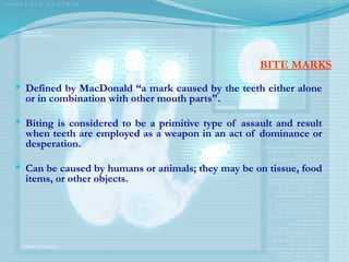
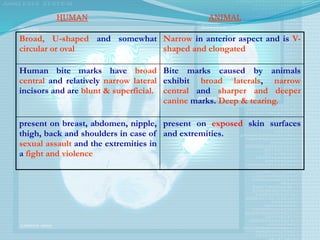
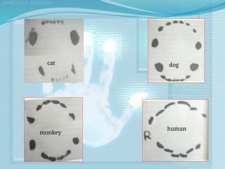
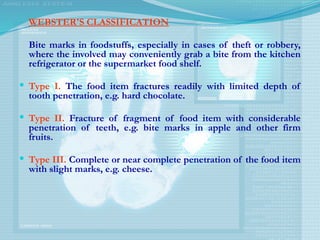
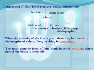
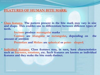
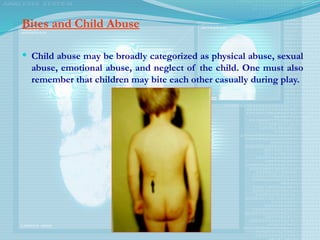
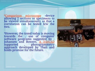
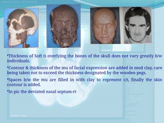
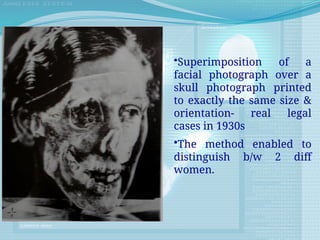
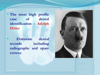
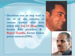
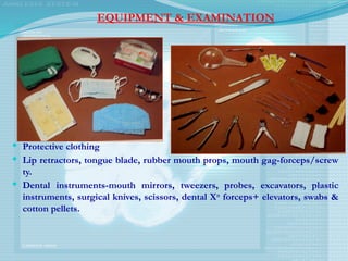
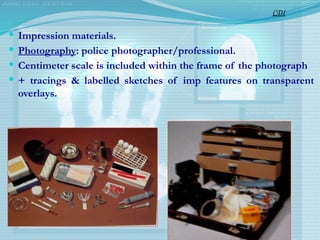
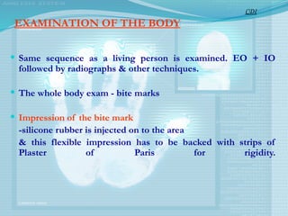
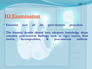
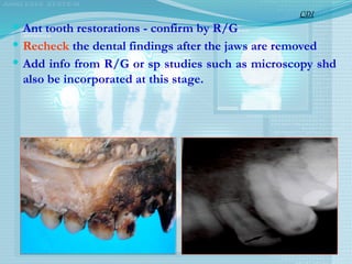
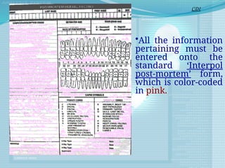
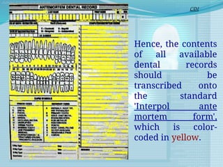
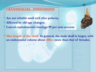
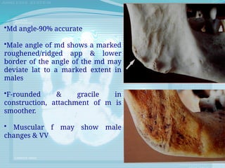
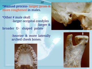
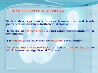
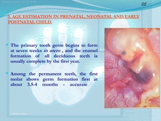
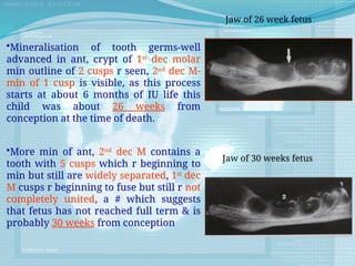
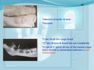
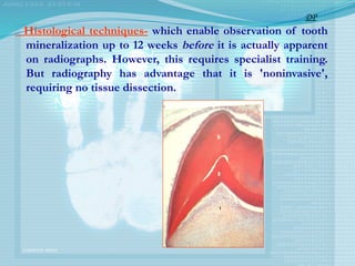
Forensic odontology is a branch of dentistry focused on the identification of individuals through dental evidence, particularly useful in mass disasters and crime investigations. It utilizes unique dental characteristics for identification, applying methods such as comparative dental identification and dental profiling to determine ethnicity, age, and sex. The document covers the history, methodology, and significance of dental identification, emphasizing its critical role in legal and humanitarian contexts.


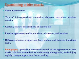
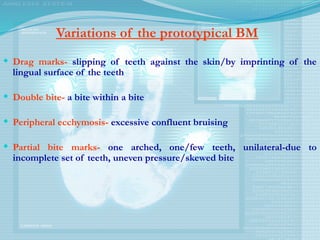
![DENTAL INDEX
In addition to absolute tooth size, tooth proportions have
been suggested for differentiating the sexes.
AITCHISON - 'INCISOR INDEX'
Ii = [MDI2
/ MDI1
] X 100
Higher in males
DP](https://image.slidesharecdn.com/forensicodontology-240930045243-a3f0101b/85/Forensic-Odontology-pptx-for-BDS-students-78-320.jpg)

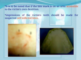
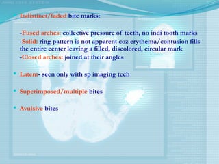
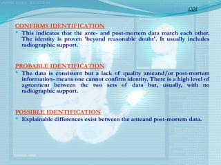
![ pulp-root length (P)
pulp-tooth length (R)
tooth-root length (T)
pulp-root width at CE] (A)
pulp-root width at mid-root level (C)
pulp
root width at mid-point between level C and A (B)
Mean value of all ratios excluding T (M)
Mean value of width ratios Band C (W)
Mean value of length ratios P and R (L).
When six teeth (right or left side) from both jaws are available, the
following regression formula can be used:
Age = 129.8-316.4(M)-66.8(W-L).
DP](https://image.slidesharecdn.com/forensicodontology-240930045243-a3f0101b/85/Forensic-Odontology-pptx-for-BDS-students-106-320.jpg)