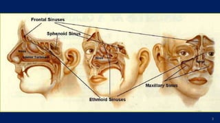
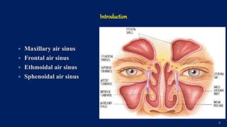
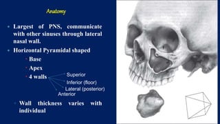
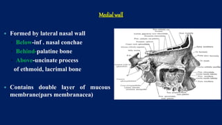
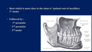
1
MAXILLARY SINUS
Introduction
 Paranasal air sinus
 Paranasal air sinuses are the air filled mucosa lined cavities which
develops in the cranial and facial bones.
 These are the spaces which communicates with the nasal airway.
 These forms the various boundaries of the nasal cavity.
 The sinuses are named for the bones in which they are located.
 Paranasal sinuses are present in a variety of animals (including
most mammals, birds, and crocodile).
2
3
Introduction
 Maxillary air sinus
 Frontal air sinus
 Ethmoidal air sinus
 Sphenoidal air sinus
4
Definition of maxillary sinus
“Maxillary sinus is the pneumatic space that is lodged inside the body of
maxilla and that communicates with the environment by way of the
middle meatus and nasal vestibule.”
Anatomy of the maxillary sinus was 1st described by Highmore in 1651.
So it is also known as “Antrum of Highmore”
5
Development
 Maxillary sinus is first of the PNS to develop.
 It starts as a shallow groove on the medial surface of maxilla during the
4th month of intrauterine life.
 Expansion occurs more rapidly until all the permanent teeth have
erupted.
 It reaches to maximum size around 18years of age.
6
AGE CHANGES
Age changes
9
Anatomy
 Largest of PNS, communicate
with other sinuses through lateral
nasal wall.
 Horizontal Pyramidal shaped
 Base
 Apex
 4 walls
◦ Wall thickness varies with
individual
Superior
Inferior (floor)
Lateral (posterior)
Anterior
Medial wall
 Formed by lateral nasal wall
◦ Below-inf . nasal conchae
◦ Behind-palatine bone
◦ Above-uncinate process
of ethmoid, lacrimal bone
 Contains double layer of mucous
membrane(pars membranacea)
Osteum:
 Opening of the maxillary sinus is called osteum.
 It opens in middle meatus at the lower part of the hiatus semilunaris.
 Lies above the level of nasal floor.
12
 The ostium lies approximately 2/3rds up the medial wall of the sinus,
making drainage of the sinus inherently difficult.
13
 In 15% to 40% of cases, a very small, accessory ostium is also
found.
 Blockage of the ostium can easily occur when there is
inflammation of the mucosal lining of the ostium.
14
Superior wall
 Forms roof of sinus and floor of orbit
 Imp structures
 Infraorbital canal
 Infraorbital foramen
 Infraorbital nerve and vessels.
Posterolateral wall
 Made of zygomatic and greater wing of sphenoid bone.
 Thick laterally, thin medially
 Imp structures
 PSA nerve
 Maxillary artery
 Pterygopalatine ganglion
 Nerve of pterygoid canal
Anterior wall
 Extends from pyriform aperture anteriorly to ZM suture & Inferior
orbital rim superiorly to alveolar process inferiorly.
 Convexity towards sinus
 Thinnest in canine fossa
 Imp structures
 Infraorbital foramen
 ASA, MSA nerves
Floor of sinus
 Formed by junction of anterior sinus wall and lateral nasal wall
 1-1.2 cm below nasal floor
 Close relationship between sinus and teeth facilitate spread of pathology.
VASCULAR SUPPLY
Arterial blood supply:-
 Greater palatine arteries
 Infraorbital artery
 Facial artery
19
Venous drainage
• Pterygoid venous plexus
• Sphenopalatine vein and
• Facial vein
20
Nerve supply
 Maxillary division of the trigeminal nerve, i.e. the posterior, middle and
anterior superior alveolar nerves, the infraorbital nerve and the anterior
palatine nerve.
21
Lymphatic Drain
22
The lymphatic drains in to submandibular lymph nodes.
Functions of the maxillary sinus
 Humidification and warming of inspired air,
 Assisting in regulating intranasal pressure,
 Lightening the skull to maintain proper head balance,
 Imparting resonance to the voice,
 Absorption of shocks to the head,
 Filtration of the inspired air.
23
HISTOLOGY
 Maxillary sinus is lined by three
layers: epithelial layer, basal lamina
and sub epithelial layer with
periostium.
 Epithelium is pseudo stratified,
columnar and ciliated.
 As cilia beats, the mucous on
epithelial surface moves from sinus
interior towards nasal cavity.
25
26
DEVELOPMENTAL ANOMALIES AND
PATHOLOGICCONDITIONS OF
MAXILLARYSINUS
Developmental anomalies
1.Aplasia
2. Agenesis
3. Hypoplasia
Aplasia
Pathologic conditions of maxillary sinus
 Maxillary Sinusitis
 Odontogenic cystic lesions of maxillary sinus
 Tumors of maxillary sinus.
28
29
Oro-antral communication
(It is a pathologic tract that connects the oral cavity to the maxillary sinus.)
•Maxillary sinus perforation occurs occasionally during the extraction of a
maxillary tooth, and it may be a cause of maxillary sinusitis or oro- antral
fistula.
•Patient complained of regurgitation of food through the nose while eating.
30
Clinical Considerations:
31
•Oro-antral communication and oro-antral fistula
•The chances of creating an oro-antral fistula in patient less than 15 yrs are
comparatively lesser than in adults due to incomplete development of sinus.
•The distance between apical end of maxillary posterior teeth and floor of
sinus is approximately 1-1.2 cm. In some cases the gap may be still lesser.
 Root which is most close to the sinus is “palatal root of maxillary
1st molar
 Followed by :
◦ 1st premolar
◦ 2nd premolar
◦ 2nd molar
32
Conclusion
 Due to close proximity of maxillary sinus to orbit, alveolar ridge,
maxillary teeth, diseases involving these structures may produce
confusing symptoms. Hence a precise information about the surgical
anatomy is essential to surgeons.
 Knowledge of the anatomical relationship between the maxillary sinus
floor and the maxillary posterior teeth is important for the preoperative
treatment planning of maxillary posterior teeth.
 Cautious when performing dental procedures involving the maxillary
posterior teeth.

maxillary sinus anatomy histology.pptx

  • 1.
  • 2.
    Introduction  Paranasal airsinus  Paranasal air sinuses are the air filled mucosa lined cavities which develops in the cranial and facial bones.  These are the spaces which communicates with the nasal airway.  These forms the various boundaries of the nasal cavity.  The sinuses are named for the bones in which they are located.  Paranasal sinuses are present in a variety of animals (including most mammals, birds, and crocodile). 2
  • 3.
  • 4.
    Introduction  Maxillary airsinus  Frontal air sinus  Ethmoidal air sinus  Sphenoidal air sinus 4
  • 5.
    Definition of maxillarysinus “Maxillary sinus is the pneumatic space that is lodged inside the body of maxilla and that communicates with the environment by way of the middle meatus and nasal vestibule.” Anatomy of the maxillary sinus was 1st described by Highmore in 1651. So it is also known as “Antrum of Highmore” 5
  • 6.
    Development  Maxillary sinusis first of the PNS to develop.  It starts as a shallow groove on the medial surface of maxilla during the 4th month of intrauterine life.  Expansion occurs more rapidly until all the permanent teeth have erupted.  It reaches to maximum size around 18years of age. 6
  • 7.
  • 9.
  • 10.
    Anatomy  Largest ofPNS, communicate with other sinuses through lateral nasal wall.  Horizontal Pyramidal shaped  Base  Apex  4 walls ◦ Wall thickness varies with individual Superior Inferior (floor) Lateral (posterior) Anterior
  • 11.
    Medial wall  Formedby lateral nasal wall ◦ Below-inf . nasal conchae ◦ Behind-palatine bone ◦ Above-uncinate process of ethmoid, lacrimal bone  Contains double layer of mucous membrane(pars membranacea)
  • 12.
    Osteum:  Opening ofthe maxillary sinus is called osteum.  It opens in middle meatus at the lower part of the hiatus semilunaris.  Lies above the level of nasal floor. 12
  • 13.
     The ostiumlies approximately 2/3rds up the medial wall of the sinus, making drainage of the sinus inherently difficult. 13
  • 14.
     In 15%to 40% of cases, a very small, accessory ostium is also found.  Blockage of the ostium can easily occur when there is inflammation of the mucosal lining of the ostium. 14
  • 15.
    Superior wall  Formsroof of sinus and floor of orbit  Imp structures  Infraorbital canal  Infraorbital foramen  Infraorbital nerve and vessels.
  • 16.
    Posterolateral wall  Madeof zygomatic and greater wing of sphenoid bone.  Thick laterally, thin medially  Imp structures  PSA nerve  Maxillary artery  Pterygopalatine ganglion  Nerve of pterygoid canal
  • 17.
    Anterior wall  Extendsfrom pyriform aperture anteriorly to ZM suture & Inferior orbital rim superiorly to alveolar process inferiorly.  Convexity towards sinus  Thinnest in canine fossa  Imp structures  Infraorbital foramen  ASA, MSA nerves
  • 18.
    Floor of sinus Formed by junction of anterior sinus wall and lateral nasal wall  1-1.2 cm below nasal floor  Close relationship between sinus and teeth facilitate spread of pathology.
  • 19.
    VASCULAR SUPPLY Arterial bloodsupply:-  Greater palatine arteries  Infraorbital artery  Facial artery 19
  • 20.
    Venous drainage • Pterygoidvenous plexus • Sphenopalatine vein and • Facial vein 20
  • 21.
    Nerve supply  Maxillarydivision of the trigeminal nerve, i.e. the posterior, middle and anterior superior alveolar nerves, the infraorbital nerve and the anterior palatine nerve. 21
  • 22.
    Lymphatic Drain 22 The lymphaticdrains in to submandibular lymph nodes.
  • 23.
    Functions of themaxillary sinus  Humidification and warming of inspired air,  Assisting in regulating intranasal pressure,  Lightening the skull to maintain proper head balance,  Imparting resonance to the voice,  Absorption of shocks to the head,  Filtration of the inspired air. 23
  • 24.
    HISTOLOGY  Maxillary sinusis lined by three layers: epithelial layer, basal lamina and sub epithelial layer with periostium.  Epithelium is pseudo stratified, columnar and ciliated.  As cilia beats, the mucous on epithelial surface moves from sinus interior towards nasal cavity.
  • 25.
  • 26.
  • 27.
  • 28.
    Pathologic conditions ofmaxillary sinus  Maxillary Sinusitis  Odontogenic cystic lesions of maxillary sinus  Tumors of maxillary sinus. 28
  • 29.
    29 Oro-antral communication (It isa pathologic tract that connects the oral cavity to the maxillary sinus.) •Maxillary sinus perforation occurs occasionally during the extraction of a maxillary tooth, and it may be a cause of maxillary sinusitis or oro- antral fistula. •Patient complained of regurgitation of food through the nose while eating.
  • 30.
  • 31.
    Clinical Considerations: 31 •Oro-antral communicationand oro-antral fistula •The chances of creating an oro-antral fistula in patient less than 15 yrs are comparatively lesser than in adults due to incomplete development of sinus. •The distance between apical end of maxillary posterior teeth and floor of sinus is approximately 1-1.2 cm. In some cases the gap may be still lesser.
  • 32.
     Root whichis most close to the sinus is “palatal root of maxillary 1st molar  Followed by : ◦ 1st premolar ◦ 2nd premolar ◦ 2nd molar 32
  • 33.
    Conclusion  Due toclose proximity of maxillary sinus to orbit, alveolar ridge, maxillary teeth, diseases involving these structures may produce confusing symptoms. Hence a precise information about the surgical anatomy is essential to surgeons.  Knowledge of the anatomical relationship between the maxillary sinus floor and the maxillary posterior teeth is important for the preoperative treatment planning of maxillary posterior teeth.  Cautious when performing dental procedures involving the maxillary posterior teeth.