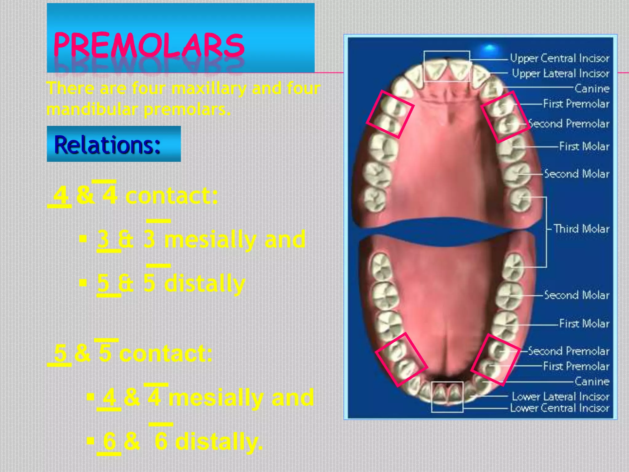
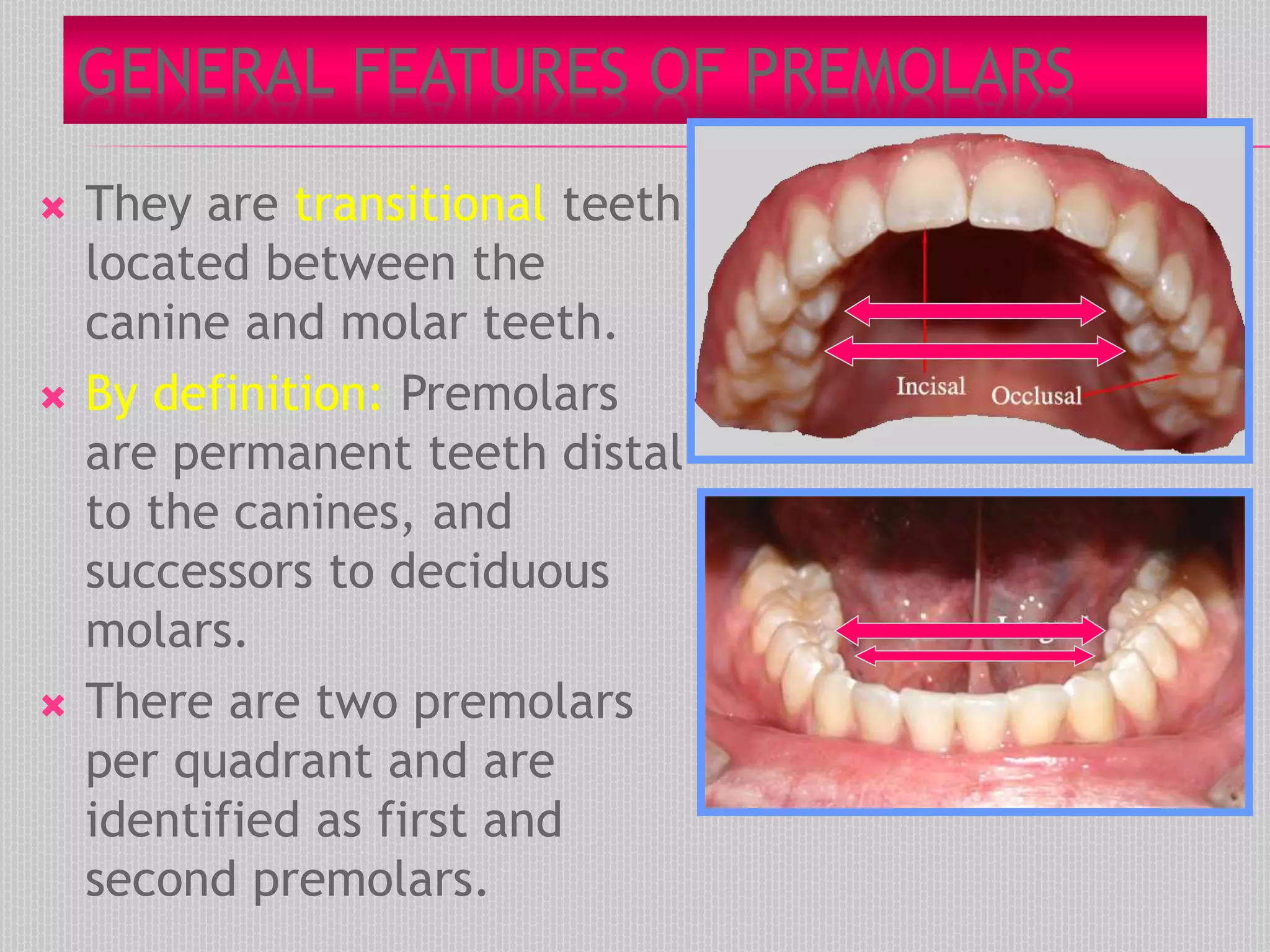
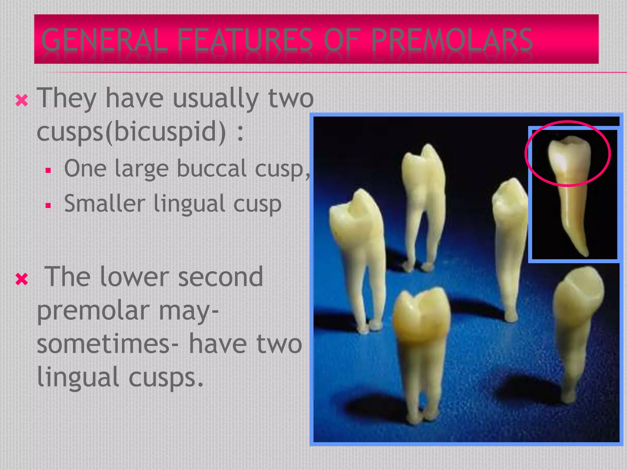
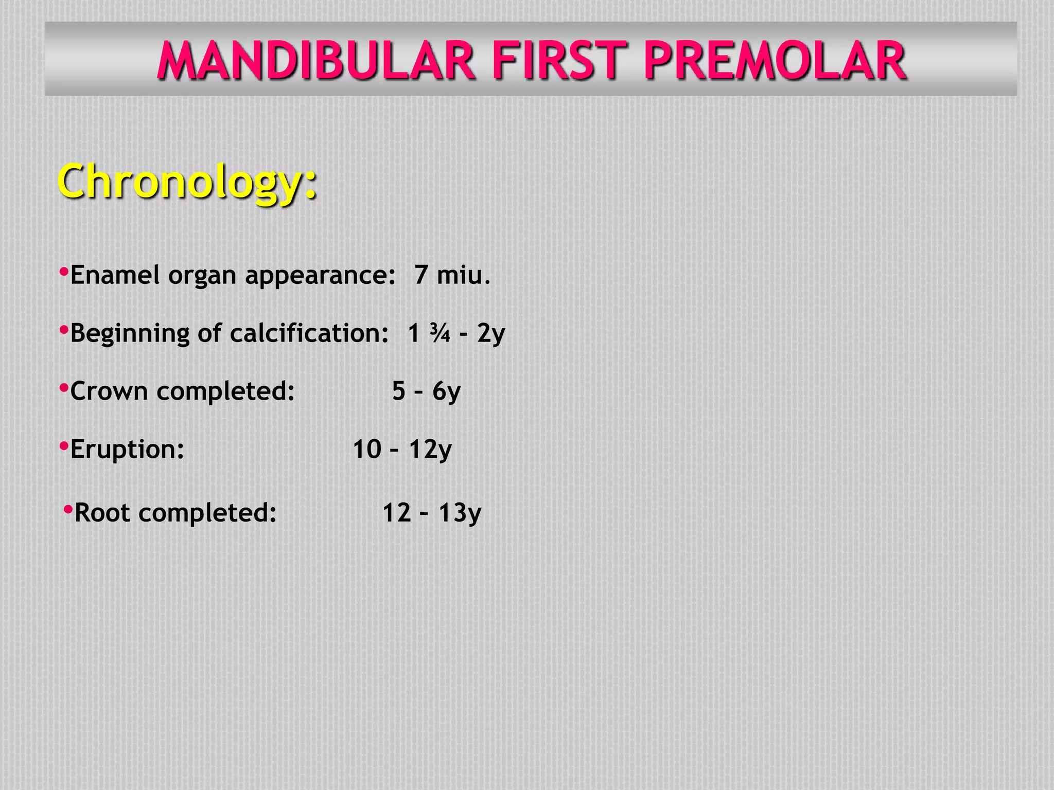
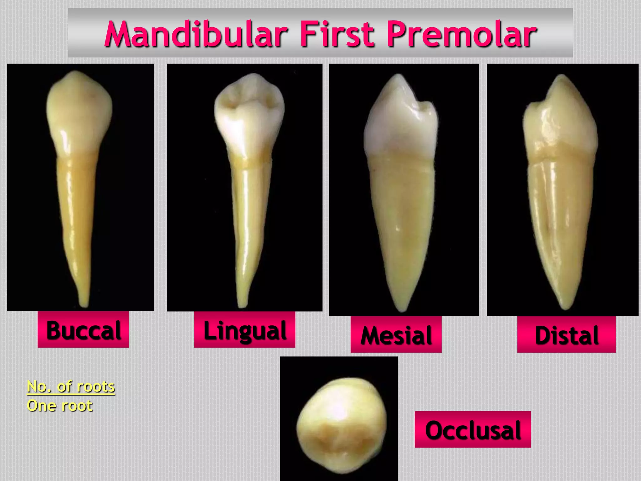
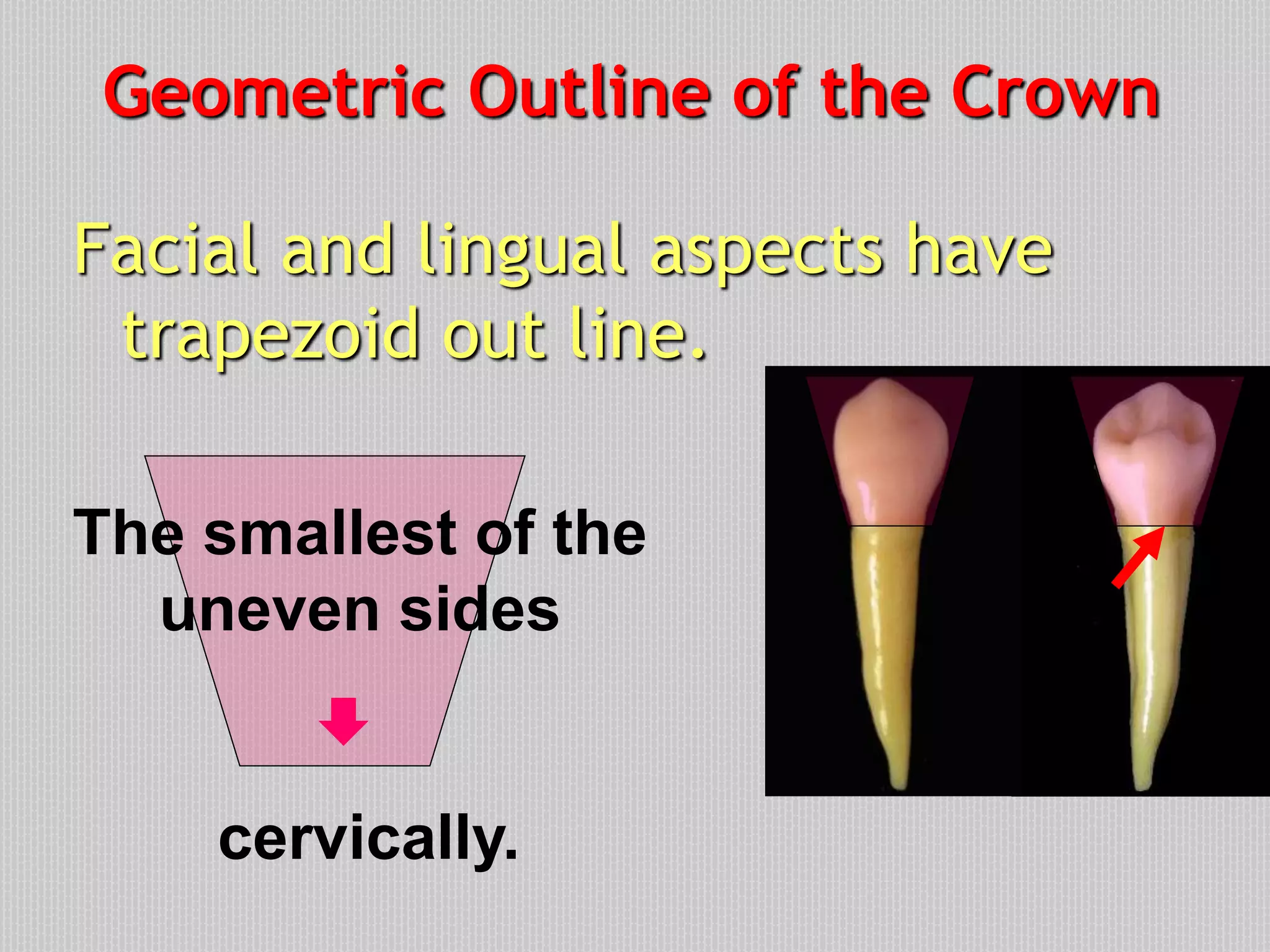
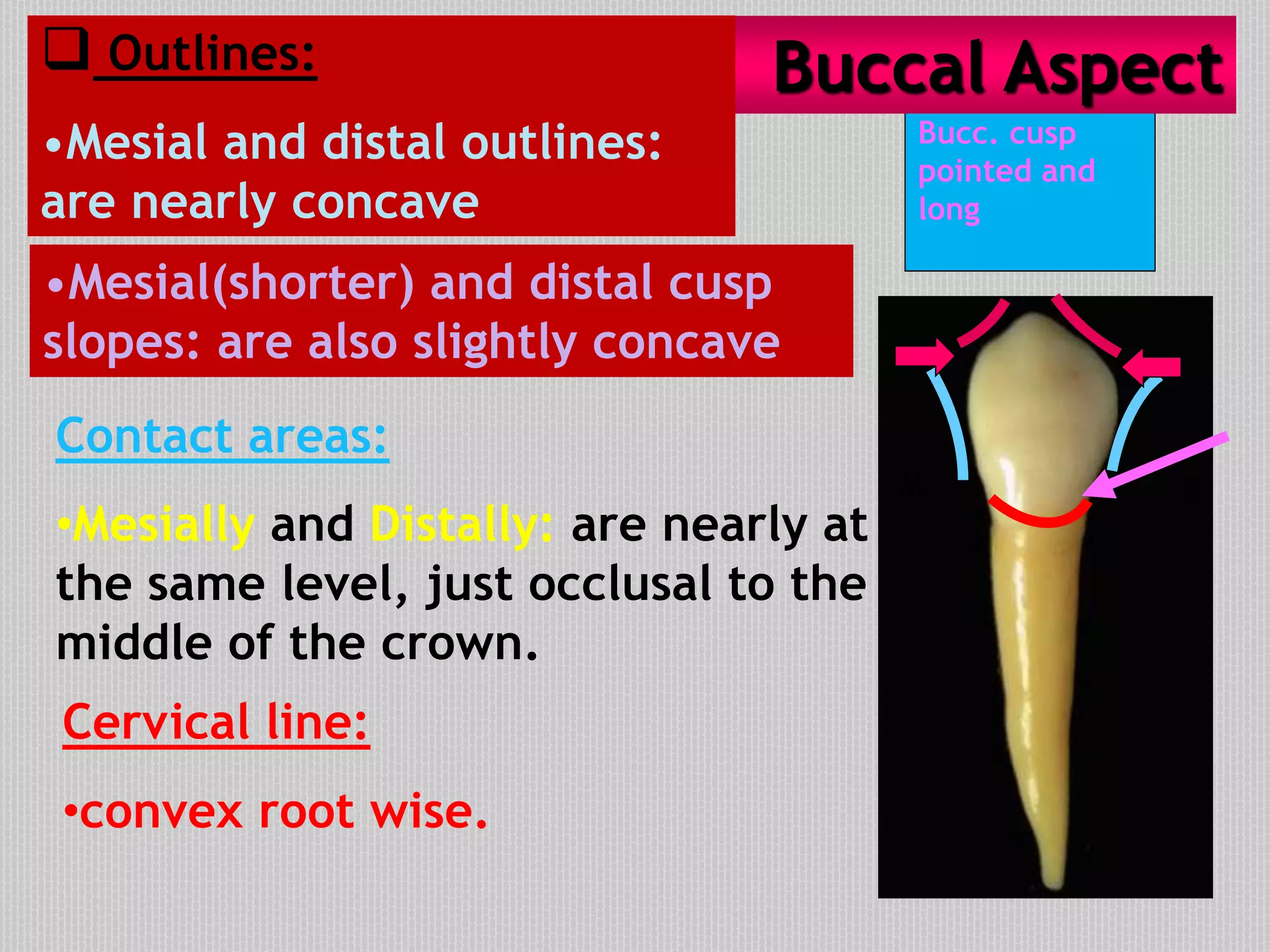
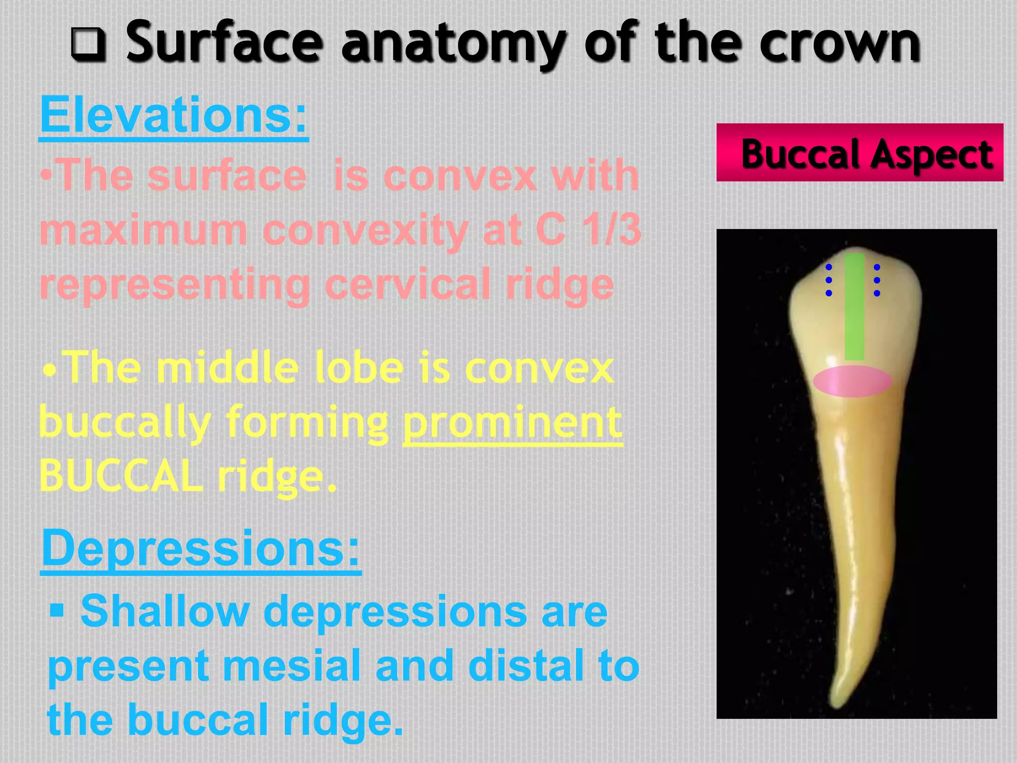
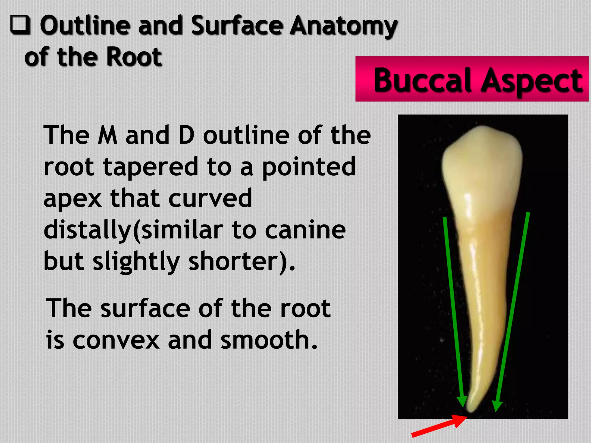
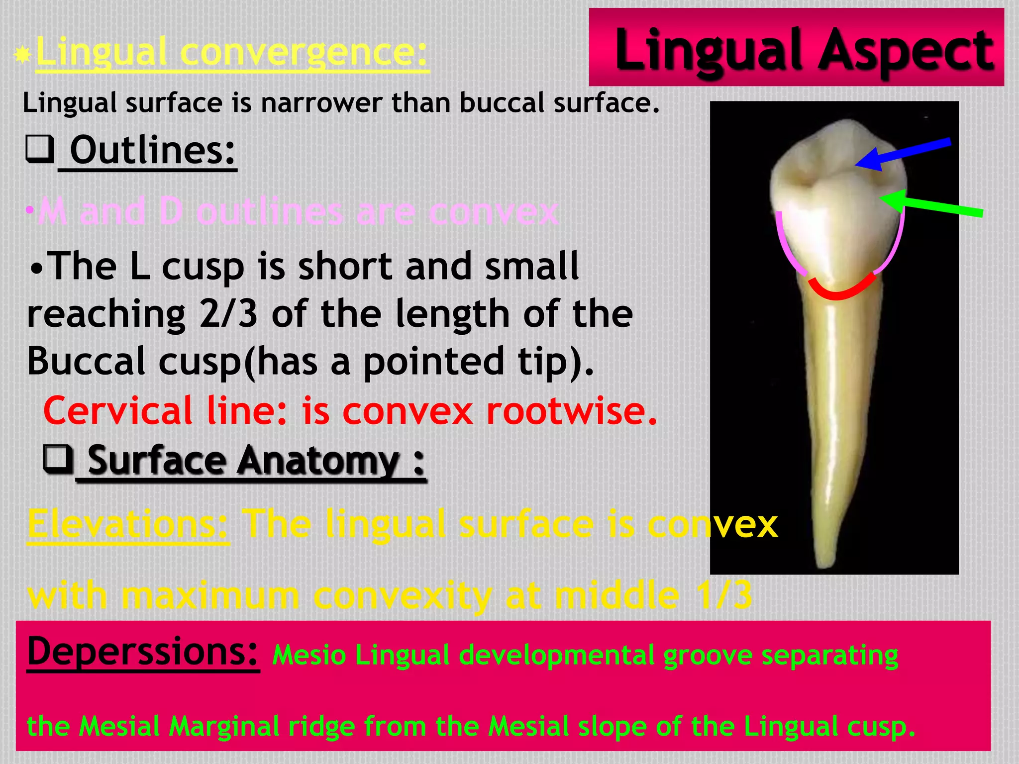
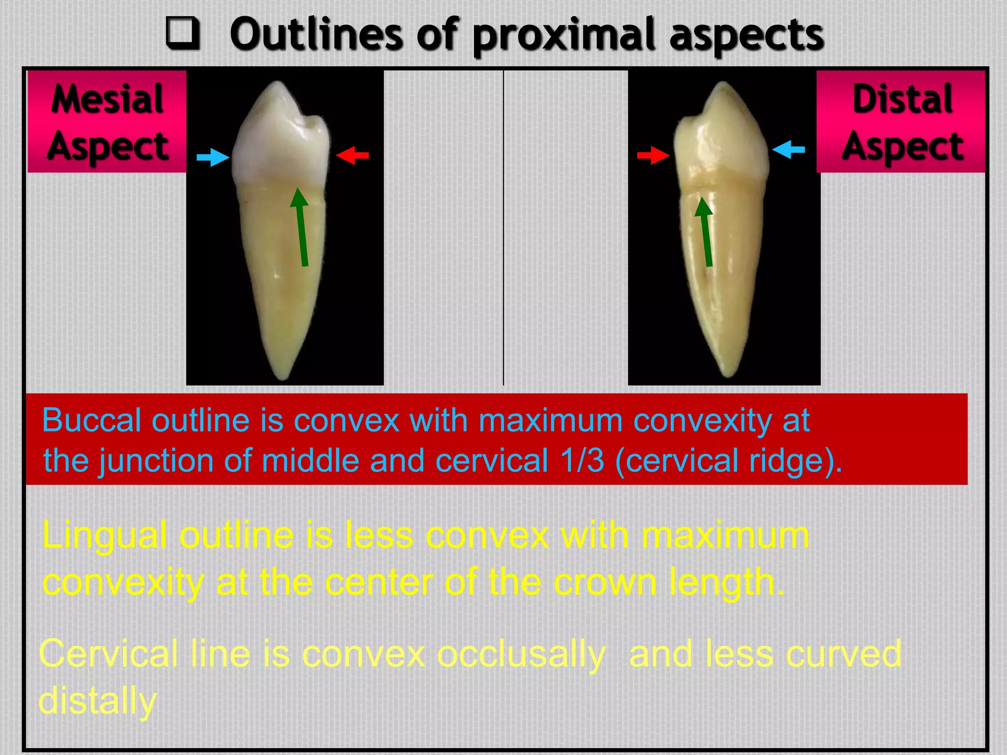
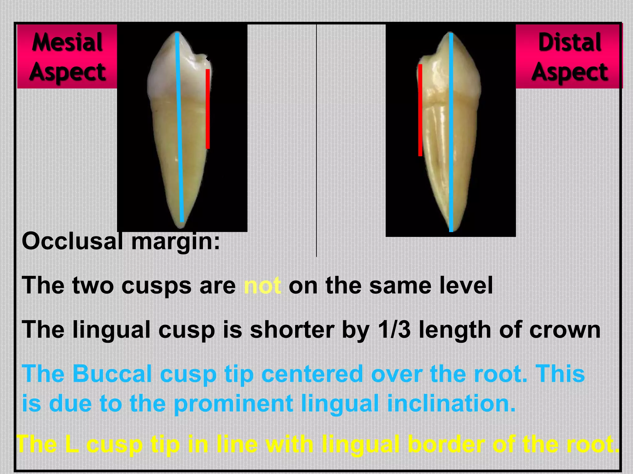
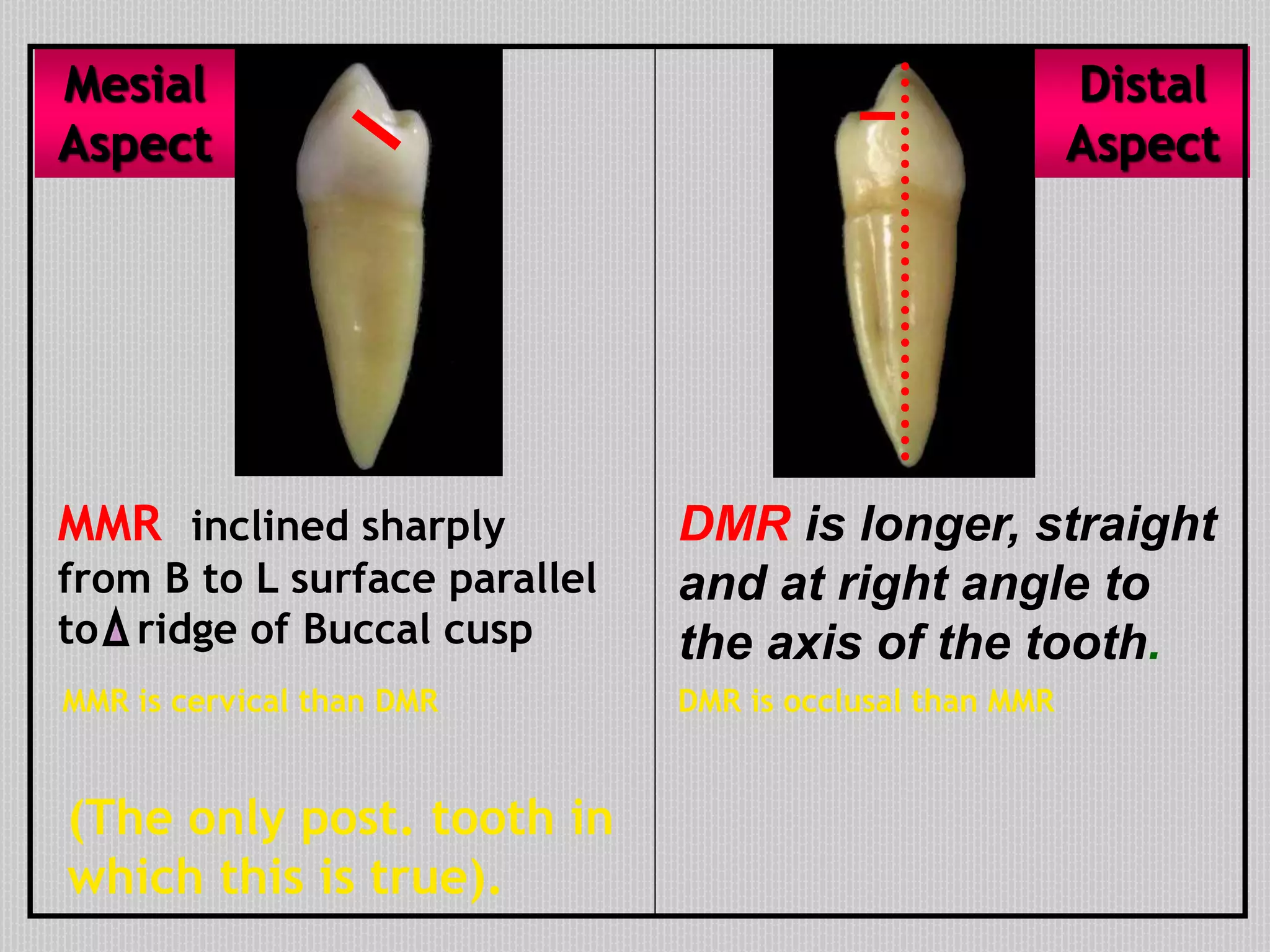
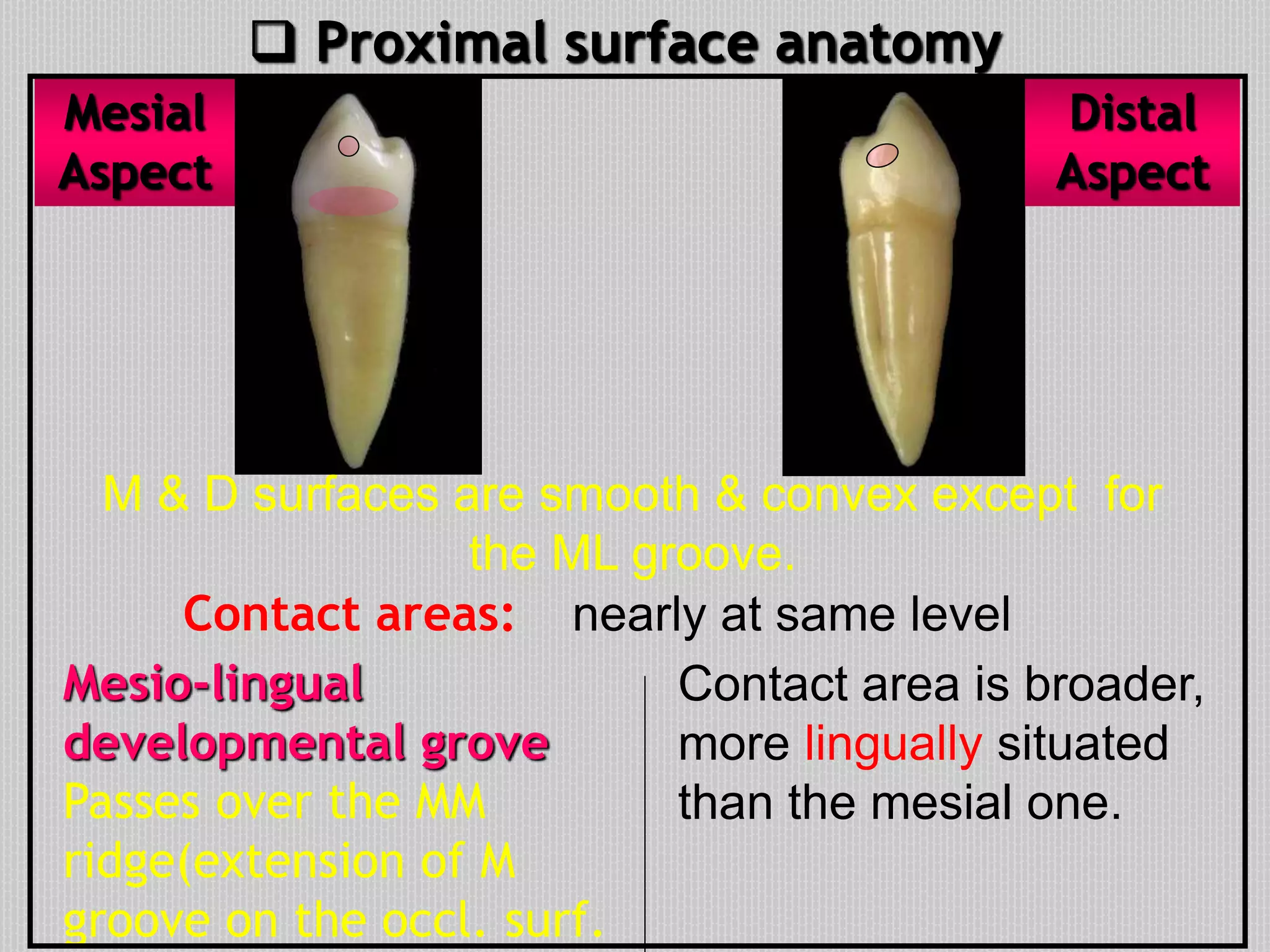
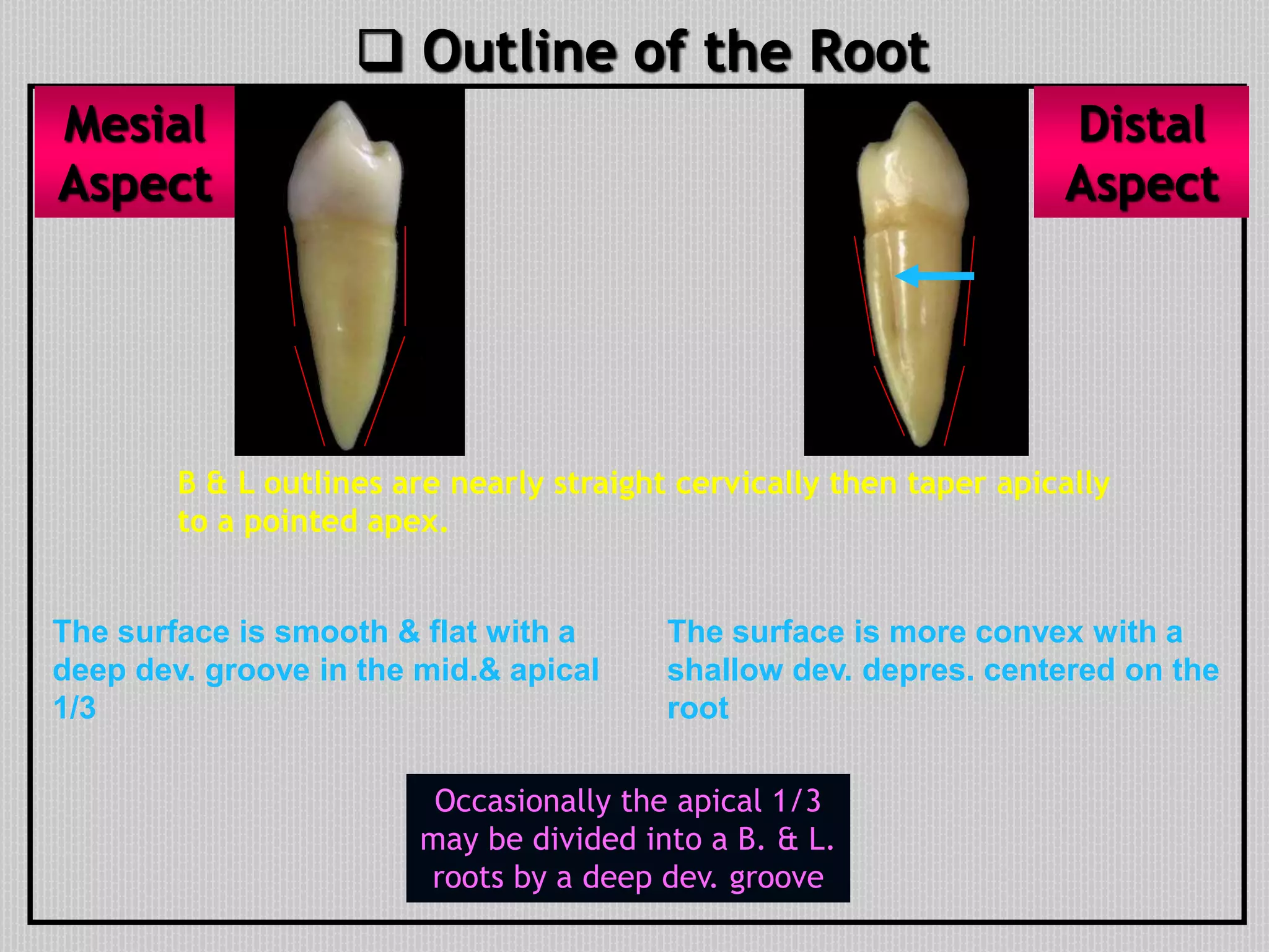
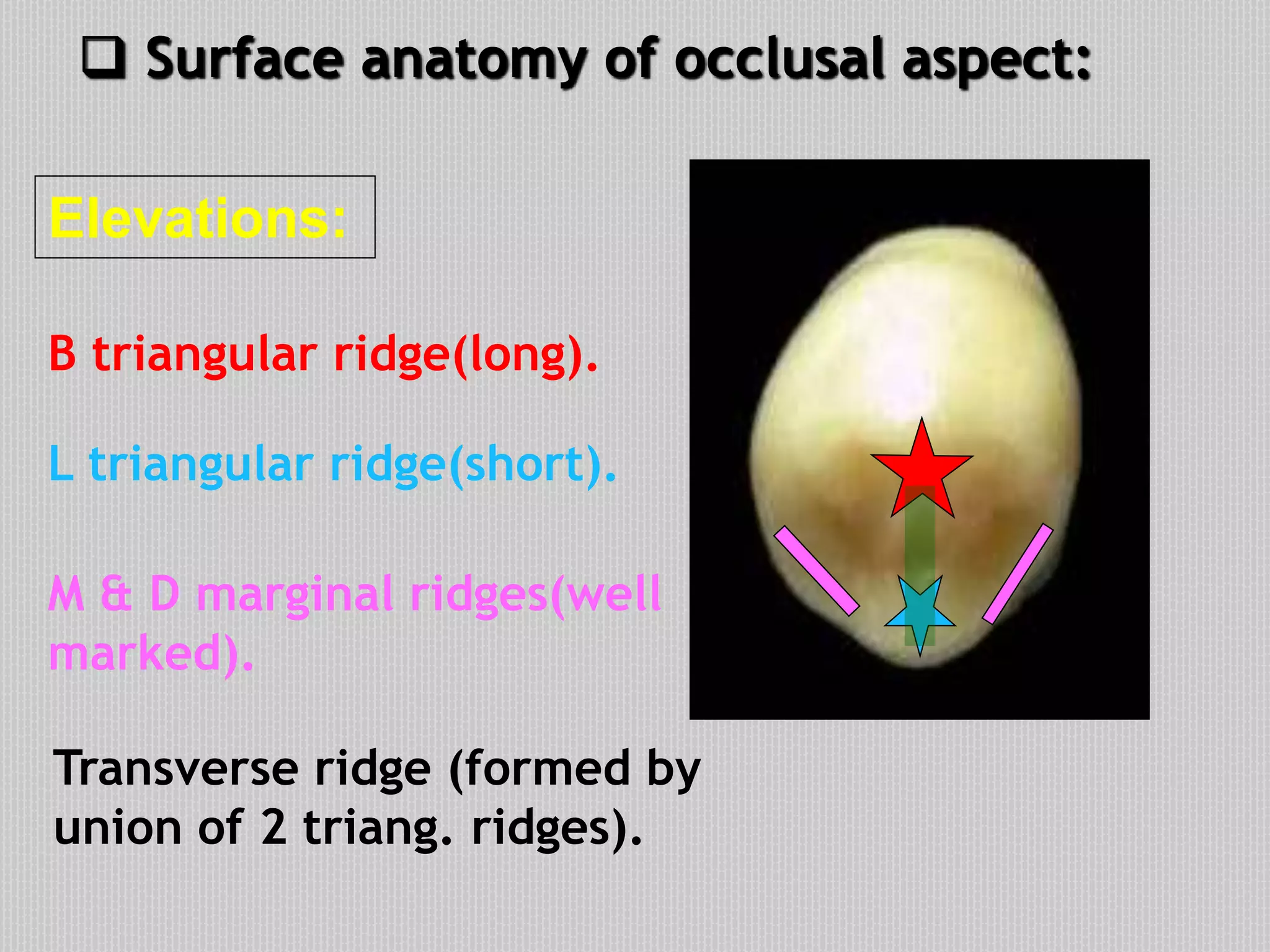
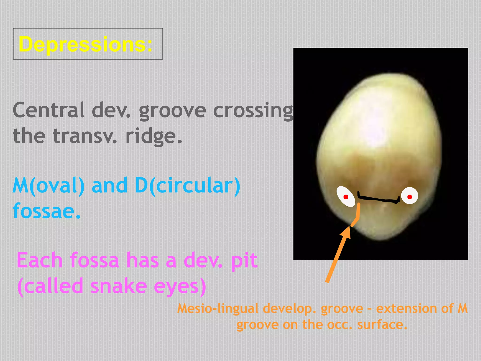
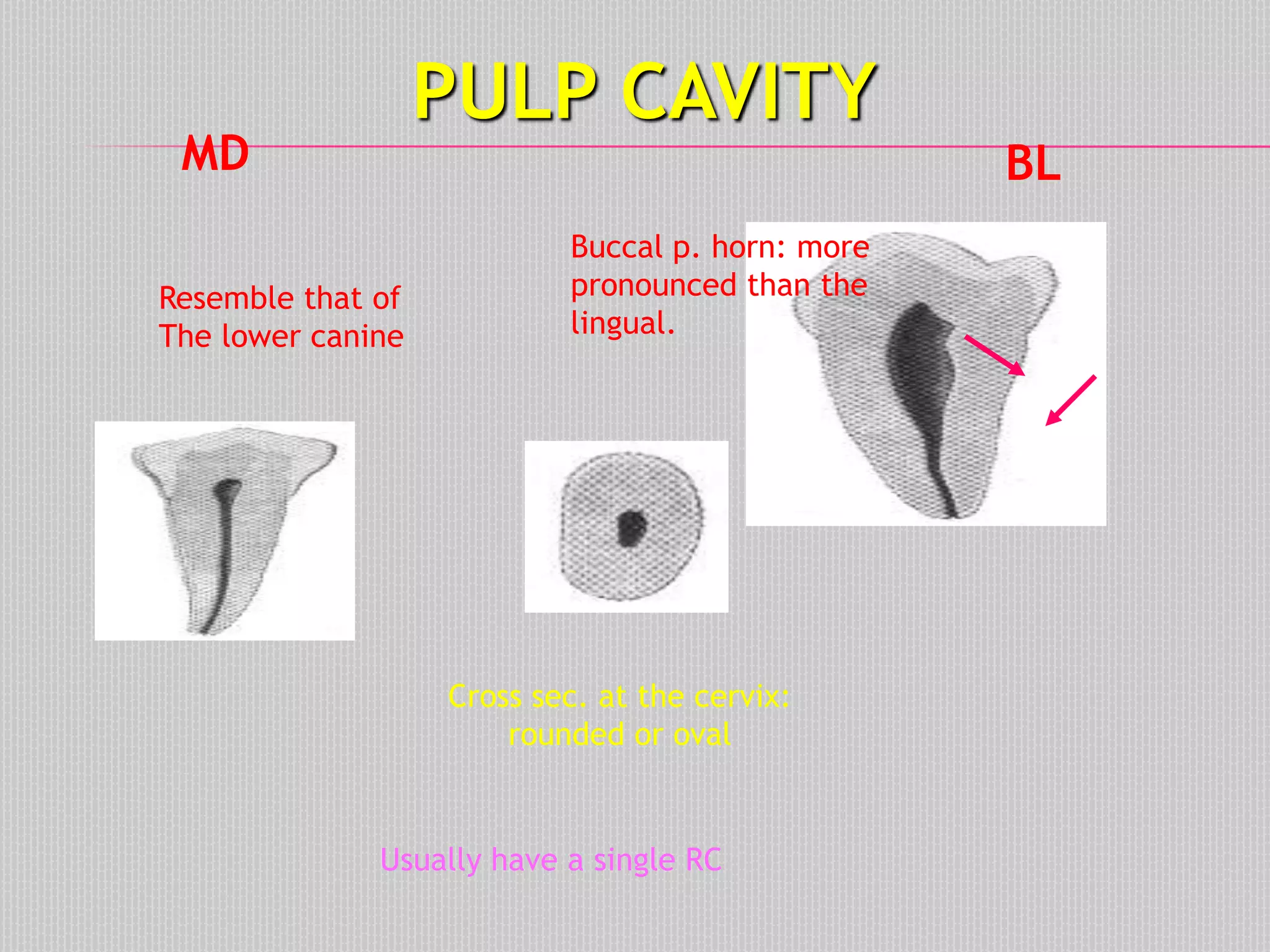
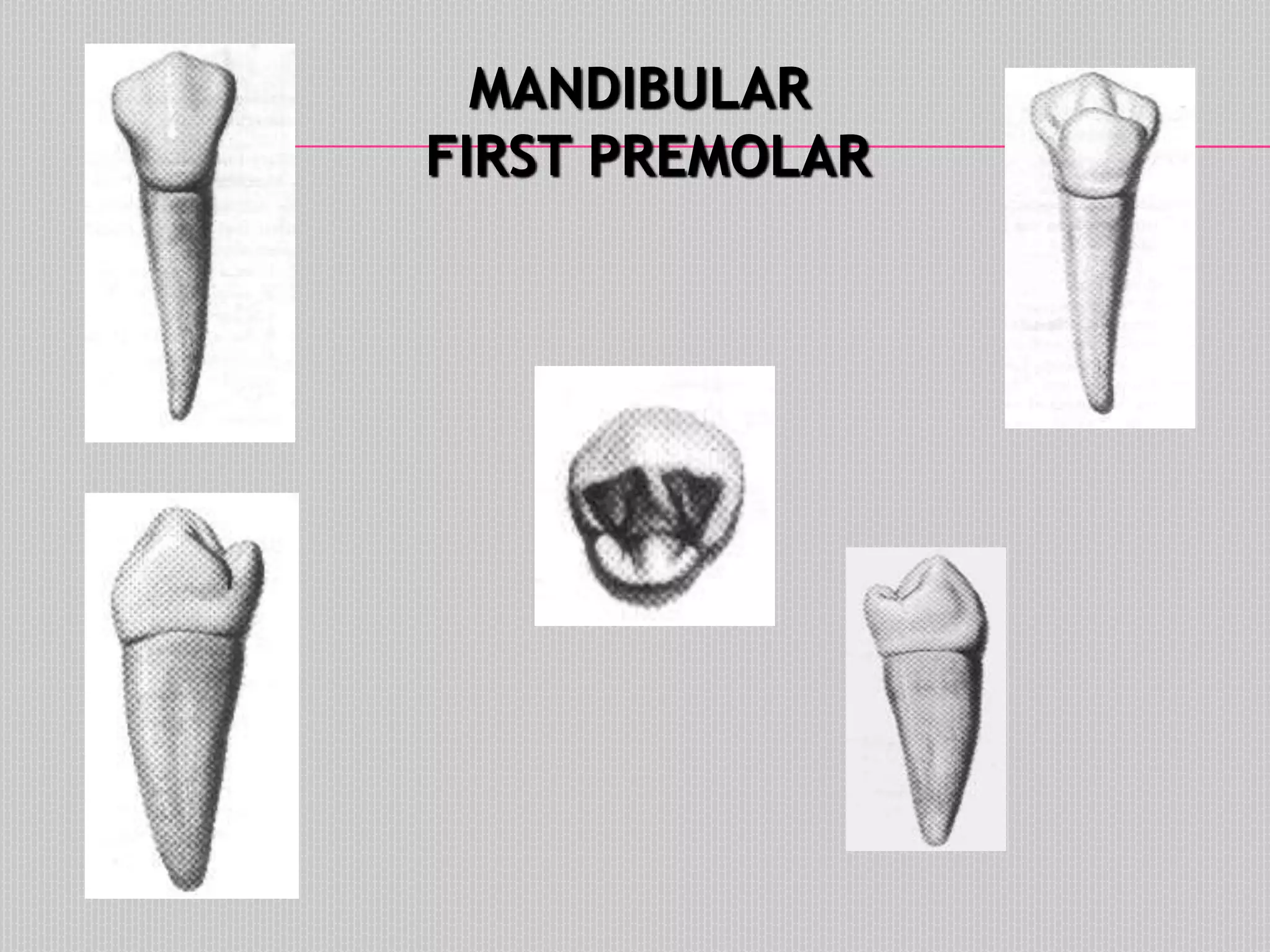
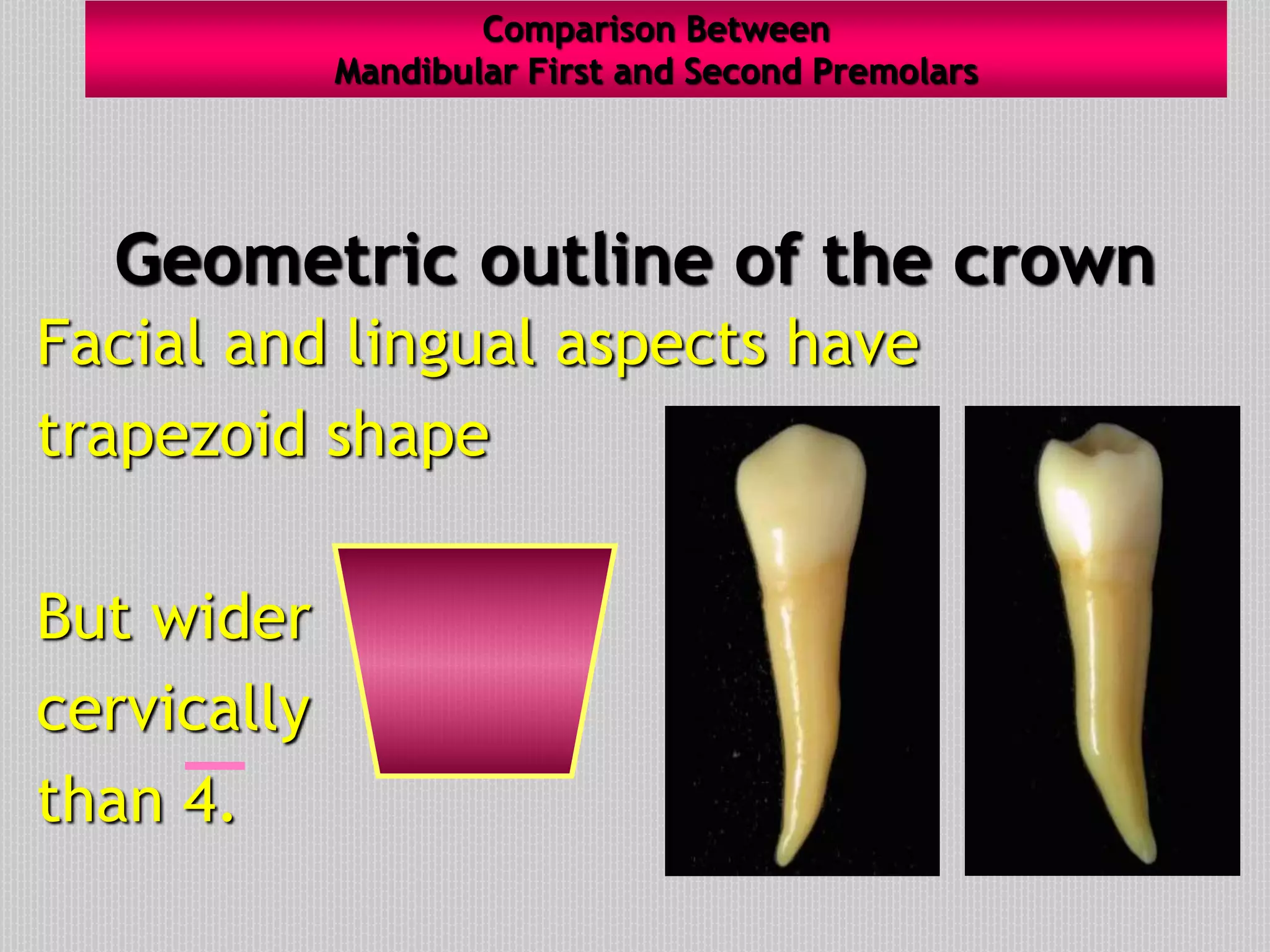
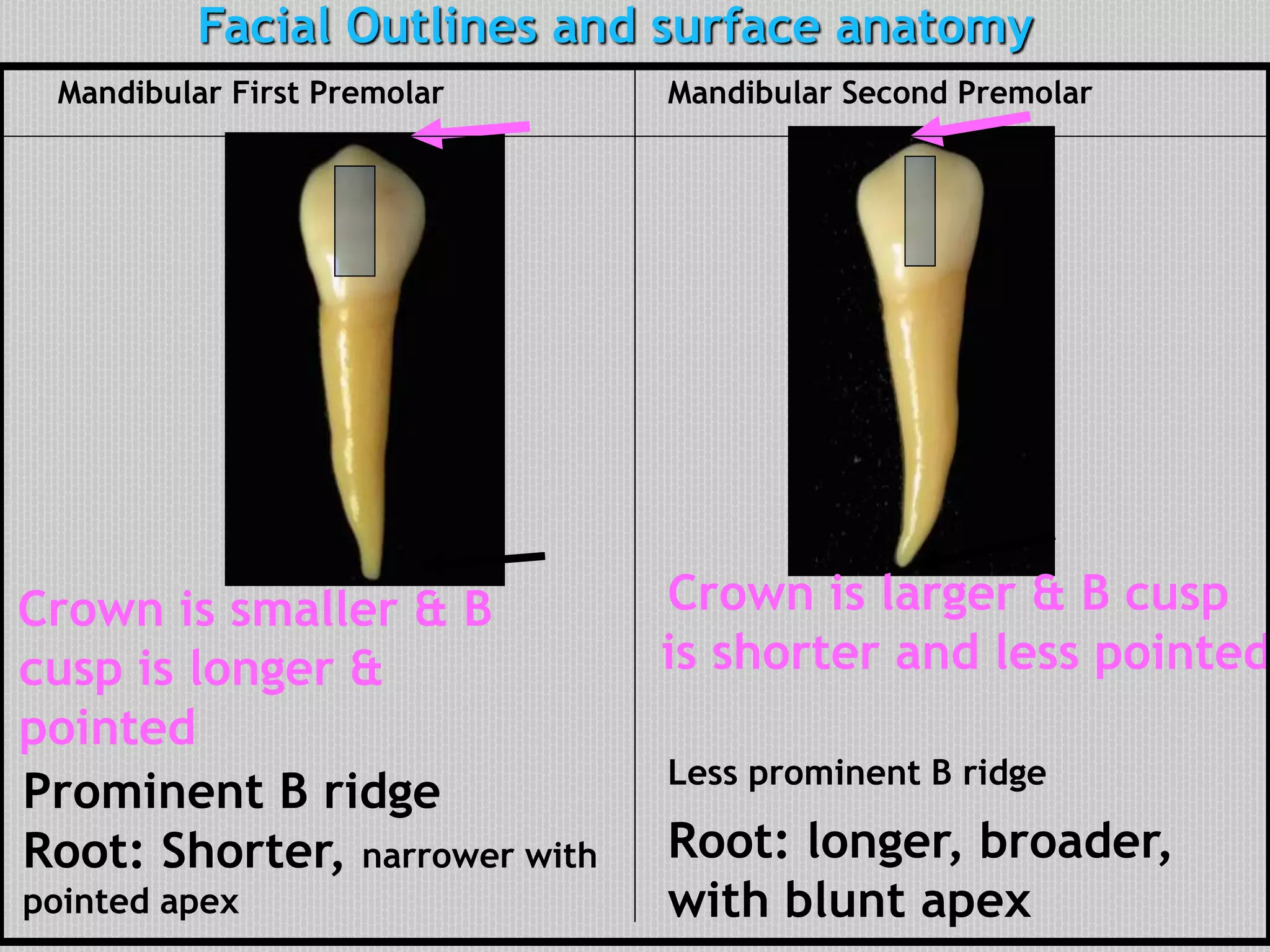
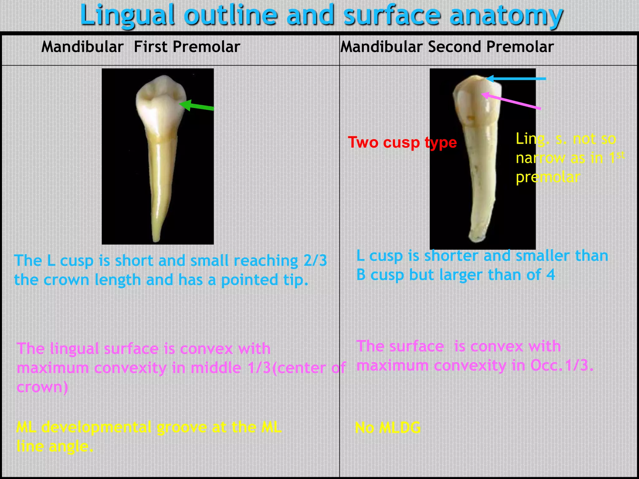
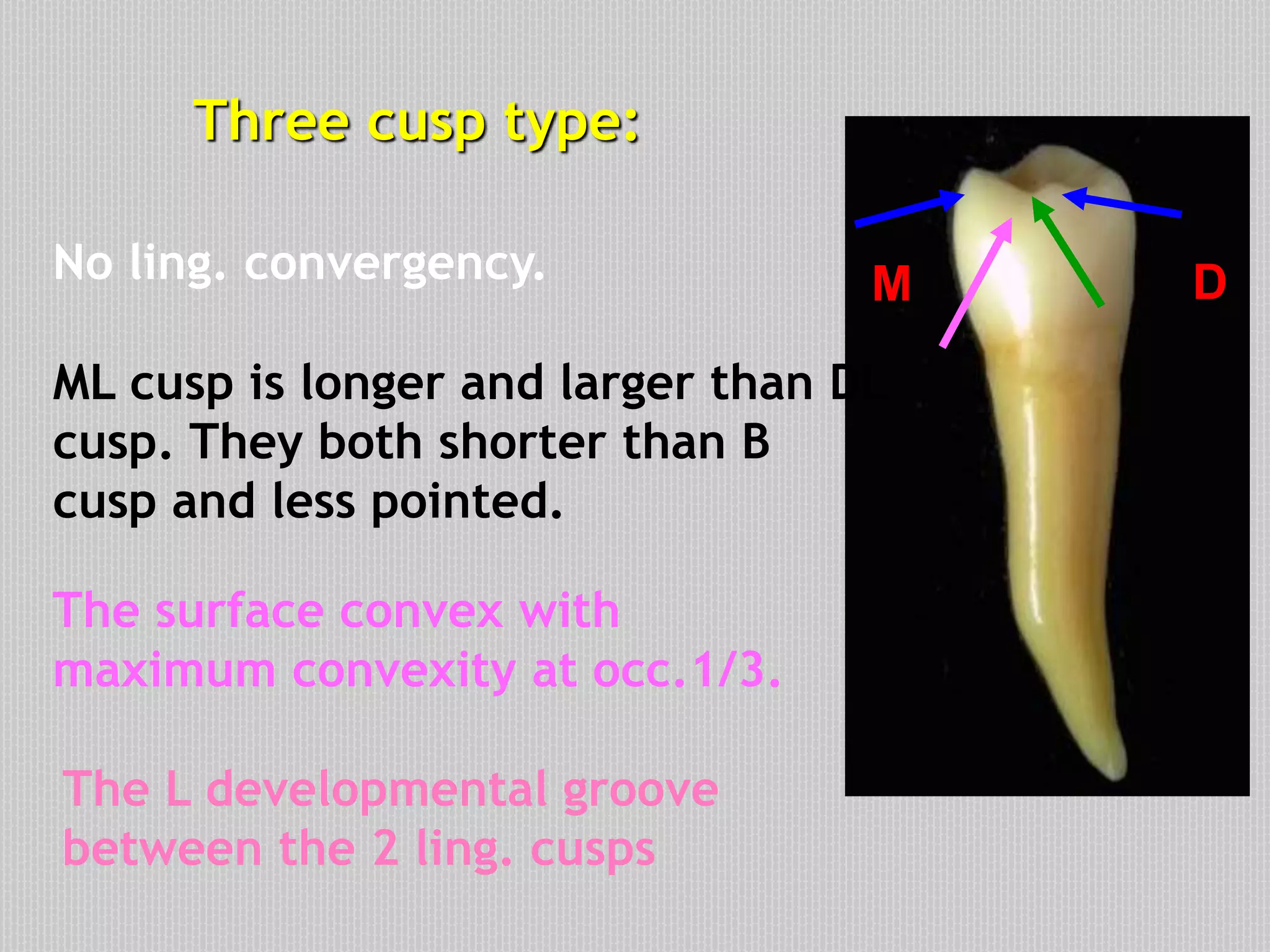
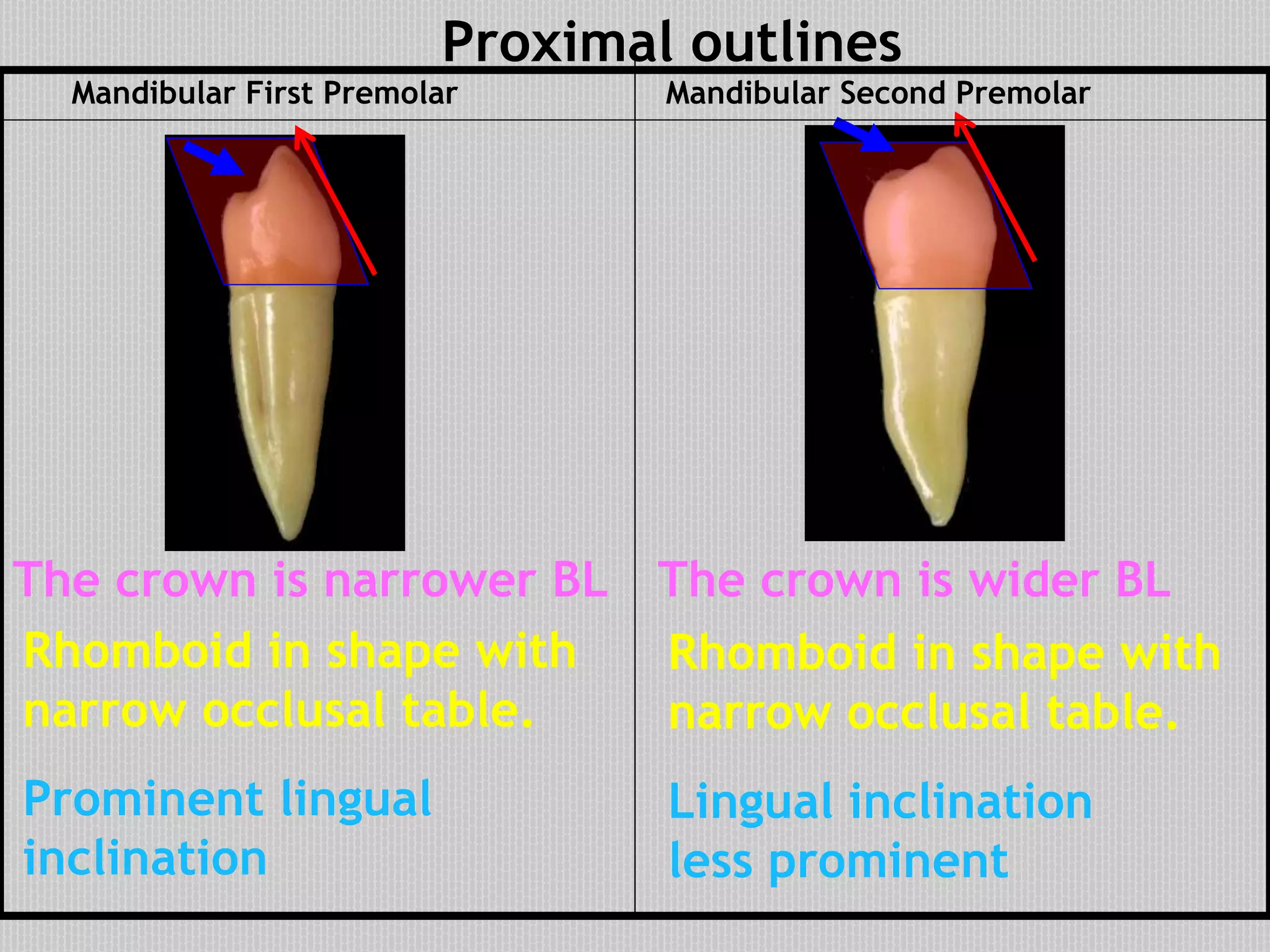
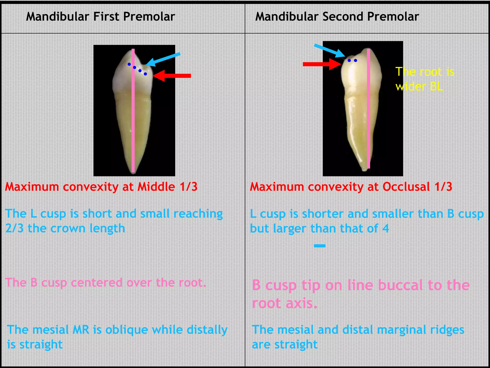
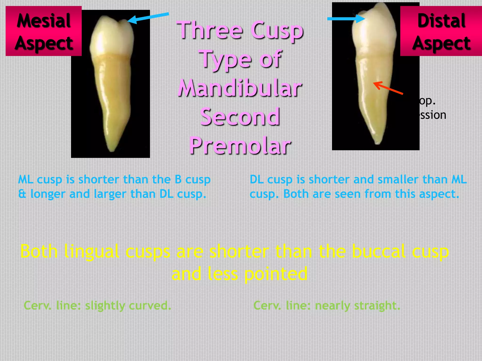
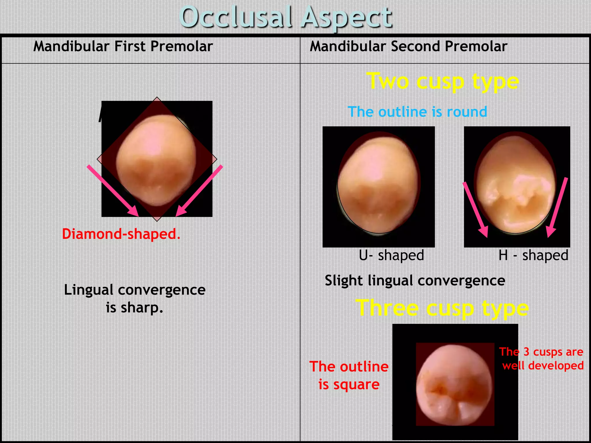
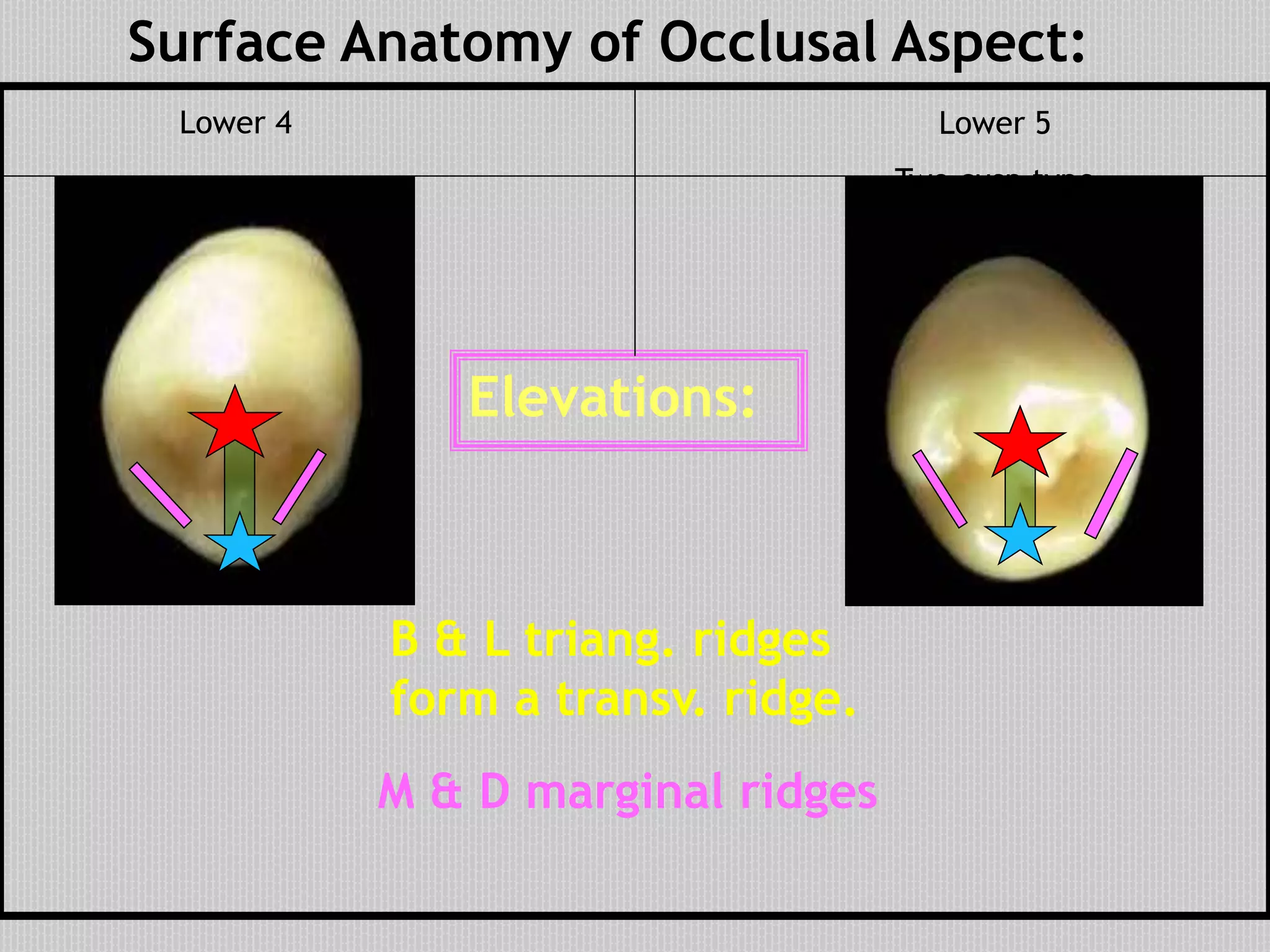
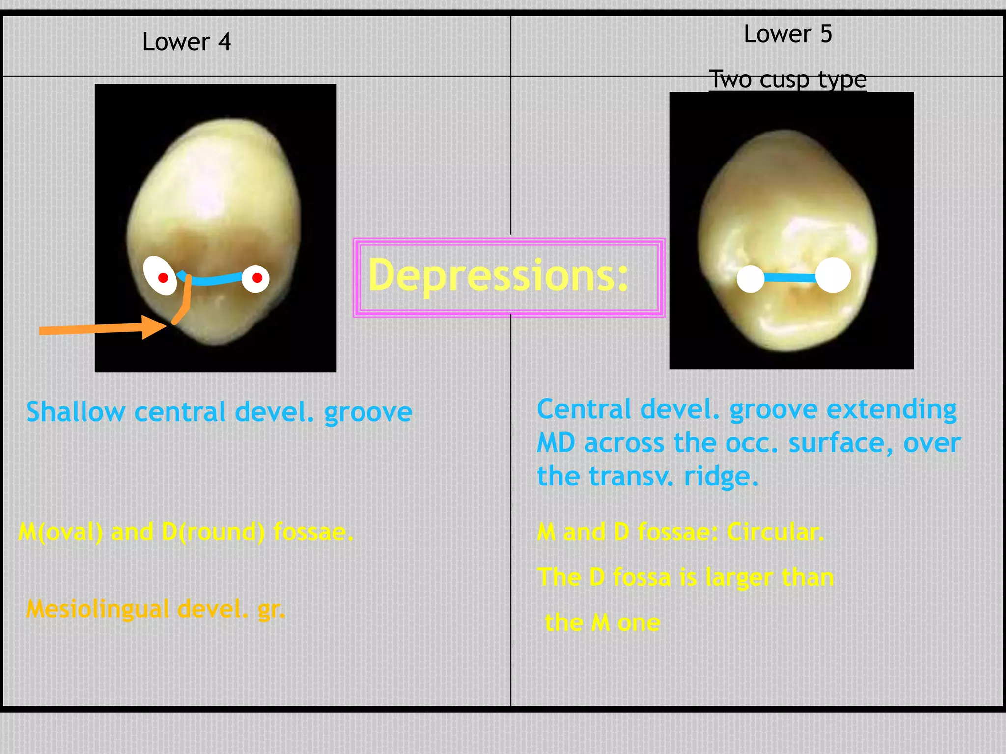
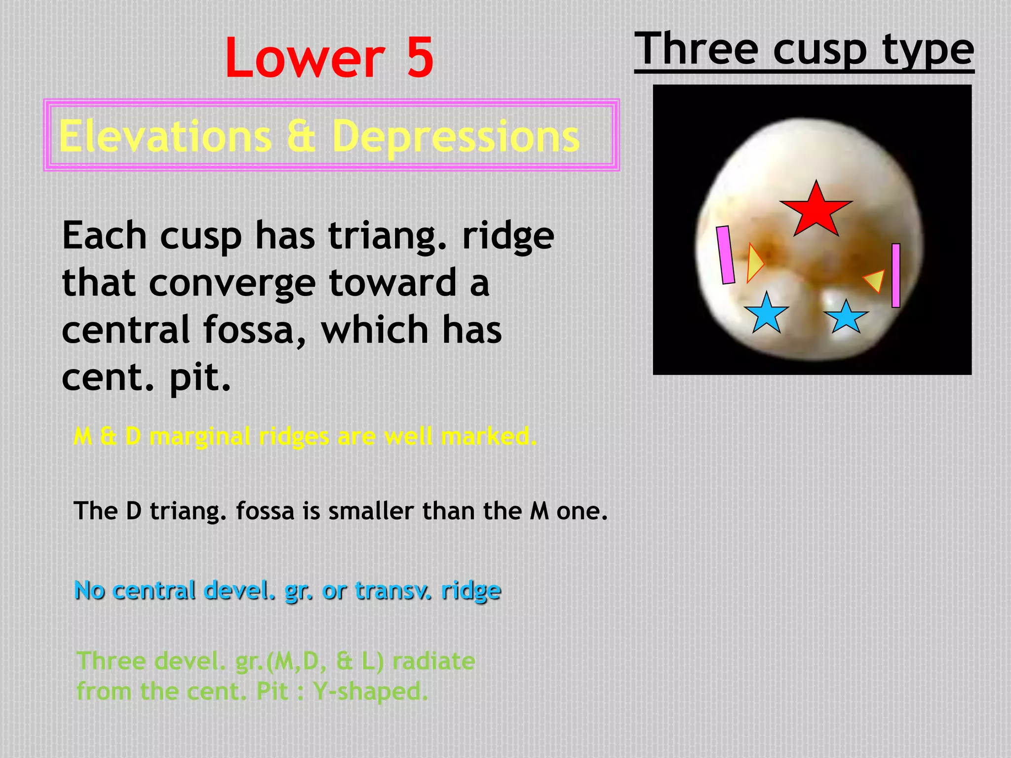
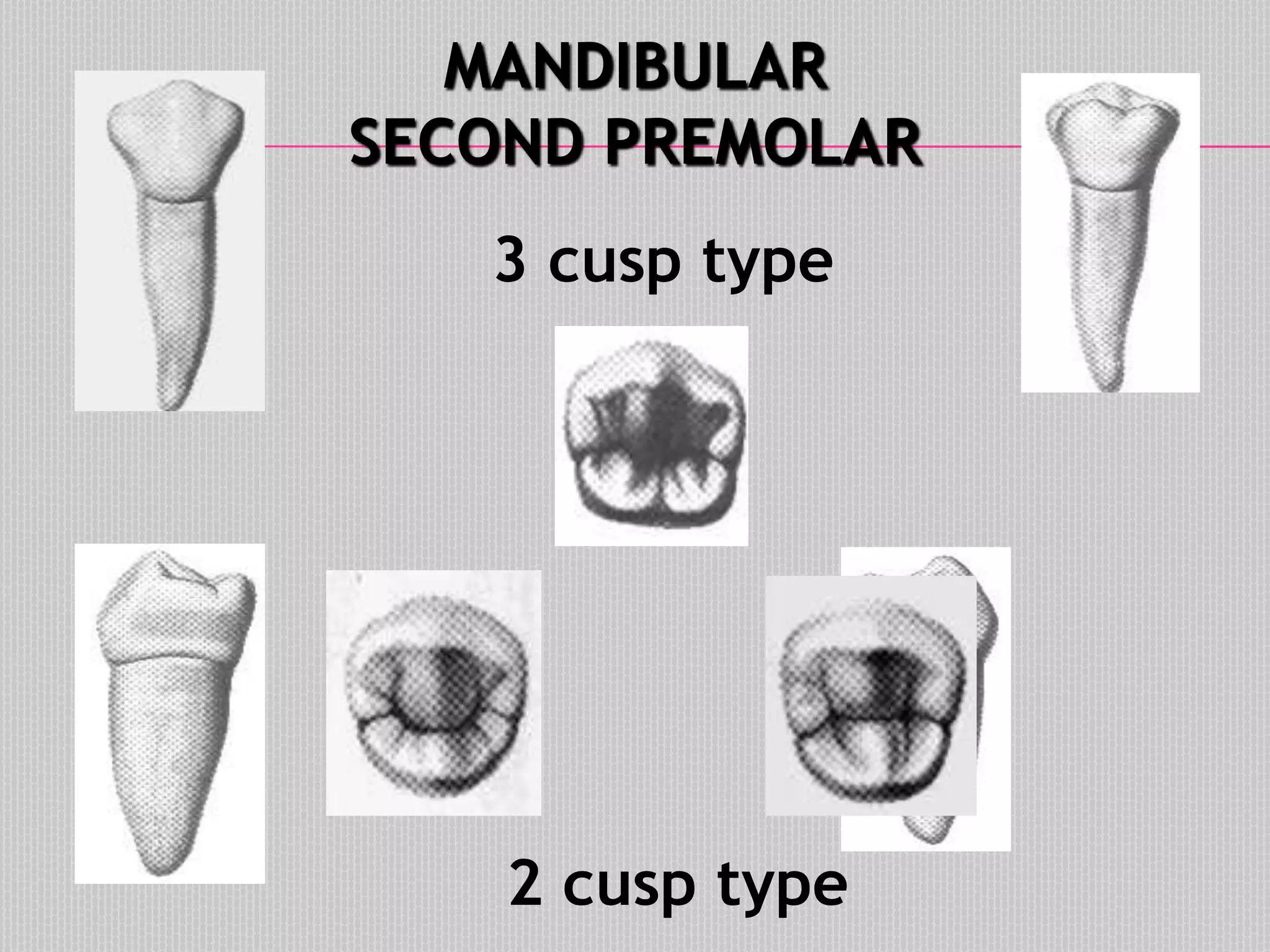
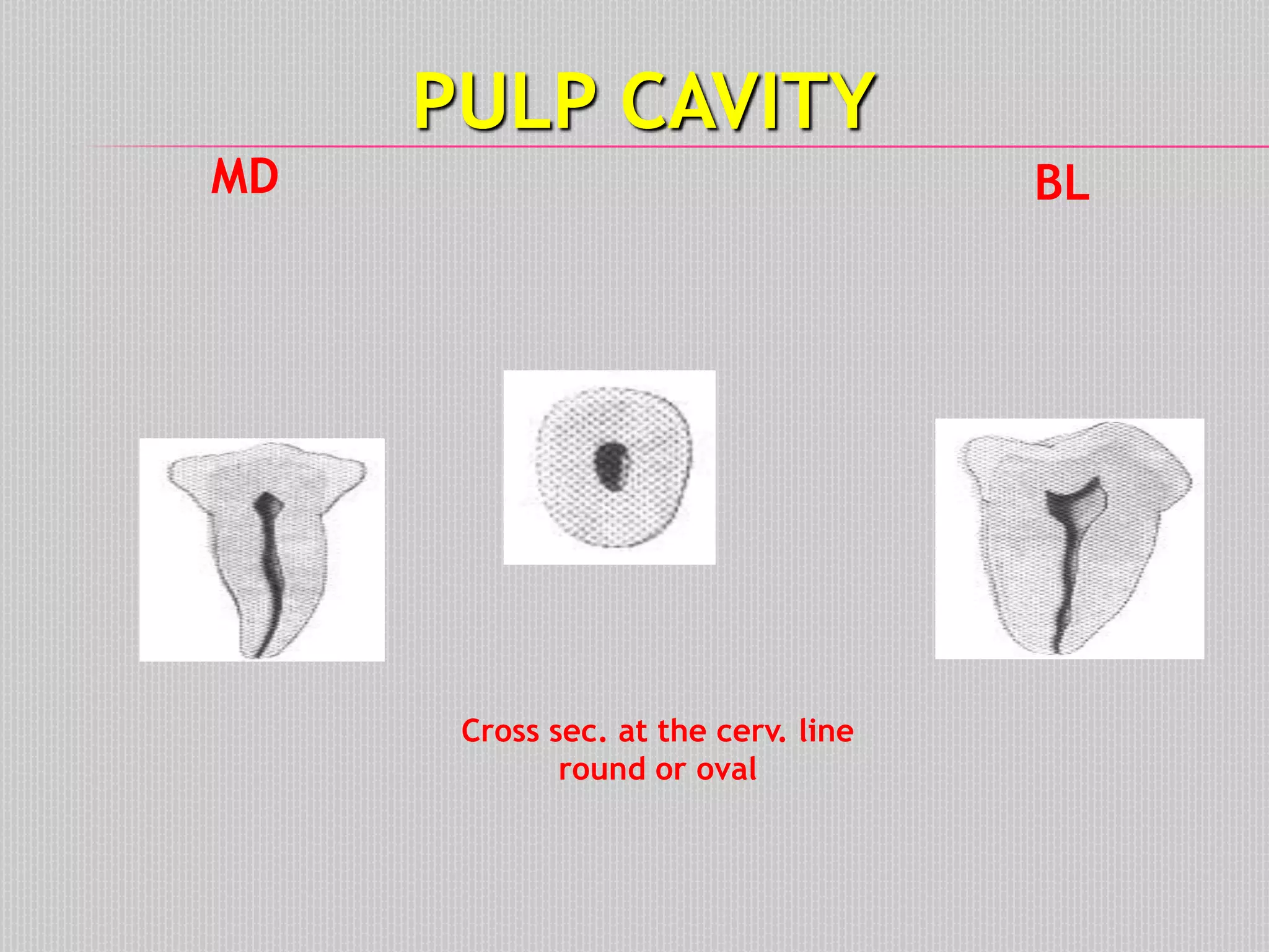
SlideShare a Scribd company logo
PERMANENT MANDIBULAR FIRST
PREMOLAR
AND
DIFFRENCES BETWEEN MANDIBULAR
FIRST AND SECOND PREMOLAR
Dr Monika Negi
MDS
Oral And Maxillofacial Pathology
PREMOLARS
There are four maxillary and four
mandibular premolars.
4&4 contact:
 3 & 3 mesially and
 5 & 5 distally
5 & 5 contact:
 4 & 4 mesially and
 6 & 6 distally.
Relations:
GENERAL FEATURES OF PREMOLARS
 They are transitional teeth
located between the
canine and molar teeth.
 By definition: Premolars
are permanent teeth distal
to the canines, and
successors to deciduous
molars.
 There are two premolars
per quadrant and are
identified as first and
second premolars.
GENERAL FEATURES OF PREMOLARS
 They have usually two
cusps(bicuspid) :
 One large buccal cusp,
 Smaller lingual cusp
 The lower second
premolar may-
sometimes- have two
lingual cusps.
MANDIBULAR FIRST PREMOLAR
Chronology:
•Enamel organ appearance: 7 miu.
•Beginning of calcification: 1 ¾ - 2y
•Crown completed: 5 – 6y
•Eruption: 10 – 12y
•Root completed: 12 – 13y
Mandibular First Premolar
Buccal Lingual Mesial
Occlusal
Distal
No. of roots
One root
Geometric Outline of the Crown
Facial and lingual aspects have
trapezoid out line.
The smallest of the
uneven sides

cervically.
Contact areas:
•Mesially and Distally: are nearly at
the same level, just occlusal to the
middle of the crown.
Cervical line:
•convex root wise.
M
Bucc. cusp
pointed and
long
Buccal Aspect Outlines:
•Mesial and distal outlines:
are nearly concave
•Mesial(shorter) and distal cusp
slopes: are also slightly concave
D
 Surface anatomy of the crown
Elevations:
Depressions:
•The middle lobe is convex
buccally forming prominent
BUCCAL ridge.
Buccal Aspect
•The surface is convex with
maximum convexity at C 1/3
representing cervical ridge
 Shallow depressions are
present mesial and distal to
the buccal ridge.
The surface of the root
is convex and smooth.
The M and D outline of the
root tapered to a pointed
apex that curved
distally(similar to canine
but slightly shorter).
Buccal Aspect
 Outline and Surface Anatomy
of the Root
 Outlines:
•M and D outlines are convex
Cervical line: is convex rootwise.
Elevations: The lingual surface is convex
with maximum convexity at middle 1/3
•The L cusp is short and small
reaching 2/3 of the length of the
Buccal cusp(has a pointed tip).
Deperssions: Mesio Lingual developmental groove separating
the Mesial Marginal ridge from the Mesial slope of the Lingual cusp.
Lingual AspectLingual convergence:
Lingual surface is narrower than buccal surface.
 Surface Anatomy :
MD
Lingual Aspect
MD
•Much of the occlusal and
proximal surfaces can be seen
from this aspect.
•The root is much narrower
than on the buccal surface. It
tapers from the cervix to a
pointed apex.
Proximal aspects have
rhomboid shape Mesial Distal
With narrow
occlusal table
Prominent lingual inclination
(much more than any other premolar)
Proximal Aspects
Lingual outline is less convex with maximum
convexity at the center of the crown length.
Cervical line is convex occlusally and less curved
distally
 Outlines of proximal aspects
Buccal outline is convex with maximum convexity at
the junction of middle and cervical 1/3 (cervical ridge).
Mesial
Aspect
Distal
Aspect
Occlusal margin:
The two cusps are not on the same level
The lingual cusp is shorter by 1/3 length of crown
The Buccal cusp tip centered over the root. This
is due to the prominent lingual inclination.
The L cusp tip in line with lingual border of the root.
Mesial
Aspect
Distal
Aspect
DMR is longer, straight
and at right angle to
the axis of the tooth.
(The only post. tooth in
which this is true).
MMR inclined sharply
from B to L surface parallel
to ridge of Buccal cusp
Mesial
Aspect
Distal
Aspect
DMR is occlusal than MMRMMR is cervical than DMR
M & D surfaces are smooth & convex except for
the ML groove.
Mesio-lingual
developmental grove
Passes over the MM
ridge(extension of M
groove on the occl. surf.
Contact areas:
 Proximal surface anatomy
nearly at same level
Contact area is broader,
more lingually situated
than the mesial one.
Mesial
Aspect
Distal
Aspect
 Outline of the Root
B & L outlines are nearly straight cervically then taper apically
to a pointed apex.
The surface is more convex with a
shallow dev. depres. centered on the
root
The surface is smooth & flat with a
deep dev. groove in the mid.& apical
1/3
Mesial
Aspect
Distal
Aspect
Occasionally the apical 1/3
may be divided into a B. & L.
roots by a deep dev. groove
It is diamond-shaped.
Lingual convergence is sharp.
Mesial outline is slightly curved.
Distal outline is more convex.
Occlusal Aspect
The B cusp is much larger than the L cusp.
M D
 Surface anatomy of occlusal aspect:
Elevations:
B triangular ridge(long).
L triangular ridge(short).
M & D marginal ridges(well
marked).
Transverse ridge (formed by
union of 2 triang. ridges).
Depressions:
Central dev. groove crossing
the transv. ridge.
M(oval) and D(circular)
fossae.
Mesio-lingual develop. groove – extension of M
groove on the occ. surface.
Each fossa has a dev. pit
(called snake eyes)
M D
..
PULP CAVITY
Buccal p. horn: more
pronounced than the
lingual.
Cross sec. at the cervix:
rounded or oval
BLMD
Usually have a single RC
Resemble that of
The lower canine
MANDIBULAR
FIRST PREMOLAR
THE 2 TYPES DIFFER MAINLY FROM
THE OCCLUSAL ASPECT. THE
OUTLINES AND GENERAL
APPEARANCE FROM ALL OTHER
ASPECTS ARE SIMILAR.
Geometric outline of the crown
Facial and lingual aspects have
trapezoid shape
But wider
cervically
than 4.
Comparison Between
Mandibular First and Second Premolars
Facial Outlines and surface anatomy
Crown is smaller & B
cusp is longer &
pointed
Prominent B ridge Less prominent B ridge
Crown is larger & B cusp
is shorter and less pointed
Root: Shorter, narrower with
pointed apex
Root: longer, broader,
with blunt apex
Mandibular First Premolar Mandibular Second Premolar
Lingual outline and surface anatomy
The lingual surface is convex with
maximum convexity in middle 1/3(center of
crown)
The L cusp is short and small reaching 2/3
the crown length and has a pointed tip.
ML developmental groove at the ML
line angle.
Two cusp type
L cusp is shorter and smaller than
B cusp but larger than of 4
The surface is convex with
maximum convexity in Occ.1/3.
No MLDG
Mandibular First Premolar Mandibular Second Premolar
Ling. s. not so
narrow as in 1st
premolar
MD D M
Three cusp type:
ML cusp is longer and larger than DL
cusp. They both shorter than B
cusp and less pointed.
DM
The surface convex with
maximum convexity at occ.1/3.
The L developmental groove
between the 2 ling. cusps
No ling. convergency.
Proximal outlines
Rhomboid in shape with
narrow occlusal table.
Prominent lingual
inclination
Rhomboid in shape with
narrow occlusal table.
Lingual inclination
less prominent
Mandibular First Premolar Mandibular Second Premolar
The crown is wider BLThe crown is narrower BL
Maximum convexity at Middle 1/3 Maximum convexity at Occlusal 1/3
B cusp tip on line buccal to the
root axis.
The mesial and distal marginal ridges
are straight
L cusp is shorter and smaller than B cusp
but larger than that of 4
The B cusp centered over the root.
The L cusp is short and small reaching
2/3 the crown length
The mesial MR is oblique while distally
is straight
Mandibular First Premolar Mandibular Second Premolar
The root is
wider BL
Three Cusp
Type of
Mandibular
Second
Premolar
ML cusp is shorter than the B cusp
& longer and larger than DL cusp.
DL cusp is shorter and smaller than ML
cusp. Both are seen from this aspect.
Both lingual cusps are shorter than the buccal cusp
and less pointed
Mesial
Aspect
Distal
Aspect
Develop.
depression
Cerv. line: slightly curved. Cerv. line: nearly straight.
Occlusal Aspect
Two cusp type
Diamond-shaped.
Lingual convergence
is sharp.
The outline is round
Slight lingual convergence
Three cusp type
The outline
is square
Mandibular First Premolar Mandibular Second Premolar
U- shaped H - shaped
M D
The 3 cusps are
well developed
M D
Surface Anatomy of Occlusal Aspect:
Elevations:
B & L triang. ridges
form a transv. ridge.
M & D marginal ridges
Lower 5
Two cusp type
Lower 4
Depressions:
Shallow central devel. groove
M(oval) and D(round) fossae.
Mesiolingual devel. gr.
Central devel. groove extending
MD across the occ. surface, over
the transv. ridge.
M and D fossae: Circular.
The D fossa is larger than
the M one
Lower 5
Two cusp type
Lower 4
. .
M D
M D
Elevations & Depressions
Each cusp has triang. ridge
that converge toward a
central fossa, which has
cent. pit.
M & D marginal ridges are well marked.
No central devel. gr. or transv. ridge
Three cusp typeLower 5
Three devel. gr.(M,D, & L) radiate
from the cent. Pit : Y-shaped.
The D triang. fossa is smaller than the M one.
M D
MANDIBULAR
SECOND PREMOLAR
3 cusp type
2 cusp type
PULP CAVITY
MD BL
Cross sec. at the cerv. line
round or oval
Permanent Mandibular First Premolar and Differences between First and Second Premolar

More Related Content

What's hot

The Permanent Maxillary First Molar
The Permanent Maxillary First MolarThe Permanent Maxillary First Molar
The Permanent Maxillary First Molar
Dr Aaron Sarwal
 
Mandibular 2nd premolars
Mandibular 2nd premolars  Mandibular 2nd premolars
Mandibular 2nd premolars
samah khaled
 
Permanent mandibular first premolar
Permanent mandibular first premolarPermanent mandibular first premolar
Permanent mandibular first premolar
Jamil Kifayatullah
 
Permanent Maxillary & Mandibular Canine
Permanent  Maxillary & Mandibular CaninePermanent  Maxillary & Mandibular Canine
Permanent Maxillary & Mandibular Canine
Abhishek Solanki
 
Permanent Maxillary Central Incisor
Permanent Maxillary Central IncisorPermanent Maxillary Central Incisor
Permanent Maxillary Central Incisor
Abhishek Solanki
 
Permanent Mandibular Lateral Incisor
Permanent Mandibular Lateral IncisorPermanent Mandibular Lateral Incisor
Permanent Mandibular Lateral Incisor
Abhishek Solanki
 
mandibular second molars
mandibular second molarsmandibular second molars
mandibular second molars
Jamil Kifayatullah
 
Upper Second Premolar
Upper Second PremolarUpper Second Premolar
Upper Second Premolar
Ahmed Elhlawany
 
Maxillary and Mandibular Canines
Maxillary and Mandibular CaninesMaxillary and Mandibular Canines
Maxillary and Mandibular Canineshchidmd
 
Permanent maxillary molars
Permanent maxillary molarsPermanent maxillary molars
Permanent maxillary molars
Basim Zwain
 
PERMANENT MANDIBULAR CENTRAL INCISOR
PERMANENT MANDIBULAR CENTRAL INCISORPERMANENT MANDIBULAR CENTRAL INCISOR
PERMANENT MANDIBULAR CENTRAL INCISOR
Ram Simsuangco
 
Peramanent Maxillary Canine
Peramanent Maxillary Canine Peramanent Maxillary Canine
Peramanent Maxillary Canine
Dr Monika Negi
 
Permanent Mandibular Central Incisor
Permanent Mandibular Central IncisorPermanent Mandibular Central Incisor
Permanent Mandibular Central Incisor
Abhishek Solanki
 
Permanent Mandibular Canine
Permanent Mandibular Canine Permanent Mandibular Canine
Permanent Mandibular Canine
Dr Monika Negi
 
Life cycle of ameloblast
Life cycle of ameloblastLife cycle of ameloblast
Life cycle of ameloblast
Haritha RK
 
Anatomical Landmarks of tooth.pptx
Anatomical Landmarks of tooth.pptxAnatomical Landmarks of tooth.pptx
Anatomical Landmarks of tooth.pptx
ridwana30
 
Trait categories helpful in describing tooth differences
Trait categories helpful in describing tooth differencesTrait categories helpful in describing tooth differences
Trait categories helpful in describing tooth differencesKuldeep Singh
 
Enamel
EnamelEnamel
morphology of maxillary & mandibular canine teeth
morphology of maxillary & mandibular canine teethmorphology of maxillary & mandibular canine teeth
morphology of maxillary & mandibular canine teeth
Akram bhuiyan
 

What's hot (20)

The Permanent Maxillary First Molar
The Permanent Maxillary First MolarThe Permanent Maxillary First Molar
The Permanent Maxillary First Molar
 
Mandibular 2nd premolars
Mandibular 2nd premolars  Mandibular 2nd premolars
Mandibular 2nd premolars
 
Permanent mandibular first premolar
Permanent mandibular first premolarPermanent mandibular first premolar
Permanent mandibular first premolar
 
Maxillary premolars
Maxillary premolarsMaxillary premolars
Maxillary premolars
 
Permanent Maxillary & Mandibular Canine
Permanent  Maxillary & Mandibular CaninePermanent  Maxillary & Mandibular Canine
Permanent Maxillary & Mandibular Canine
 
Permanent Maxillary Central Incisor
Permanent Maxillary Central IncisorPermanent Maxillary Central Incisor
Permanent Maxillary Central Incisor
 
Permanent Mandibular Lateral Incisor
Permanent Mandibular Lateral IncisorPermanent Mandibular Lateral Incisor
Permanent Mandibular Lateral Incisor
 
mandibular second molars
mandibular second molarsmandibular second molars
mandibular second molars
 
Upper Second Premolar
Upper Second PremolarUpper Second Premolar
Upper Second Premolar
 
Maxillary and Mandibular Canines
Maxillary and Mandibular CaninesMaxillary and Mandibular Canines
Maxillary and Mandibular Canines
 
Permanent maxillary molars
Permanent maxillary molarsPermanent maxillary molars
Permanent maxillary molars
 
PERMANENT MANDIBULAR CENTRAL INCISOR
PERMANENT MANDIBULAR CENTRAL INCISORPERMANENT MANDIBULAR CENTRAL INCISOR
PERMANENT MANDIBULAR CENTRAL INCISOR
 
Peramanent Maxillary Canine
Peramanent Maxillary Canine Peramanent Maxillary Canine
Peramanent Maxillary Canine
 
Permanent Mandibular Central Incisor
Permanent Mandibular Central IncisorPermanent Mandibular Central Incisor
Permanent Mandibular Central Incisor
 
Permanent Mandibular Canine
Permanent Mandibular Canine Permanent Mandibular Canine
Permanent Mandibular Canine
 
Life cycle of ameloblast
Life cycle of ameloblastLife cycle of ameloblast
Life cycle of ameloblast
 
Anatomical Landmarks of tooth.pptx
Anatomical Landmarks of tooth.pptxAnatomical Landmarks of tooth.pptx
Anatomical Landmarks of tooth.pptx
 
Trait categories helpful in describing tooth differences
Trait categories helpful in describing tooth differencesTrait categories helpful in describing tooth differences
Trait categories helpful in describing tooth differences
 
Enamel
EnamelEnamel
Enamel
 
morphology of maxillary & mandibular canine teeth
morphology of maxillary & mandibular canine teethmorphology of maxillary & mandibular canine teeth
morphology of maxillary & mandibular canine teeth
 

Similar to Permanent Mandibular First Premolar and Differences between First and Second Premolar

Mandibular 2nd premolar
Mandibular 2nd premolar Mandibular 2nd premolar
Mandibular 2nd premolar
dentistry
 
3 premolars
3 premolars3 premolars
3 premolars
mahmod3mmar
 
Naji Premolars.pptx
Naji Premolars.pptxNaji Premolars.pptx
Naji Premolars.pptx
NajiZArandi
 
Maillary molar
Maillary molarMaillary molar
Maillary molar
Ajish Saji
 
Max. first molar 2020
Max. first molar 2020Max. first molar 2020
Max. first molar 2020
HeshamAhmed104
 
Maxillary first molar
Maxillary first molarMaxillary first molar
Maxillary first molar
Hesham Dameer
 
upper5-150520204446-lva1-app6891.pdf
upper5-150520204446-lva1-app6891.pdfupper5-150520204446-lva1-app6891.pdf
upper5-150520204446-lva1-app6891.pdf
xMGMx1
 
Premolars Revision - Dentition
Premolars Revision - DentitionPremolars Revision - Dentition
Premolars Revision - DentitionCU Dentistry 2019
 
premolar
premolar premolar
maxillary first molar
maxillary first molar maxillary first molar
maxillary first molar
mahmodammar
 
5 maxillary first molar
5 maxillary  first molar5 maxillary  first molar
5 maxillary first molar
mahmodammar
 
Permanent Maxillary First Molar of Oral Cavity
Permanent Maxillary First Molar of Oral CavityPermanent Maxillary First Molar of Oral Cavity
Permanent Maxillary First Molar of Oral Cavity
UmaDatar
 
Mand. molars
Mand. molarsMand. molars
Mand. molarsReko Kemo
 
Mandibular 2nd premolars 2020
Mandibular 2nd premolars 2020Mandibular 2nd premolars 2020
Mandibular 2nd premolars 2020
HeshamAhmed104
 
Maxillary molars Dental Anatomy
Maxillary molars Dental AnatomyMaxillary molars Dental Anatomy
Maxillary molars Dental Anatomy
SiddharthRoy52
 
Maxillary Permanent Premolars
Maxillary Permanent  PremolarsMaxillary Permanent  Premolars
Maxillary Permanent Premolars
Dr Monika Negi
 
06. Mandibular Molars - Dentition
06. Mandibular Molars - Dentition06. Mandibular Molars - Dentition
06. Mandibular Molars - DentitionCU Dentistry 2019
 
7264369-Morphology-of-Deciduous-Dentition.ppt
7264369-Morphology-of-Deciduous-Dentition.ppt7264369-Morphology-of-Deciduous-Dentition.ppt
7264369-Morphology-of-Deciduous-Dentition.ppt
nona798438
 

Similar to Permanent Mandibular First Premolar and Differences between First and Second Premolar (20)

Mandibular 2nd premolar
Mandibular 2nd premolar Mandibular 2nd premolar
Mandibular 2nd premolar
 
3 premolars
3 premolars3 premolars
3 premolars
 
Naji Premolars.pptx
Naji Premolars.pptxNaji Premolars.pptx
Naji Premolars.pptx
 
04. Premolars - Dentition
04. Premolars - Dentition04. Premolars - Dentition
04. Premolars - Dentition
 
Maillary molar
Maillary molarMaillary molar
Maillary molar
 
Max. first molar 2020
Max. first molar 2020Max. first molar 2020
Max. first molar 2020
 
Maxillary first molar
Maxillary first molarMaxillary first molar
Maxillary first molar
 
upper5-150520204446-lva1-app6891.pdf
upper5-150520204446-lva1-app6891.pdfupper5-150520204446-lva1-app6891.pdf
upper5-150520204446-lva1-app6891.pdf
 
Premolars Revision - Dentition
Premolars Revision - DentitionPremolars Revision - Dentition
Premolars Revision - Dentition
 
premolar
premolar premolar
premolar
 
maxillary first molar
maxillary first molar maxillary first molar
maxillary first molar
 
Lower 4
Lower 4Lower 4
Lower 4
 
5 maxillary first molar
5 maxillary  first molar5 maxillary  first molar
5 maxillary first molar
 
Permanent Maxillary First Molar of Oral Cavity
Permanent Maxillary First Molar of Oral CavityPermanent Maxillary First Molar of Oral Cavity
Permanent Maxillary First Molar of Oral Cavity
 
Mand. molars
Mand. molarsMand. molars
Mand. molars
 
Mandibular 2nd premolars 2020
Mandibular 2nd premolars 2020Mandibular 2nd premolars 2020
Mandibular 2nd premolars 2020
 
Maxillary molars Dental Anatomy
Maxillary molars Dental AnatomyMaxillary molars Dental Anatomy
Maxillary molars Dental Anatomy
 
Maxillary Permanent Premolars
Maxillary Permanent  PremolarsMaxillary Permanent  Premolars
Maxillary Permanent Premolars
 
06. Mandibular Molars - Dentition
06. Mandibular Molars - Dentition06. Mandibular Molars - Dentition
06. Mandibular Molars - Dentition
 
7264369-Morphology-of-Deciduous-Dentition.ppt
7264369-Morphology-of-Deciduous-Dentition.ppt7264369-Morphology-of-Deciduous-Dentition.ppt
7264369-Morphology-of-Deciduous-Dentition.ppt
 

More from Dr Monika Negi

Gangrene
GangreneGangrene
Gangrene
Dr Monika Negi
 
Diseases Of Bone Manifested in Jaw
Diseases Of Bone Manifested in Jaw Diseases Of Bone Manifested in Jaw
Diseases Of Bone Manifested in Jaw
Dr Monika Negi
 
Allergic and Immunologic Diseases of Oral Cavity
Allergic and Immunologic Diseases of Oral CavityAllergic and Immunologic Diseases of Oral Cavity
Allergic and Immunologic Diseases of Oral Cavity
Dr Monika Negi
 
Spread of Oral Infection
Spread of Oral Infection Spread of Oral Infection
Spread of Oral Infection
Dr Monika Negi
 
Form and function of orofacial complex
Form and function of orofacial complexForm and function of orofacial complex
Form and function of orofacial complex
Dr Monika Negi
 
Permanent Mandibular Central Incisor
Permanent Mandibular Central IncisorPermanent Mandibular Central Incisor
Permanent Mandibular Central Incisor
Dr Monika Negi
 
Permanent Mandibular Lateral Incisor
Permanent Mandibular Lateral IncisorPermanent Mandibular Lateral Incisor
Permanent Mandibular Lateral Incisor
Dr Monika Negi
 
Diseases of Nerves
Diseases of NervesDiseases of Nerves
Diseases of Nerves
Dr Monika Negi
 
Dentinogenesis
DentinogenesisDentinogenesis
Dentinogenesis
Dr Monika Negi
 
Mycotic Infections of the Oral cavity . ( Candidiasis )
Mycotic Infections of the Oral cavity . ( Candidiasis )Mycotic Infections of the Oral cavity . ( Candidiasis )
Mycotic Infections of the Oral cavity . ( Candidiasis )
Dr Monika Negi
 
Biopsy and Exfoliative Cytology
Biopsy  and Exfoliative CytologyBiopsy  and Exfoliative Cytology
Biopsy and Exfoliative Cytology
Dr Monika Negi
 
Occlusion
OcclusionOcclusion
Occlusion
Dr Monika Negi
 
Permanent Maxillary Lateral Incisor
Permanent Maxillary Lateral IncisorPermanent Maxillary Lateral Incisor
Permanent Maxillary Lateral Incisor
Dr Monika Negi
 
Temporomandibular Joint (TMJ )
Temporomandibular Joint  (TMJ )Temporomandibular Joint  (TMJ )
Temporomandibular Joint (TMJ )
Dr Monika Negi
 
Tooth Development & Molecular aspect
Tooth Development & Molecular aspectTooth Development & Molecular aspect
Tooth Development & Molecular aspect
Dr Monika Negi
 

More from Dr Monika Negi (15)

Gangrene
GangreneGangrene
Gangrene
 
Diseases Of Bone Manifested in Jaw
Diseases Of Bone Manifested in Jaw Diseases Of Bone Manifested in Jaw
Diseases Of Bone Manifested in Jaw
 
Allergic and Immunologic Diseases of Oral Cavity
Allergic and Immunologic Diseases of Oral CavityAllergic and Immunologic Diseases of Oral Cavity
Allergic and Immunologic Diseases of Oral Cavity
 
Spread of Oral Infection
Spread of Oral Infection Spread of Oral Infection
Spread of Oral Infection
 
Form and function of orofacial complex
Form and function of orofacial complexForm and function of orofacial complex
Form and function of orofacial complex
 
Permanent Mandibular Central Incisor
Permanent Mandibular Central IncisorPermanent Mandibular Central Incisor
Permanent Mandibular Central Incisor
 
Permanent Mandibular Lateral Incisor
Permanent Mandibular Lateral IncisorPermanent Mandibular Lateral Incisor
Permanent Mandibular Lateral Incisor
 
Diseases of Nerves
Diseases of NervesDiseases of Nerves
Diseases of Nerves
 
Dentinogenesis
DentinogenesisDentinogenesis
Dentinogenesis
 
Mycotic Infections of the Oral cavity . ( Candidiasis )
Mycotic Infections of the Oral cavity . ( Candidiasis )Mycotic Infections of the Oral cavity . ( Candidiasis )
Mycotic Infections of the Oral cavity . ( Candidiasis )
 
Biopsy and Exfoliative Cytology
Biopsy  and Exfoliative CytologyBiopsy  and Exfoliative Cytology
Biopsy and Exfoliative Cytology
 
Occlusion
OcclusionOcclusion
Occlusion
 
Permanent Maxillary Lateral Incisor
Permanent Maxillary Lateral IncisorPermanent Maxillary Lateral Incisor
Permanent Maxillary Lateral Incisor
 
Temporomandibular Joint (TMJ )
Temporomandibular Joint  (TMJ )Temporomandibular Joint  (TMJ )
Temporomandibular Joint (TMJ )
 
Tooth Development & Molecular aspect
Tooth Development & Molecular aspectTooth Development & Molecular aspect
Tooth Development & Molecular aspect
 

Recently uploaded

263778731218 Abortion Clinic /Pills In Harare ,
263778731218 Abortion Clinic /Pills In Harare ,263778731218 Abortion Clinic /Pills In Harare ,
263778731218 Abortion Clinic /Pills In Harare ,
sisternakatoto
 
Surat @ℂall @Girls ꧁❤8527049040❤꧂@ℂall @Girls Service Vip Top Model Safe
Surat @ℂall @Girls ꧁❤8527049040❤꧂@ℂall @Girls Service Vip Top Model SafeSurat @ℂall @Girls ꧁❤8527049040❤꧂@ℂall @Girls Service Vip Top Model Safe
Surat @ℂall @Girls ꧁❤8527049040❤꧂@ℂall @Girls Service Vip Top Model Safe
Savita Shen $i11
 
basicmodesofventilation2022-220313203758.pdf
basicmodesofventilation2022-220313203758.pdfbasicmodesofventilation2022-220313203758.pdf
basicmodesofventilation2022-220313203758.pdf
aljamhori teaching hospital
 
Non-respiratory Functions of the Lungs.pdf
Non-respiratory Functions of the Lungs.pdfNon-respiratory Functions of the Lungs.pdf
Non-respiratory Functions of the Lungs.pdf
MedicoseAcademics
 
KDIGO 2024 guidelines for diabetologists
KDIGO 2024 guidelines for diabetologistsKDIGO 2024 guidelines for diabetologists
KDIGO 2024 guidelines for diabetologists
د.محمود نجيب
 
How to Give Better Lectures: Some Tips for Doctors
How to Give Better Lectures: Some Tips for DoctorsHow to Give Better Lectures: Some Tips for Doctors
How to Give Better Lectures: Some Tips for Doctors
LanceCatedral
 
Superficial & Deep Fascia of the NECK.pptx
Superficial & Deep Fascia of the NECK.pptxSuperficial & Deep Fascia of the NECK.pptx
Superficial & Deep Fascia of the NECK.pptx
Dr. Rabia Inam Gandapore
 
Pharynx and Clinical Correlations BY Dr.Rabia Inam Gandapore.pptx
Pharynx and Clinical Correlations BY Dr.Rabia Inam Gandapore.pptxPharynx and Clinical Correlations BY Dr.Rabia Inam Gandapore.pptx
Pharynx and Clinical Correlations BY Dr.Rabia Inam Gandapore.pptx
Dr. Rabia Inam Gandapore
 
TEST BANK for Operations Management, 14th Edition by William J. Stevenson, Ve...
TEST BANK for Operations Management, 14th Edition by William J. Stevenson, Ve...TEST BANK for Operations Management, 14th Edition by William J. Stevenson, Ve...
TEST BANK for Operations Management, 14th Edition by William J. Stevenson, Ve...
kevinkariuki227
 
Charaka Samhita Sutra sthana Chapter 15 Upakalpaniyaadhyaya
Charaka Samhita Sutra sthana Chapter 15 UpakalpaniyaadhyayaCharaka Samhita Sutra sthana Chapter 15 Upakalpaniyaadhyaya
Charaka Samhita Sutra sthana Chapter 15 Upakalpaniyaadhyaya
Dr KHALID B.M
 
ARTIFICIAL INTELLIGENCE IN HEALTHCARE.pdf
ARTIFICIAL INTELLIGENCE IN  HEALTHCARE.pdfARTIFICIAL INTELLIGENCE IN  HEALTHCARE.pdf
ARTIFICIAL INTELLIGENCE IN HEALTHCARE.pdf
Anujkumaranit
 
For Better Surat #ℂall #Girl Service ❤85270-49040❤ Surat #ℂall #Girls
For Better Surat #ℂall #Girl Service ❤85270-49040❤ Surat #ℂall #GirlsFor Better Surat #ℂall #Girl Service ❤85270-49040❤ Surat #ℂall #Girls
For Better Surat #ℂall #Girl Service ❤85270-49040❤ Surat #ℂall #Girls
Savita Shen $i11
 
NVBDCP.pptx Nation vector borne disease control program
NVBDCP.pptx Nation vector borne disease control programNVBDCP.pptx Nation vector borne disease control program
NVBDCP.pptx Nation vector borne disease control program
Sapna Thakur
 
Novas diretrizes da OMS para os cuidados perinatais de mais qualidade
Novas diretrizes da OMS para os cuidados perinatais de mais qualidadeNovas diretrizes da OMS para os cuidados perinatais de mais qualidade
Novas diretrizes da OMS para os cuidados perinatais de mais qualidade
Prof. Marcus Renato de Carvalho
 
Flu Vaccine Alert in Bangalore Karnataka
Flu Vaccine Alert in Bangalore KarnatakaFlu Vaccine Alert in Bangalore Karnataka
Flu Vaccine Alert in Bangalore Karnataka
addon Scans
 
Maxilla, Mandible & Hyoid Bone & Clinical Correlations by Dr. RIG.pptx
Maxilla, Mandible & Hyoid Bone & Clinical Correlations by Dr. RIG.pptxMaxilla, Mandible & Hyoid Bone & Clinical Correlations by Dr. RIG.pptx
Maxilla, Mandible & Hyoid Bone & Clinical Correlations by Dr. RIG.pptx
Dr. Rabia Inam Gandapore
 
BENIGN PROSTATIC HYPERPLASIA.BPH. BPHpdf
BENIGN PROSTATIC HYPERPLASIA.BPH. BPHpdfBENIGN PROSTATIC HYPERPLASIA.BPH. BPHpdf
BENIGN PROSTATIC HYPERPLASIA.BPH. BPHpdf
DR SETH JOTHAM
 
Ophthalmology Clinical Tests for OSCE exam
Ophthalmology Clinical Tests for OSCE examOphthalmology Clinical Tests for OSCE exam
Ophthalmology Clinical Tests for OSCE exam
KafrELShiekh University
 
Knee anatomy and clinical tests 2024.pdf
Knee anatomy and clinical tests 2024.pdfKnee anatomy and clinical tests 2024.pdf
Knee anatomy and clinical tests 2024.pdf
vimalpl1234
 
heat stroke and heat exhaustion in children
heat stroke and heat exhaustion in childrenheat stroke and heat exhaustion in children
heat stroke and heat exhaustion in children
SumeraAhmad5
 

Recently uploaded (20)

263778731218 Abortion Clinic /Pills In Harare ,
263778731218 Abortion Clinic /Pills In Harare ,263778731218 Abortion Clinic /Pills In Harare ,
263778731218 Abortion Clinic /Pills In Harare ,
 
Surat @ℂall @Girls ꧁❤8527049040❤꧂@ℂall @Girls Service Vip Top Model Safe
Surat @ℂall @Girls ꧁❤8527049040❤꧂@ℂall @Girls Service Vip Top Model SafeSurat @ℂall @Girls ꧁❤8527049040❤꧂@ℂall @Girls Service Vip Top Model Safe
Surat @ℂall @Girls ꧁❤8527049040❤꧂@ℂall @Girls Service Vip Top Model Safe
 
basicmodesofventilation2022-220313203758.pdf
basicmodesofventilation2022-220313203758.pdfbasicmodesofventilation2022-220313203758.pdf
basicmodesofventilation2022-220313203758.pdf
 
Non-respiratory Functions of the Lungs.pdf
Non-respiratory Functions of the Lungs.pdfNon-respiratory Functions of the Lungs.pdf
Non-respiratory Functions of the Lungs.pdf
 
KDIGO 2024 guidelines for diabetologists
KDIGO 2024 guidelines for diabetologistsKDIGO 2024 guidelines for diabetologists
KDIGO 2024 guidelines for diabetologists
 
How to Give Better Lectures: Some Tips for Doctors
How to Give Better Lectures: Some Tips for DoctorsHow to Give Better Lectures: Some Tips for Doctors
How to Give Better Lectures: Some Tips for Doctors
 
Superficial & Deep Fascia of the NECK.pptx
Superficial & Deep Fascia of the NECK.pptxSuperficial & Deep Fascia of the NECK.pptx
Superficial & Deep Fascia of the NECK.pptx
 
Pharynx and Clinical Correlations BY Dr.Rabia Inam Gandapore.pptx
Pharynx and Clinical Correlations BY Dr.Rabia Inam Gandapore.pptxPharynx and Clinical Correlations BY Dr.Rabia Inam Gandapore.pptx
Pharynx and Clinical Correlations BY Dr.Rabia Inam Gandapore.pptx
 
TEST BANK for Operations Management, 14th Edition by William J. Stevenson, Ve...
TEST BANK for Operations Management, 14th Edition by William J. Stevenson, Ve...TEST BANK for Operations Management, 14th Edition by William J. Stevenson, Ve...
TEST BANK for Operations Management, 14th Edition by William J. Stevenson, Ve...
 
Charaka Samhita Sutra sthana Chapter 15 Upakalpaniyaadhyaya
Charaka Samhita Sutra sthana Chapter 15 UpakalpaniyaadhyayaCharaka Samhita Sutra sthana Chapter 15 Upakalpaniyaadhyaya
Charaka Samhita Sutra sthana Chapter 15 Upakalpaniyaadhyaya
 
ARTIFICIAL INTELLIGENCE IN HEALTHCARE.pdf
ARTIFICIAL INTELLIGENCE IN  HEALTHCARE.pdfARTIFICIAL INTELLIGENCE IN  HEALTHCARE.pdf
ARTIFICIAL INTELLIGENCE IN HEALTHCARE.pdf
 
For Better Surat #ℂall #Girl Service ❤85270-49040❤ Surat #ℂall #Girls
For Better Surat #ℂall #Girl Service ❤85270-49040❤ Surat #ℂall #GirlsFor Better Surat #ℂall #Girl Service ❤85270-49040❤ Surat #ℂall #Girls
For Better Surat #ℂall #Girl Service ❤85270-49040❤ Surat #ℂall #Girls
 
NVBDCP.pptx Nation vector borne disease control program
NVBDCP.pptx Nation vector borne disease control programNVBDCP.pptx Nation vector borne disease control program
NVBDCP.pptx Nation vector borne disease control program
 
Novas diretrizes da OMS para os cuidados perinatais de mais qualidade
Novas diretrizes da OMS para os cuidados perinatais de mais qualidadeNovas diretrizes da OMS para os cuidados perinatais de mais qualidade
Novas diretrizes da OMS para os cuidados perinatais de mais qualidade
 
Flu Vaccine Alert in Bangalore Karnataka
Flu Vaccine Alert in Bangalore KarnatakaFlu Vaccine Alert in Bangalore Karnataka
Flu Vaccine Alert in Bangalore Karnataka
 
Maxilla, Mandible & Hyoid Bone & Clinical Correlations by Dr. RIG.pptx
Maxilla, Mandible & Hyoid Bone & Clinical Correlations by Dr. RIG.pptxMaxilla, Mandible & Hyoid Bone & Clinical Correlations by Dr. RIG.pptx
Maxilla, Mandible & Hyoid Bone & Clinical Correlations by Dr. RIG.pptx
 
BENIGN PROSTATIC HYPERPLASIA.BPH. BPHpdf
BENIGN PROSTATIC HYPERPLASIA.BPH. BPHpdfBENIGN PROSTATIC HYPERPLASIA.BPH. BPHpdf
BENIGN PROSTATIC HYPERPLASIA.BPH. BPHpdf
 
Ophthalmology Clinical Tests for OSCE exam
Ophthalmology Clinical Tests for OSCE examOphthalmology Clinical Tests for OSCE exam
Ophthalmology Clinical Tests for OSCE exam
 
Knee anatomy and clinical tests 2024.pdf
Knee anatomy and clinical tests 2024.pdfKnee anatomy and clinical tests 2024.pdf
Knee anatomy and clinical tests 2024.pdf
 
heat stroke and heat exhaustion in children
heat stroke and heat exhaustion in childrenheat stroke and heat exhaustion in children
heat stroke and heat exhaustion in children
 

Permanent Mandibular First Premolar and Differences between First and Second Premolar

  • 1. PERMANENT MANDIBULAR FIRST PREMOLAR AND DIFFRENCES BETWEEN MANDIBULAR FIRST AND SECOND PREMOLAR Dr Monika Negi MDS Oral And Maxillofacial Pathology
  • 2. PREMOLARS There are four maxillary and four mandibular premolars. 4&4 contact:  3 & 3 mesially and  5 & 5 distally 5 & 5 contact:  4 & 4 mesially and  6 & 6 distally. Relations:
  • 3. GENERAL FEATURES OF PREMOLARS  They are transitional teeth located between the canine and molar teeth.  By definition: Premolars are permanent teeth distal to the canines, and successors to deciduous molars.  There are two premolars per quadrant and are identified as first and second premolars.
  • 4. GENERAL FEATURES OF PREMOLARS  They have usually two cusps(bicuspid) :  One large buccal cusp,  Smaller lingual cusp  The lower second premolar may- sometimes- have two lingual cusps.
  • 5. MANDIBULAR FIRST PREMOLAR Chronology: •Enamel organ appearance: 7 miu. •Beginning of calcification: 1 ¾ - 2y •Crown completed: 5 – 6y •Eruption: 10 – 12y •Root completed: 12 – 13y
  • 6. Mandibular First Premolar Buccal Lingual Mesial Occlusal Distal No. of roots One root
  • 7. Geometric Outline of the Crown Facial and lingual aspects have trapezoid out line. The smallest of the uneven sides  cervically.
  • 8. Contact areas: •Mesially and Distally: are nearly at the same level, just occlusal to the middle of the crown. Cervical line: •convex root wise. M Bucc. cusp pointed and long Buccal Aspect Outlines: •Mesial and distal outlines: are nearly concave •Mesial(shorter) and distal cusp slopes: are also slightly concave D
  • 9.  Surface anatomy of the crown Elevations: Depressions: •The middle lobe is convex buccally forming prominent BUCCAL ridge. Buccal Aspect •The surface is convex with maximum convexity at C 1/3 representing cervical ridge  Shallow depressions are present mesial and distal to the buccal ridge.
  • 10. The surface of the root is convex and smooth. The M and D outline of the root tapered to a pointed apex that curved distally(similar to canine but slightly shorter). Buccal Aspect  Outline and Surface Anatomy of the Root
  • 11.  Outlines: •M and D outlines are convex Cervical line: is convex rootwise. Elevations: The lingual surface is convex with maximum convexity at middle 1/3 •The L cusp is short and small reaching 2/3 of the length of the Buccal cusp(has a pointed tip). Deperssions: Mesio Lingual developmental groove separating the Mesial Marginal ridge from the Mesial slope of the Lingual cusp. Lingual AspectLingual convergence: Lingual surface is narrower than buccal surface.  Surface Anatomy : MD
  • 12. Lingual Aspect MD •Much of the occlusal and proximal surfaces can be seen from this aspect. •The root is much narrower than on the buccal surface. It tapers from the cervix to a pointed apex.
  • 13. Proximal aspects have rhomboid shape Mesial Distal With narrow occlusal table Prominent lingual inclination (much more than any other premolar) Proximal Aspects
  • 14. Lingual outline is less convex with maximum convexity at the center of the crown length. Cervical line is convex occlusally and less curved distally  Outlines of proximal aspects Buccal outline is convex with maximum convexity at the junction of middle and cervical 1/3 (cervical ridge). Mesial Aspect Distal Aspect
  • 15. Occlusal margin: The two cusps are not on the same level The lingual cusp is shorter by 1/3 length of crown The Buccal cusp tip centered over the root. This is due to the prominent lingual inclination. The L cusp tip in line with lingual border of the root. Mesial Aspect Distal Aspect
  • 16. DMR is longer, straight and at right angle to the axis of the tooth. (The only post. tooth in which this is true). MMR inclined sharply from B to L surface parallel to ridge of Buccal cusp Mesial Aspect Distal Aspect DMR is occlusal than MMRMMR is cervical than DMR
  • 17. M & D surfaces are smooth & convex except for the ML groove. Mesio-lingual developmental grove Passes over the MM ridge(extension of M groove on the occl. surf. Contact areas:  Proximal surface anatomy nearly at same level Contact area is broader, more lingually situated than the mesial one. Mesial Aspect Distal Aspect
  • 18.  Outline of the Root B & L outlines are nearly straight cervically then taper apically to a pointed apex. The surface is more convex with a shallow dev. depres. centered on the root The surface is smooth & flat with a deep dev. groove in the mid.& apical 1/3 Mesial Aspect Distal Aspect Occasionally the apical 1/3 may be divided into a B. & L. roots by a deep dev. groove
  • 19. It is diamond-shaped. Lingual convergence is sharp. Mesial outline is slightly curved. Distal outline is more convex. Occlusal Aspect The B cusp is much larger than the L cusp. M D
  • 20.  Surface anatomy of occlusal aspect: Elevations: B triangular ridge(long). L triangular ridge(short). M & D marginal ridges(well marked). Transverse ridge (formed by union of 2 triang. ridges).
  • 21. Depressions: Central dev. groove crossing the transv. ridge. M(oval) and D(circular) fossae. Mesio-lingual develop. groove – extension of M groove on the occ. surface. Each fossa has a dev. pit (called snake eyes) M D ..
  • 22. PULP CAVITY Buccal p. horn: more pronounced than the lingual. Cross sec. at the cervix: rounded or oval BLMD Usually have a single RC Resemble that of The lower canine
  • 24. THE 2 TYPES DIFFER MAINLY FROM THE OCCLUSAL ASPECT. THE OUTLINES AND GENERAL APPEARANCE FROM ALL OTHER ASPECTS ARE SIMILAR.
  • 25. Geometric outline of the crown Facial and lingual aspects have trapezoid shape But wider cervically than 4. Comparison Between Mandibular First and Second Premolars
  • 26. Facial Outlines and surface anatomy Crown is smaller & B cusp is longer & pointed Prominent B ridge Less prominent B ridge Crown is larger & B cusp is shorter and less pointed Root: Shorter, narrower with pointed apex Root: longer, broader, with blunt apex Mandibular First Premolar Mandibular Second Premolar
  • 27. Lingual outline and surface anatomy The lingual surface is convex with maximum convexity in middle 1/3(center of crown) The L cusp is short and small reaching 2/3 the crown length and has a pointed tip. ML developmental groove at the ML line angle. Two cusp type L cusp is shorter and smaller than B cusp but larger than of 4 The surface is convex with maximum convexity in Occ.1/3. No MLDG Mandibular First Premolar Mandibular Second Premolar Ling. s. not so narrow as in 1st premolar MD D M
  • 28. Three cusp type: ML cusp is longer and larger than DL cusp. They both shorter than B cusp and less pointed. DM The surface convex with maximum convexity at occ.1/3. The L developmental groove between the 2 ling. cusps No ling. convergency.
  • 29. Proximal outlines Rhomboid in shape with narrow occlusal table. Prominent lingual inclination Rhomboid in shape with narrow occlusal table. Lingual inclination less prominent Mandibular First Premolar Mandibular Second Premolar The crown is wider BLThe crown is narrower BL
  • 30. Maximum convexity at Middle 1/3 Maximum convexity at Occlusal 1/3 B cusp tip on line buccal to the root axis. The mesial and distal marginal ridges are straight L cusp is shorter and smaller than B cusp but larger than that of 4 The B cusp centered over the root. The L cusp is short and small reaching 2/3 the crown length The mesial MR is oblique while distally is straight Mandibular First Premolar Mandibular Second Premolar The root is wider BL
  • 31. Three Cusp Type of Mandibular Second Premolar ML cusp is shorter than the B cusp & longer and larger than DL cusp. DL cusp is shorter and smaller than ML cusp. Both are seen from this aspect. Both lingual cusps are shorter than the buccal cusp and less pointed Mesial Aspect Distal Aspect Develop. depression Cerv. line: slightly curved. Cerv. line: nearly straight.
  • 32. Occlusal Aspect Two cusp type Diamond-shaped. Lingual convergence is sharp. The outline is round Slight lingual convergence Three cusp type The outline is square Mandibular First Premolar Mandibular Second Premolar U- shaped H - shaped M D The 3 cusps are well developed M D
  • 33. Surface Anatomy of Occlusal Aspect: Elevations: B & L triang. ridges form a transv. ridge. M & D marginal ridges Lower 5 Two cusp type Lower 4
  • 34. Depressions: Shallow central devel. groove M(oval) and D(round) fossae. Mesiolingual devel. gr. Central devel. groove extending MD across the occ. surface, over the transv. ridge. M and D fossae: Circular. The D fossa is larger than the M one Lower 5 Two cusp type Lower 4 . . M D M D
  • 35. Elevations & Depressions Each cusp has triang. ridge that converge toward a central fossa, which has cent. pit. M & D marginal ridges are well marked. No central devel. gr. or transv. ridge Three cusp typeLower 5 Three devel. gr.(M,D, & L) radiate from the cent. Pit : Y-shaped. The D triang. fossa is smaller than the M one. M D
  • 37. PULP CAVITY MD BL Cross sec. at the cerv. line round or oval