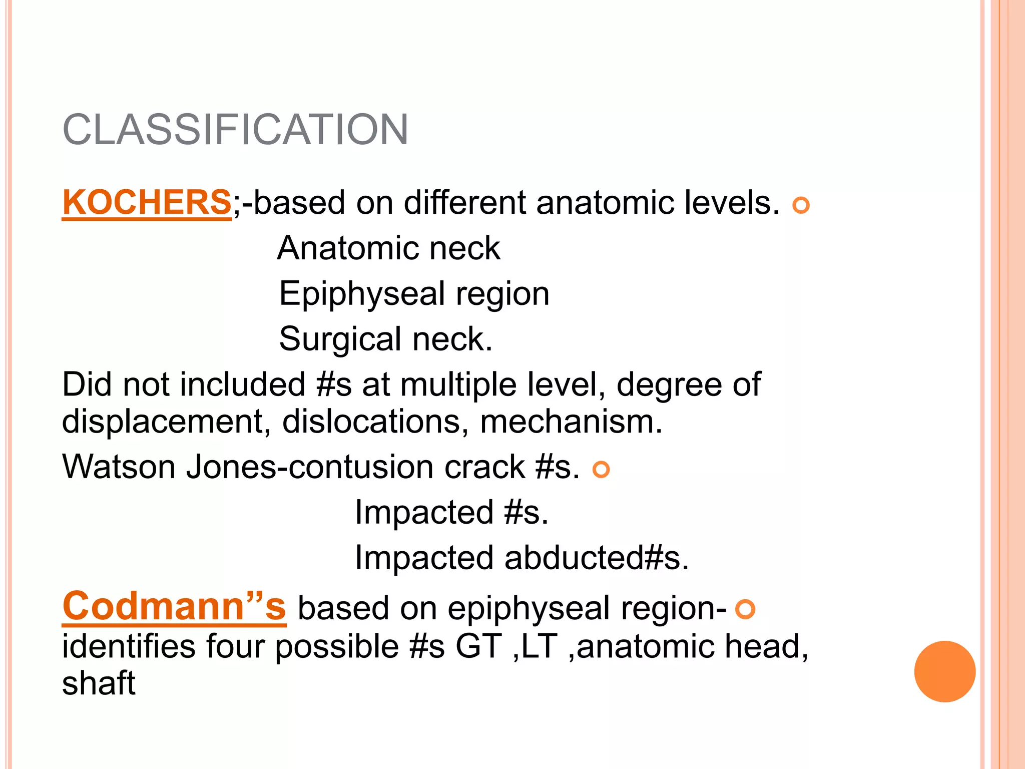
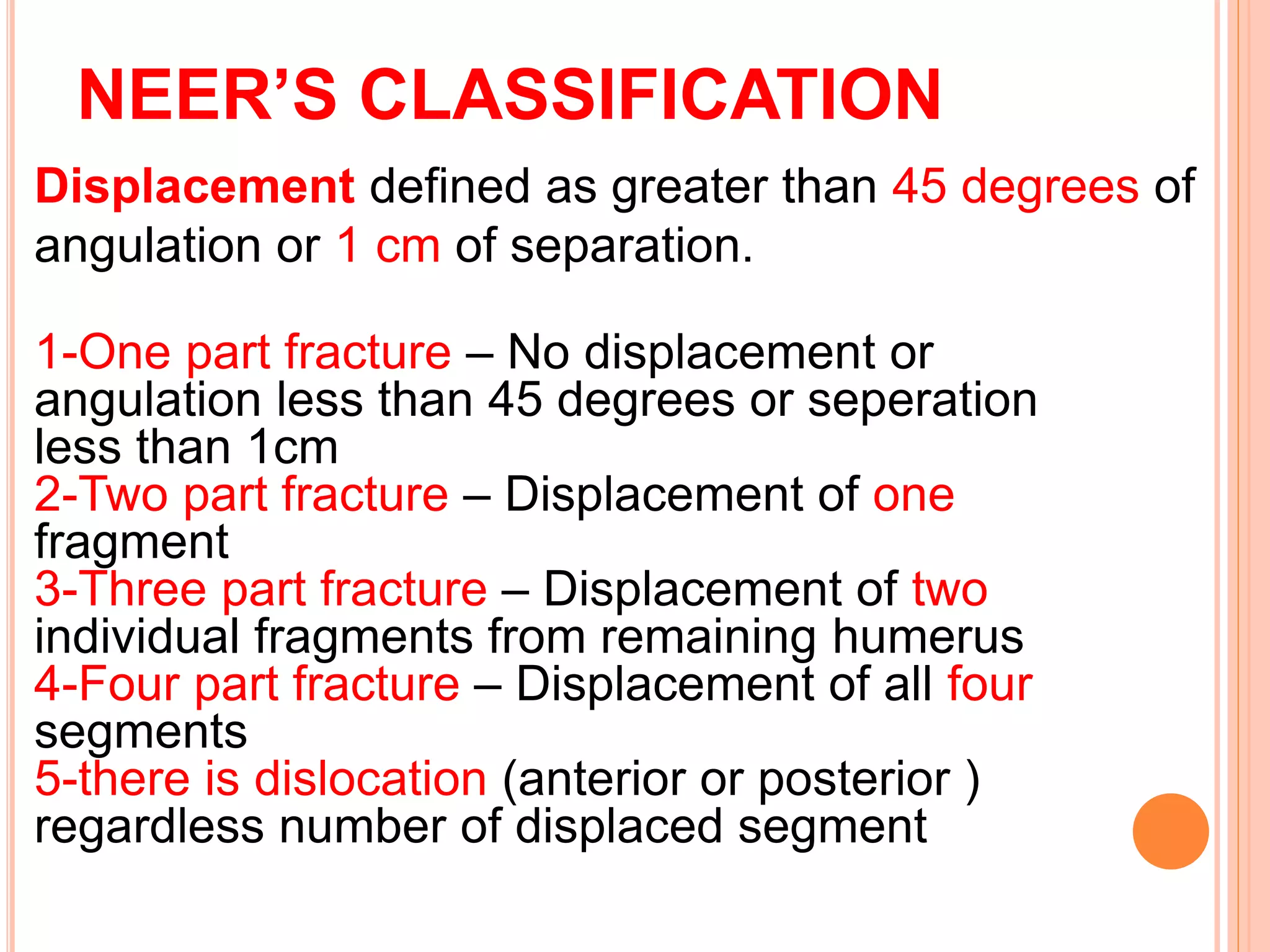
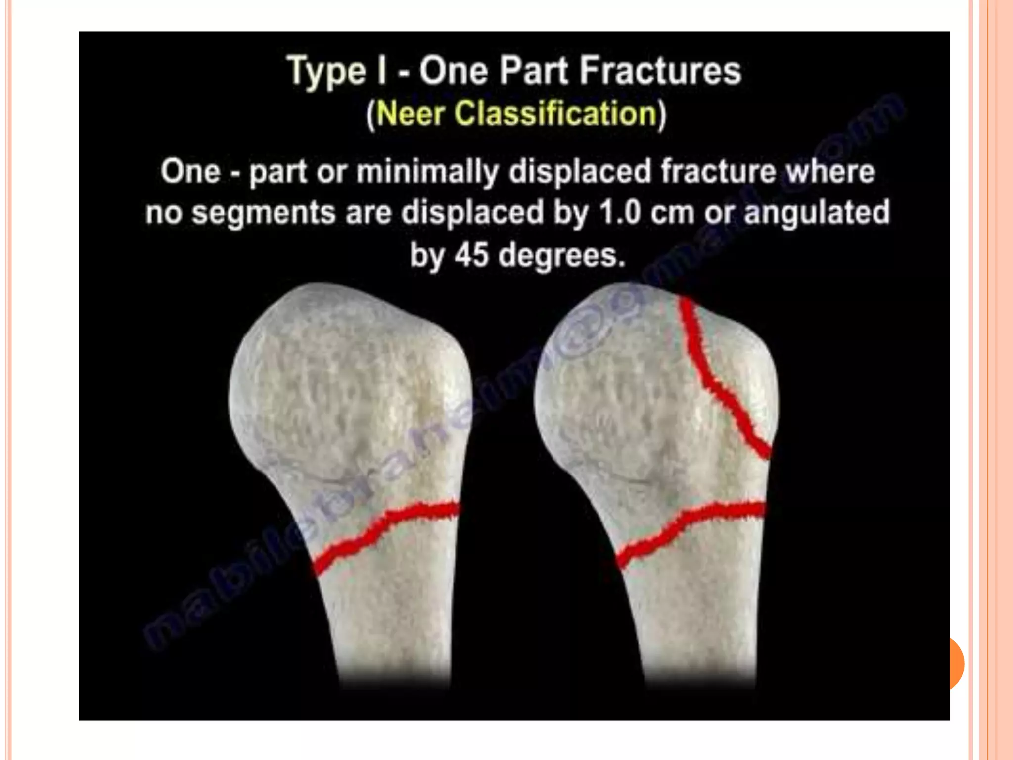
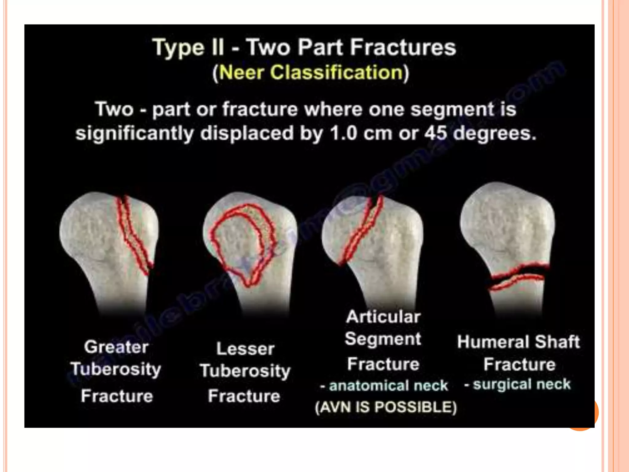
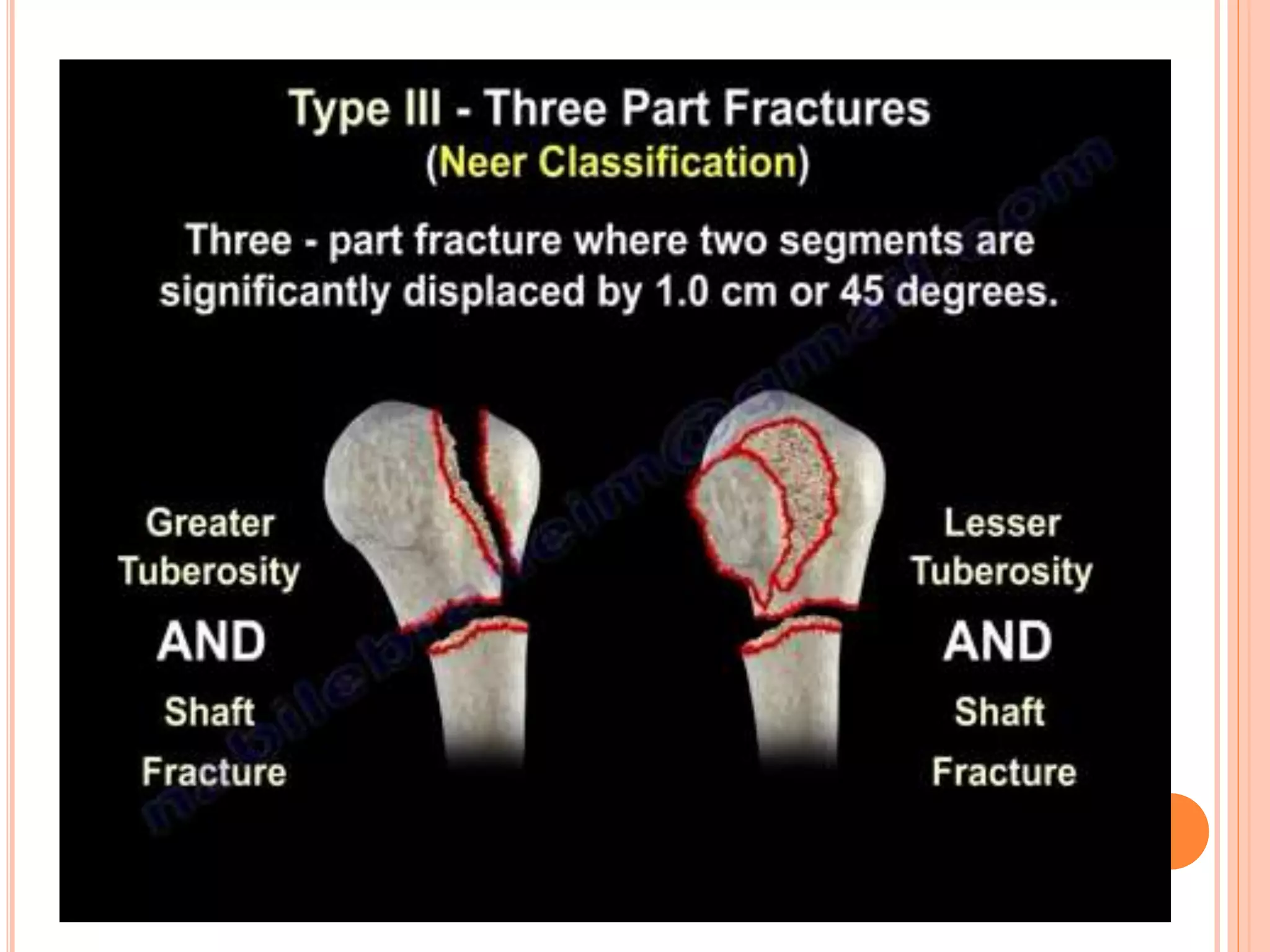
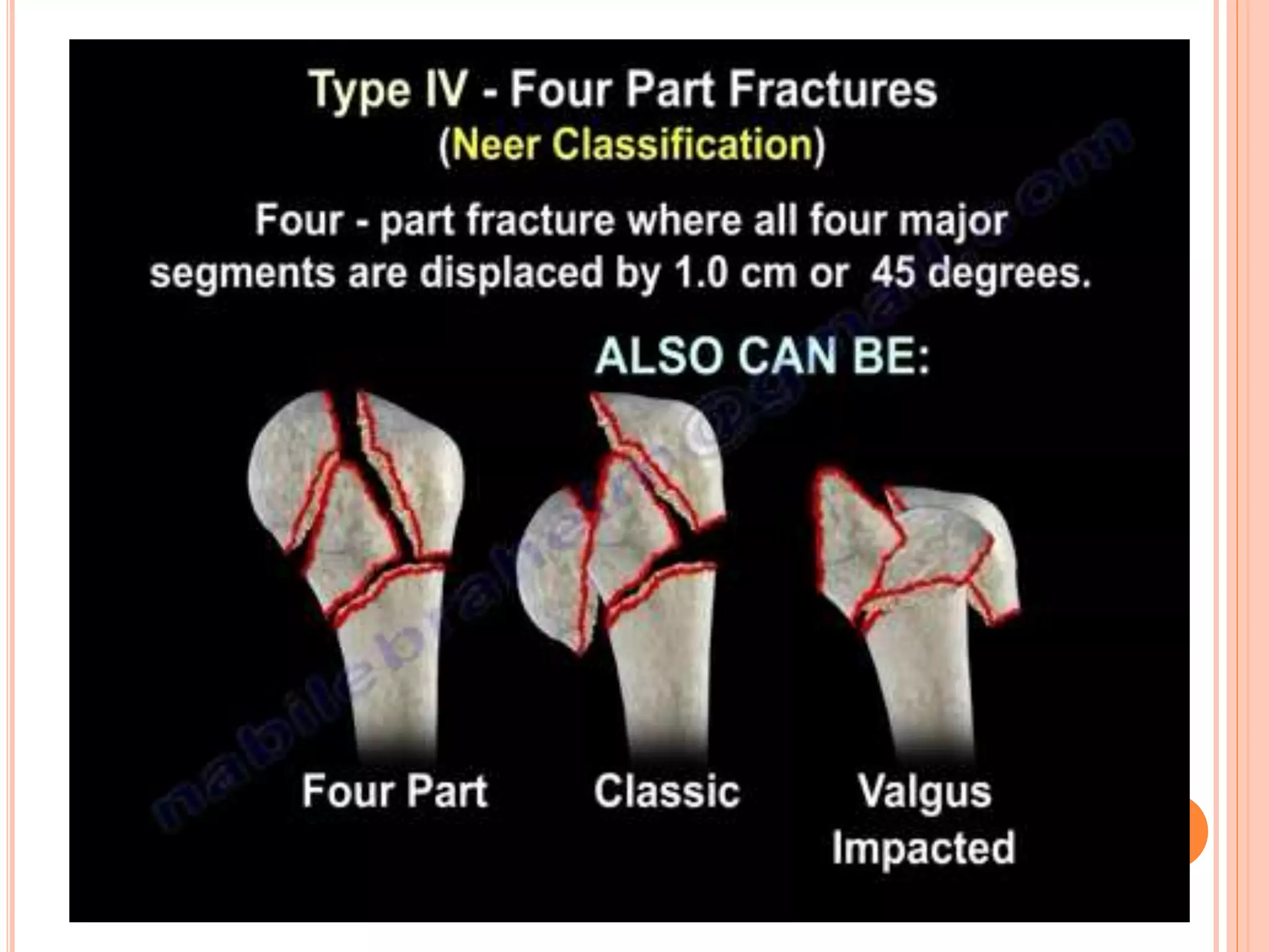
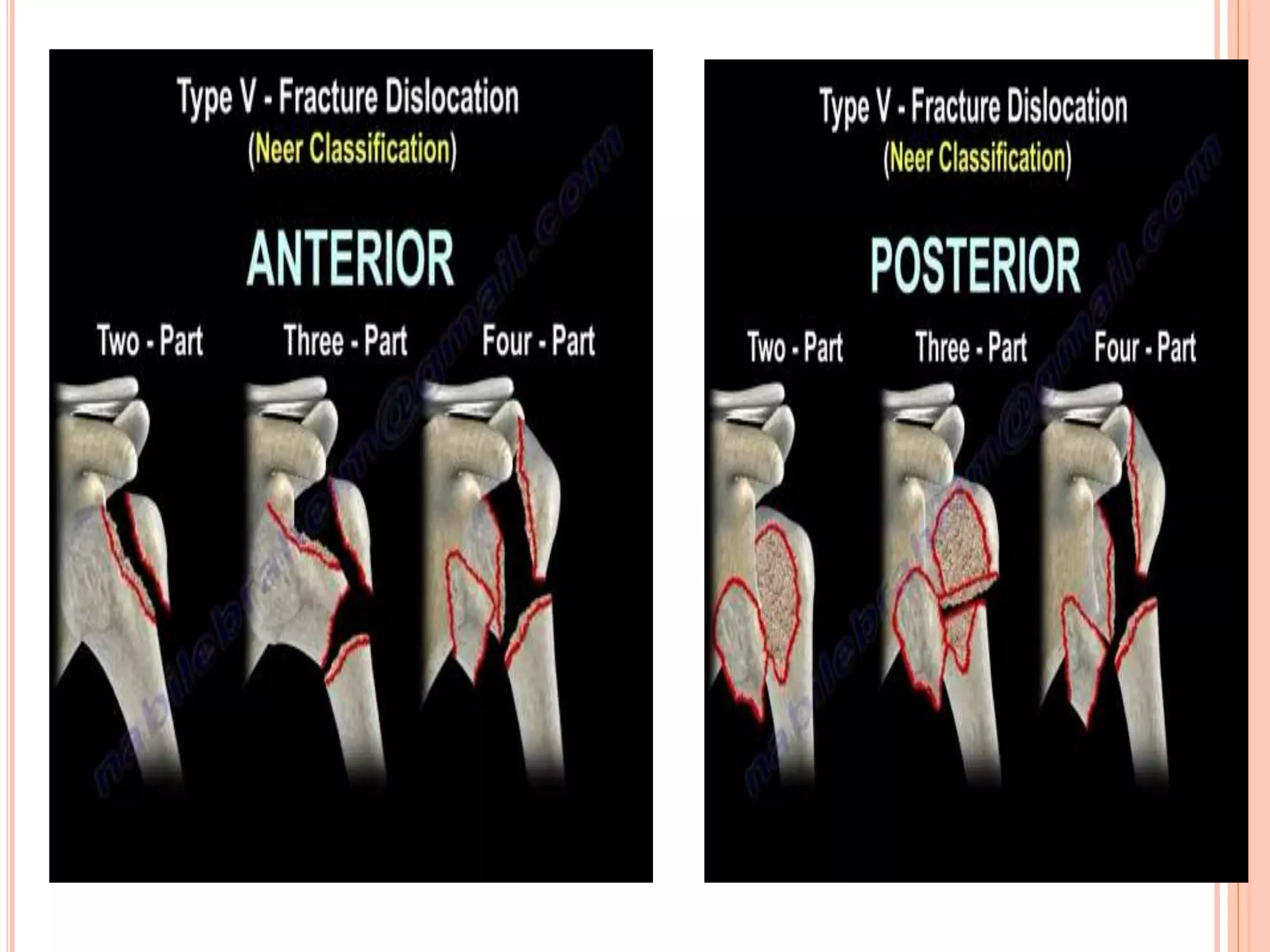
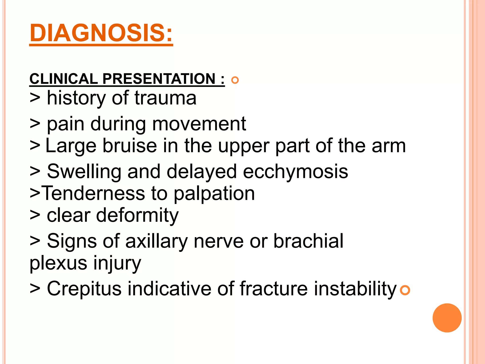
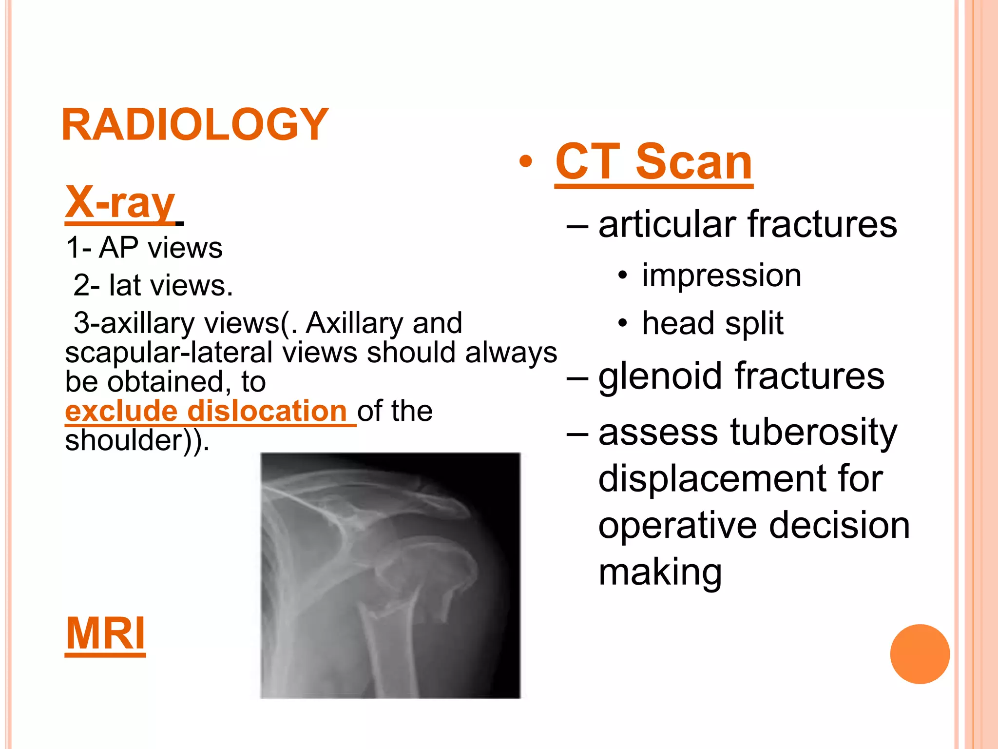
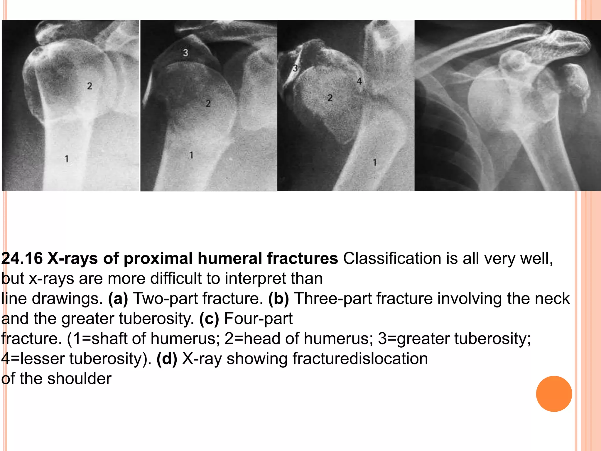
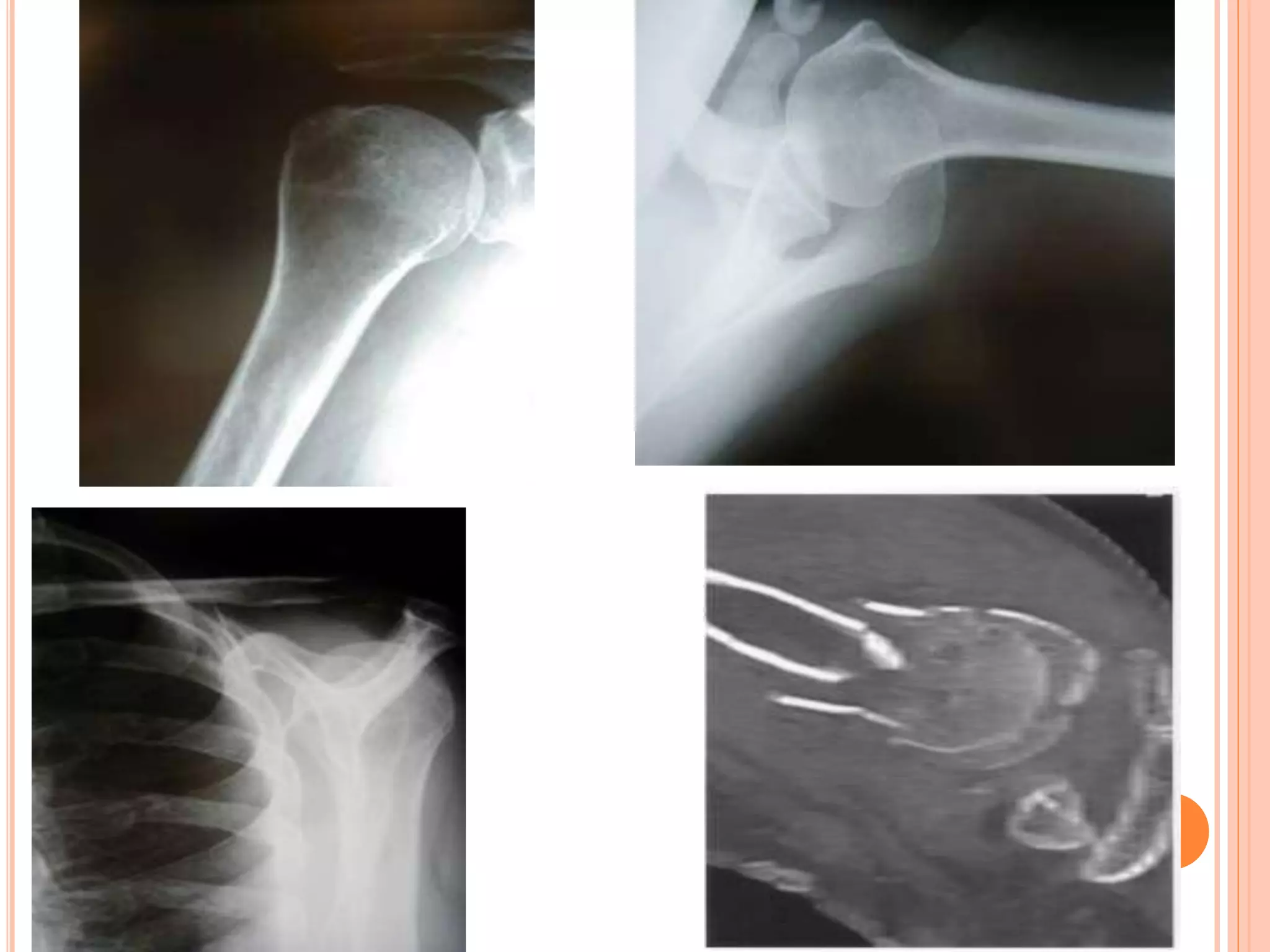
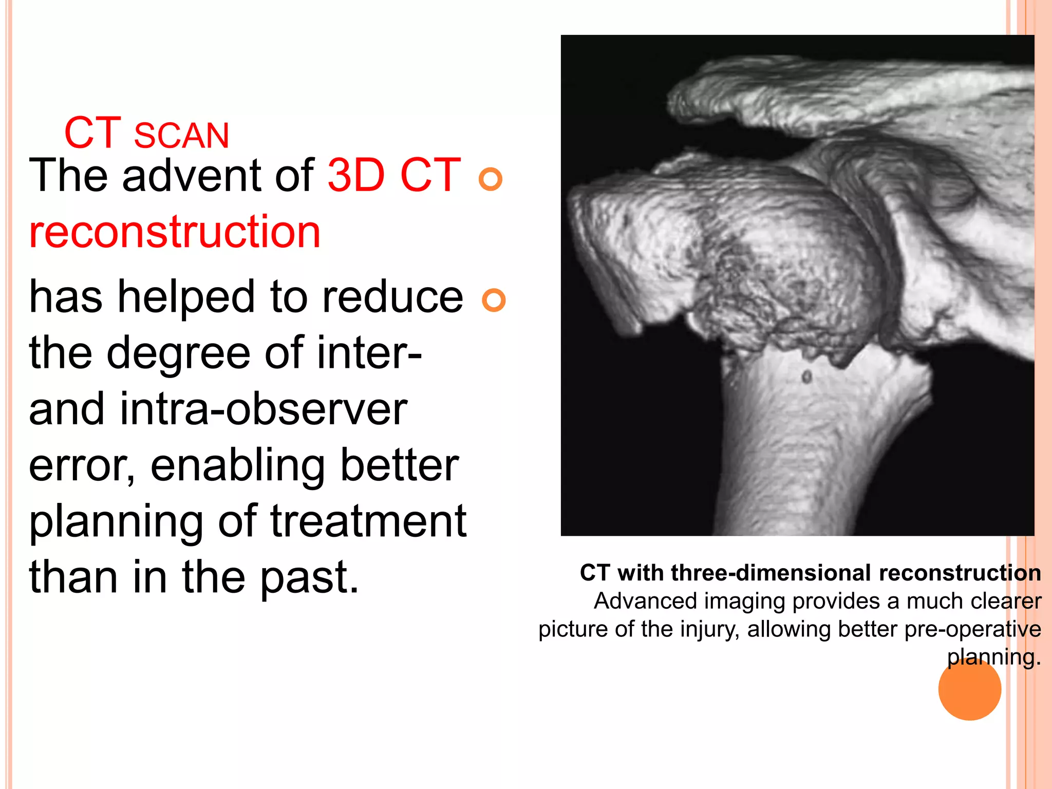
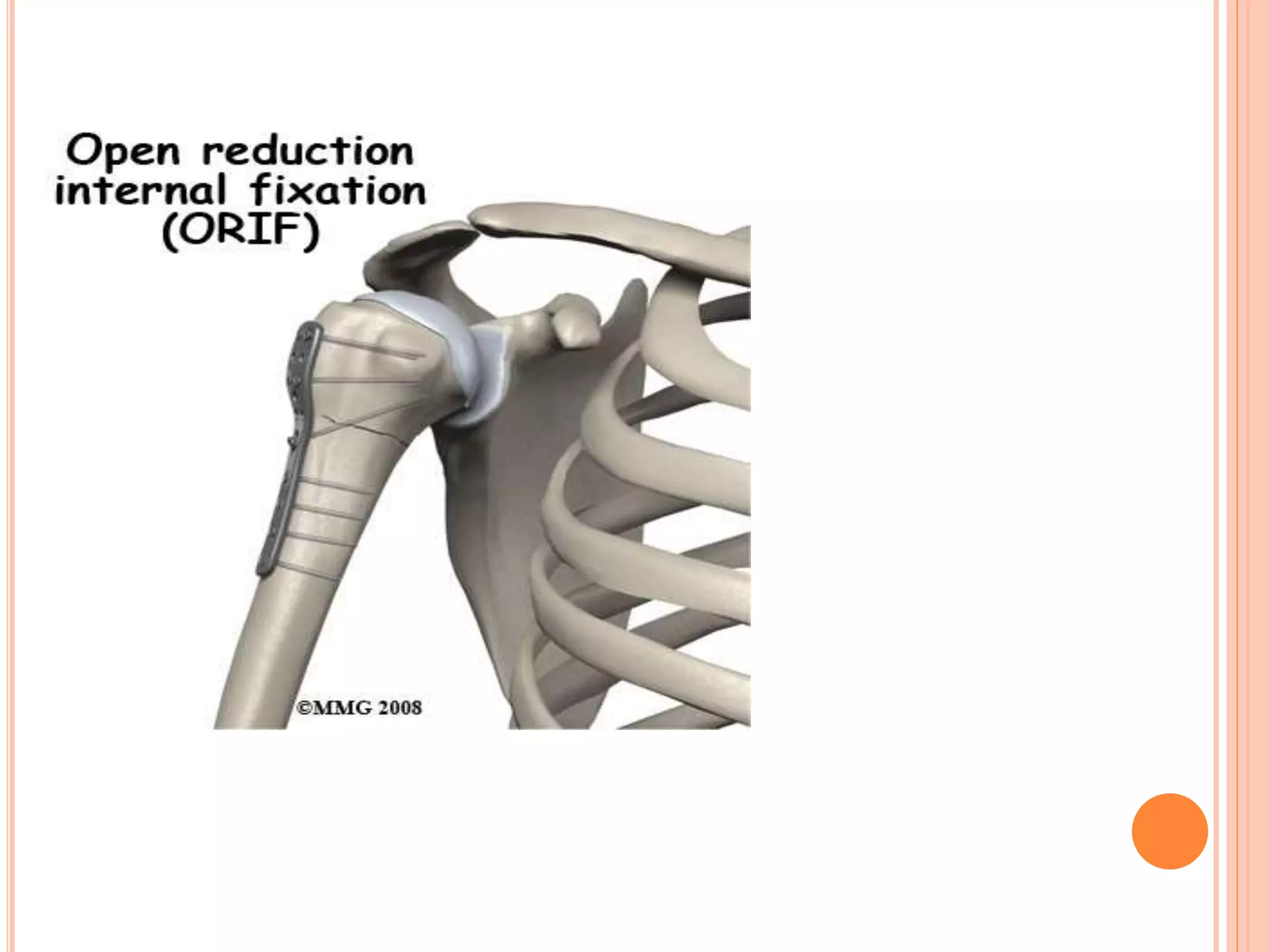
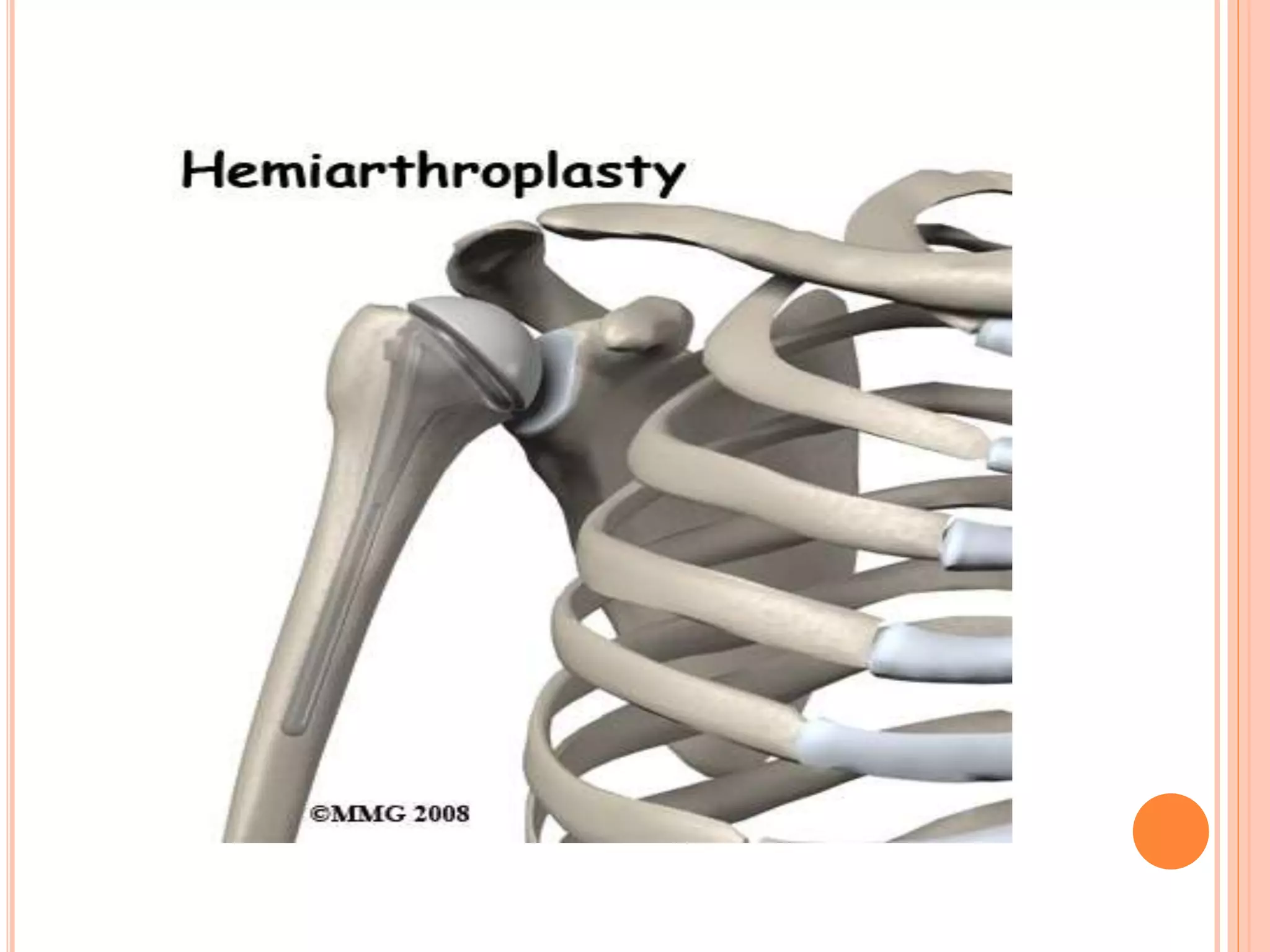
Proximal fracture # of the&
humers
fracture of humeral shaft
ANATOMY OF HUMERUS
VASCULAR SUPPLY
NERVE SUPPLY
SITES
Common))-surgical neck
-anatomical neck
-both tubercities
CLINICAL PICTURE:
# History
# Symptoms
Upper extremity held closed to the chest by
contra lat.hand.
Pain,swelling,crepitus, painfull
# signs:
look
palpation
moove
(RANGE OF THE MOTION( ROM
FLEXION 180
ABDUCTION 180
ADDUCTION 75
EXTENTION 50
ETERNAL ROTATION 65
INTERNAL ROTATION 90
CLASSIFICATION
KOCHERS;-based on different anatomic levels.
Anatomic neck
Epiphyseal region
Surgical neck.
Did not included #s at multiple level, degree of
displacement, dislocations, mechanism.
Watson Jones-contusion crack #s.
Impacted #s.
Impacted abducted#s.
Codmann”s based on epiphyseal region-
identifies four possible #s GT ,LT ,anatomic head,
shaft
CLASSIFICATION:
NEER’S CLASSIFICATION
(5-TYPE)
THE MOST COMMONLY USED CLASSIFICATION SYSTEM FOR PROXIMAL
HUMERAL
HEAD FRACTURES WAS DEVELOPED IN 1970 BY DR CHARLES NEER.
THE BASIS OF THE SYSTEM ACCORDING TO
1-DISPLACEMENT
2-ANATOMICAL LINES OF EPIPHYSEAL UNION,
INCLUDING THE
HUMERAL HEAD, GREATER TUBEROSITY
, LESSER TUBEROSITY, AND THE SHAFT (SURGICAL NECK).
NEER’S CLASSIFICATION
Displacement defined as greater than 45 degrees of
angulation or 1 cm of separation.
1-One part fracture – No displacement or
angulation less than 45 degrees or seperation
less than 1cm
2-Two part fracture – Displacement of one
fragment
3-Three part fracture – Displacement of two
individual fragments from remaining humerus
4-Four part fracture – Displacement of all four
segments
5-there is dislocation (anterior or posterior )
regardless number of displaced segment
DIAGNOSIS:
CLINICAL PRESENTATION :
> history of trauma
> pain during movement
> Large bruise in the upper part of the arm
> Swelling and delayed ecchymosis
>Tenderness to palpation
> clear deformity
> Signs of axillary nerve or brachial
plexus injury
> Crepitus indicative of fracture instability
RADIOLOGY
X-ray
1- AP views
2- lat views.
3-axillary views(. Axillary and
scapular-lateral views should always
be obtained, to
exclude dislocation of the
shoulder)).
MRI
• CT Scan
– articular fractures
• impression
• head split
– glenoid fractures
– assess tuberosity
displacement for
operative decision
making
24.16 X-rays of proximal humeral fractures Classification is all very well,
but x-rays are more difficult to interpret than
line drawings. (a) Two-part fracture. (b) Three-part fracture involving the neck
and the greater tuberosity. (c) Four-part
fracture. (1=shaft of humerus; 2=head of humerus; 3=greater tuberosity;
4=lesser tuberosity). (d) X-ray showing fracturedislocation
of the shoulder
CT SCAN
The advent of 3D CT
reconstruction
has helped to reduce
the degree of inter-
and intra-observer
error, enabling better
planning of treatment
than in the past. CT with three-dimensional reconstruction
Advanced imaging provides a much clearer
picture of the injury, allowing better pre-operative
planning.
TREATMENT
1-One part fracture
These comprise the vast
majority. They need no
treatment apart from a week
or two period of rest with the
arm in a sling until the pain
subsides, and then gentle
passive movements of the shoulder. Once the fracture has
united (usually after 6 weeks), active exercises are encouraged;
the hand is, of course, actively exercised from the start.
2-TWO PART FRACTURE
STABLE Closed reduction then sling for about
four weeks or until the fracture feels stable and the
x-ray shows some signs of
healing. Elbow and hand exercises are encouraged
throughout this period; shoulder exercises are
commenced at about four weeks.
if the fracture cannot be reduced closed or if the
fracture is very unstable after closed reduction,
then external fixation is Required Options
.include percutaneous pins, bone sutures,
intramedullary pins
3-THREE PART FRACTURE
4-Four part fracture
5-there is dislocation
they are extremely difficult to reduce closed.
In active individuals this injury is best
managed by open reduction and internal
fixation.
Proximal humerus fractures – treatment
(a) Three-part fracture, treated by
(b) locked nail fixation.
(c) Four-part fracture fixed with a locked plate; the intra-operative
picture
(d) shows how the plate was positioned
INTRA-MEDULLARY K WIRE FIXATION
COMPLICATIONS:
Early complication:
*Rotator cuff syndrom
* Vascular injury .
* Nerve injury.
* Biceps tendon
rupture
* Thoracic injury
late complication:
•* stiffness of the
shoulder.
* malunion.
* infection
* Avascular
necrosis.
HUMERUS SHAFT FRACTURES
THE HUMERUS IS THE LONG,
TUBULAR BONE THAT MAKES UP THE
UPPER ARM. THE HUMERAL SHAFT IS
THE MIDDLE PORTION OF THE BONE
WITH THE SHOULDER JOINT AT THE
TOP END AND THE ELBOW JOINT AT
THE BOTTOM. ONE OF THE NERVES
THAT TRAVELS FROM THE NECK TO
THE HAND, THE RADIAL NERVE,
SPIRALS AROUND THE HUMERAL
SHAFT LYING VERY CLOSE TO THE
BONE ABOUT TWO THIRDS OF THE
WAY TO THE ELBOW. FRACTURES OF
THE HUMERAL SHAFT ARE IMPORTANT
BECAUSE THEY CAN INJURE THE
RADIAL NERVE RESULTING IN THE
INABILITY TO EXTEND (BEND) THE
WRIST AND FINGERS BACKWARDS
ANATOMY
AO/OTA CLASSIFICATION
CLINICAL:
DIAGNOSIS
TETMENT
INDICATIONS FOR ORIF -
INJURY FACTORS
Failed closed treatment
Loss of reduction
Poor patient tolerance/compliance
(Open fractures)
Vascular injury/
Change in neuro exam (radial n.)
Floating elbow
COMPLICATIONS OF HUMERAL
SHAFT FRACTURES
Radial nerve injury
Vascular injury
Nonunion
COMLICATION
Proximal humerus fractures

Proximal humerus fractures

  • 1.
    Proximal fracture #of the& humers fracture of humeral shaft
  • 3.
  • 6.
  • 7.
  • 10.
  • 11.
    CLINICAL PICTURE: # History #Symptoms Upper extremity held closed to the chest by contra lat.hand. Pain,swelling,crepitus, painfull # signs: look palpation moove
  • 12.
    (RANGE OF THEMOTION( ROM
  • 13.
    FLEXION 180 ABDUCTION 180 ADDUCTION75 EXTENTION 50 ETERNAL ROTATION 65 INTERNAL ROTATION 90
  • 14.
    CLASSIFICATION KOCHERS;-based on differentanatomic levels. Anatomic neck Epiphyseal region Surgical neck. Did not included #s at multiple level, degree of displacement, dislocations, mechanism. Watson Jones-contusion crack #s. Impacted #s. Impacted abducted#s. Codmann”s based on epiphyseal region- identifies four possible #s GT ,LT ,anatomic head, shaft
  • 15.
    CLASSIFICATION: NEER’S CLASSIFICATION (5-TYPE) THE MOSTCOMMONLY USED CLASSIFICATION SYSTEM FOR PROXIMAL HUMERAL HEAD FRACTURES WAS DEVELOPED IN 1970 BY DR CHARLES NEER. THE BASIS OF THE SYSTEM ACCORDING TO 1-DISPLACEMENT 2-ANATOMICAL LINES OF EPIPHYSEAL UNION, INCLUDING THE HUMERAL HEAD, GREATER TUBEROSITY , LESSER TUBEROSITY, AND THE SHAFT (SURGICAL NECK).
  • 16.
    NEER’S CLASSIFICATION Displacement definedas greater than 45 degrees of angulation or 1 cm of separation. 1-One part fracture – No displacement or angulation less than 45 degrees or seperation less than 1cm 2-Two part fracture – Displacement of one fragment 3-Three part fracture – Displacement of two individual fragments from remaining humerus 4-Four part fracture – Displacement of all four segments 5-there is dislocation (anterior or posterior ) regardless number of displaced segment
  • 24.
    DIAGNOSIS: CLINICAL PRESENTATION : >history of trauma > pain during movement > Large bruise in the upper part of the arm > Swelling and delayed ecchymosis >Tenderness to palpation > clear deformity > Signs of axillary nerve or brachial plexus injury > Crepitus indicative of fracture instability
  • 25.
    RADIOLOGY X-ray 1- AP views 2-lat views. 3-axillary views(. Axillary and scapular-lateral views should always be obtained, to exclude dislocation of the shoulder)). MRI • CT Scan – articular fractures • impression • head split – glenoid fractures – assess tuberosity displacement for operative decision making
  • 26.
    24.16 X-rays ofproximal humeral fractures Classification is all very well, but x-rays are more difficult to interpret than line drawings. (a) Two-part fracture. (b) Three-part fracture involving the neck and the greater tuberosity. (c) Four-part fracture. (1=shaft of humerus; 2=head of humerus; 3=greater tuberosity; 4=lesser tuberosity). (d) X-ray showing fracturedislocation of the shoulder
  • 28.
    CT SCAN The adventof 3D CT reconstruction has helped to reduce the degree of inter- and intra-observer error, enabling better planning of treatment than in the past. CT with three-dimensional reconstruction Advanced imaging provides a much clearer picture of the injury, allowing better pre-operative planning.
  • 29.
    TREATMENT 1-One part fracture Thesecomprise the vast majority. They need no treatment apart from a week or two period of rest with the arm in a sling until the pain subsides, and then gentle passive movements of the shoulder. Once the fracture has united (usually after 6 weeks), active exercises are encouraged; the hand is, of course, actively exercised from the start.
  • 30.
    2-TWO PART FRACTURE STABLEClosed reduction then sling for about four weeks or until the fracture feels stable and the x-ray shows some signs of healing. Elbow and hand exercises are encouraged throughout this period; shoulder exercises are commenced at about four weeks. if the fracture cannot be reduced closed or if the fracture is very unstable after closed reduction, then external fixation is Required Options .include percutaneous pins, bone sutures, intramedullary pins
  • 31.
    3-THREE PART FRACTURE 4-Fourpart fracture 5-there is dislocation they are extremely difficult to reduce closed. In active individuals this injury is best managed by open reduction and internal fixation.
  • 33.
    Proximal humerus fractures– treatment (a) Three-part fracture, treated by (b) locked nail fixation. (c) Four-part fracture fixed with a locked plate; the intra-operative picture (d) shows how the plate was positioned
  • 34.
  • 36.
    COMPLICATIONS: Early complication: *Rotator cuffsyndrom * Vascular injury . * Nerve injury. * Biceps tendon rupture * Thoracic injury late complication: •* stiffness of the shoulder. * malunion. * infection * Avascular necrosis.
  • 39.
  • 40.
    THE HUMERUS ISTHE LONG, TUBULAR BONE THAT MAKES UP THE UPPER ARM. THE HUMERAL SHAFT IS THE MIDDLE PORTION OF THE BONE WITH THE SHOULDER JOINT AT THE TOP END AND THE ELBOW JOINT AT THE BOTTOM. ONE OF THE NERVES THAT TRAVELS FROM THE NECK TO THE HAND, THE RADIAL NERVE, SPIRALS AROUND THE HUMERAL SHAFT LYING VERY CLOSE TO THE BONE ABOUT TWO THIRDS OF THE WAY TO THE ELBOW. FRACTURES OF THE HUMERAL SHAFT ARE IMPORTANT BECAUSE THEY CAN INJURE THE RADIAL NERVE RESULTING IN THE INABILITY TO EXTEND (BEND) THE WRIST AND FINGERS BACKWARDS
  • 41.
  • 43.
  • 44.
  • 45.
  • 46.
  • 48.
    INDICATIONS FOR ORIF- INJURY FACTORS Failed closed treatment Loss of reduction Poor patient tolerance/compliance (Open fractures) Vascular injury/ Change in neuro exam (radial n.) Floating elbow
  • 53.
    COMPLICATIONS OF HUMERAL SHAFTFRACTURES Radial nerve injury Vascular injury Nonunion
  • 56.