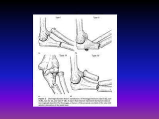
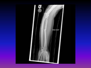
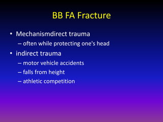
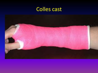
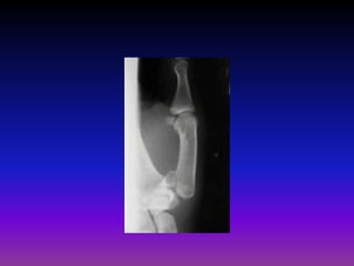
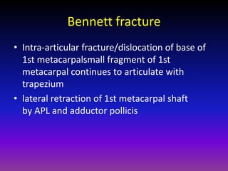
UPPER LIMB FRACTURES

                   D VA U SH R A
                    r RN AM
    RESIDENT IN ORTHOPEDICS AND TRAUMATOLOGY
                     OSMANIA GENERAL HOSPITAL
Topics
•   Fractures of proximal end of radius.
•   Fractures of proximal end of ulna.
•   Fractures of both bones of the forearm.
•   Fractures around wrist
•   Injuries of hand.
Pulled elbow
• Traumatic subluxation
• 2-6yrs age
• Jerk on forearm
• Pain in elbow n tenderness in proximal radius
• Radiographs normal
• Rx simple manipulation of FA into supination with
  elbow stabilised
• Palpable click, pain decreases n normal
  movement restored.
•
Proximal End of RADIUS
Types
• Fracture Head.
• Fracture neck.
• Epiphyseal injuries.
Fracture head of radius
• Mechanism: Forcible valgus strain, head
  against capitellum, loose fragments into joint.
• Masons classification
Symptoms
• Pain on the outside of the elbow
• Swelling in the elbow joint
• Difficulty in bending or straightening the
  elbow accompanied by pain
• Inability or difficulty in turning the forearm
  (palm up to palm down or vice versa)
Treatment
• Type I Fractures
• Type I fractures are generally small, like cracks,
  and the bone pieces remain fitted together.
• The fracture may not be visible on initial X-rays,
  but can usually be seen if the X-ray is taken three
  weeks after the injury.
• Nonsurgical treatment involves using a splint or
  sling for a few days, followed by early motion.
• If too much motion is attempted too quickly, the
  bones may shift and become displaced.
Treatment
• Type II Fractures
• Type II fractures are slightly displaced and involve a larger piece of
  bone.
• If displacement is minimal, splinting for one to two weeks, followed
  by range of motion exercises, is usually successful.
• Small fragments may be surgically removed.
• If the fragment is large and can be fitted back to the bone, the
  orthopaedic surgeon will first attempt to fix it with pins or screws. If
  this is not possible, however, the surgeon will remove the broken
  pieces or the radial head.
• For older, less active individuals, the surgeon may simply remove
  the broken piece, or perhaps the entire radial head.
• The surgeon will also correct any other soft-tissue injury, such as a
  torn ligament.
• Type III Fractures
• Type III fractures have multiple broken pieces of bone,
  which cannot be fitted back together for healing.
• Usually, there is also significant damage to the joint
  and ligaments.
• Surgery is always required to remove the broken bits of
  bone, including the radial head, and repair the soft-
  tissue damage.
• Early movement to stretch and bend the elbow is
  necessary to avoid stiffness.
• A prosthesis (artificial radial head) can be used to
  prevent deformity if elbow instability is severe.
Complications
• Myositis ossificans
Olecranon fracture
• Mechanism
• A direct blow. This can happen in a fall (landing
  directly on the elbow) or by being struck by a
  hard object (baseball bat, dashboard of a car
  during a crash).
• An indirect fracture. This can happen by landing
  on an outstretched arm. The person lands on the
  wrist with the elbow locked out straight. The
  triceps muscle on the back of the upper arm help
  "pull" the olecranon off of the ulna.
Symptoms

•   Sudden, intense pain
•   Inability to straighten elbow
•   Swelling over the bone site
•   Bruising around the elbow
•   Tenderness to the touch
•   Numbness in one or more fingers
•   Pain with movement of the joint
• Radiographs
  – recommended views
       • AP/lateral radiographs

           – true lateral essential for determination of fracture pattern
  – additional views
       • radiocapitellar may be helpful for
           – radial head fracture
           – capitellar shear fracture
• CT
  – may be useful for preoperative planning in
    comminuted fractures
Nonoperative

• immobilization
  – indications
     • nondisplaced fractures
     • displaced fracture is low demand, elderly individuals
• Technique
• immobilization in 45-90 degrees of flexion for
  3 weeks
• begin motion at 3 weeks
Operative
•   tension band technique
     – indications
           •   transverse fracture with no comminution
     – outcomes
           •   excellent results with appropriate indications
•   technique
     – converts distraction force of triceps into a compressive force
     – engaging anterior cortex of ulna with Kirschner wires may prevent wire migration
     – avoid overpenetration of wires through anterior cortex
           •   may injury anterior interosseous nerve (AIN)
           •   may lead to decreased forearm rotation
     – use 18-gauge wire in figure-of-eight fashion through drill holes in ulna
•   cons
     – high % of second surgeries for hardware removal (40-80%)
     – does not provide axial stability in comminuted fractures
Olecranon Fracture


     ORIF with Tension Band Wiring
• plate and screw fixation indications
  – comminuted fractures
  – Monteggia fractures
  – fracture-dislocations
  – oblique fractures that extend distal to coronoid
• technique
  – place plate on dorsal (tension) side
  – oblique fractures benefit from lag screws in addition to
    plate fixation
  – one-third tubular plates may not provide sufficient
    strength in comminuted fractures
  – may advance distal triceps tendon over plate to avoid
    hardware prominence
• pros
  – more stable than tension band technique
• cons
  – 20% need second surgery for plate removal
• intramedullary fixation
indications
   – transverse fracture with no comminution (same as tension band
     technique)
• excision and triceps advancement
indications
   – elderly patients with osteoporotic bone
   – fracture must involve <50% of joint surface
   – nonunions
• outcomes
   – salvage procedure that leads to decreased extension strength
   – may result in instability if ligamentous injury is not diagnosed
     before operation
Complications
• Symptomatic hardware
   – most frequent reported complication
• Stiffness
   – occurs in ~50% of patients
   – usually doesn't alter functional capabilities
• Heterotopic ossification
   – more common with associated head injury
• Posttraumatic arthritis
• Nonunion
   – rare
• Ulnar nerve symptoms
• Anterior interosseous nerve injury
• Loss of extension strength
Terrible triad of elbow
• A traumatic injury pattern of the elbow
  characterized by
  – posterolateral dislocation
  – radial head fx
  – coronoid fracture
• Mechanism is fall on extended arm that leads to
  – valgus stress produces posterolateral dislocation
  – structures of elbow fail from lateral to medial
     • anterior bundle of MCL last to fail
     • LCL disrupted in most cases
• Nonoperativeimmobilize in 90 deg of flexion
  for 7-10 days
• Operativeacute radial head
  stabilization, coronoid ORIF, and LCL
  reconstruction, MCL reconstuction if needed
Monteggia fracture
• Injury defined as
  – proximal 1/3 ulnar fracture with associated radial
    head dislocation
• Epidemiology
  – rare in adults
  – more common in children with peak incidence
    between 4 and 10 years of age
     • different treatment protocol for children
classification
• Symptoms
   – pain and swelling at elbow joint
• Physical exam
   – inspection
       • may or may not be obvious dislocation at radiocapitellar joint
       • should include skin integrity
   – ROM & instability
       • may be loss of ROM at elbow due to dislocation
   – neurovascular exam
       • PIN neuropathy
           –   radial deviation of hand with wrist extension
           –   weakness of thumb extension
           –   weakness of MCP extension
           –   most likely nerve injury
• Radiographs
  – recommended view
     • AP and Lateral of elbow, wrist, and forearm
• CT scan
  – helpful in fractures involving
    coronoid, olecranon, and radial head
Rx
• Operative
• ORIF of ulna shaft fracture
• ORIF of ulna shaft fracture, open reduction of
  radial head
• IM Nailing of ulna
complications
• PIN neuropathy
  – up to 10% in acute injuries
  – treatment
     • observation for 2-3 months
        – spontaneously resolves in most cases
        – if no improvement obtain nerve conduction studies

• Malunion with radial head dislocation
  – caused by failure to obtain anatomic alignment of ulna
  – treatment
     • ulnar osteotomy and open reduction of the radial head
Fracture both bones of forearm
BB FA Fracture
• Mechanismdirect trauma
  – often while protecting one's head
• indirect trauma
  – motor vehicle accidents
  – falls from height
  – athletic competition
•   closed versus open
•   location
•   comminuted, segmental, multifragmented
•   displacement
•   angulation
•   rotational alignment
• Radiographsrecommended views
  – AP and lateral views of the forearm
• additional views
  – oblique forearm views for further fracture
    definition
  – ipsilateral wrist and elbow
     • to evaluate for associated fractures or dislocation
     • radial head must be aligned with the capitellum on all
       views
Nonoperative
• functional fx brace with good interosseous
  mold
  – indications
     • isolated nondisplaced or distal 2/3 ulna shaft
       fx (nightstick fx) with
        – < 50% displacement and
        – < 10° of angulation
  – outcomes
     • union rates > 96%
     • acceptable to fix surgically due to long time to union
Operative
•   ORIF without bone grafting
•   ORIF with bone grafting
•   external fixation
•   IM nailing
ORIF with DCP
CRIF or ORIF with Rush/ Square Nail
Complications
•   Synostosis
•   Infection
•   Compartment syndrome
•   Nonunion
•   Malunion
•   Neurovascular injury
    Refracture
Galeazzi Fractures
• Defined as
• distal 1/3 radius shaft fx AND
• associated distal radioulnar joint (DRUJ) injury
• Mechanism
• direct wrist trauma
  – typically dorsolateral aspect
• fall onto outstretched hand with forearm in
  pronation
• Symptoms
  – pain, swelling, deformity
• Physical exam
  – point tenderness over fracture site
  – ROM
     • test forearm supination and pronation for instability
  – DRUJ stress
     • causes wrist or midline forearm pain
• Radiographsrecomended views
  – AP and lateral views of forearm, elbow, and wrist
• findings
  – signs of DRUJ injury
     •   ulnar styloid fx
     •   widening of joint on AP view
     •   dorsal or volar displacement on lateral view
     •   radial shortening (≥5mm)
Rx
• Operative
• ORIF of radius with reduction and
  stabilization of DRUJ
  – indications
     • all cases, as anatomic reduction of DRUJ is required
     • acute operative treatment far superior to late
       reconstruction
Complications
• Compartment syndrome

• Neurovascular injury

• Refracture

• Nonunion
• Malunion
• DRUJ subluxation
Monteggia Fracture Dislocation
        Dislocation


                      Fracture
Dislocation


                        Fracture


GALEAZZI FRACTURE DISLOCATION
Distal Radius Fractures

• Most common orthopaedic injury with a bimodal
  distribution
   – younger patients - high energy
   – older patients - low energy / falls
• 50% intra-articular
• Associated injuries
   – DRUJ injuries must be evaluated
   – radial styloid fx - indication of higher energy
• Osteoporosis
   – high incidence of distal radius fractures in women >50
   – distal radius fractures are a predictor of subsequent fractures
       • DEXA scan is recommended in woman with a distal radius fracture
Dinner fork Deformity
Colles’ Fracture

• Transverse fracture at the
  cortico-cancellous junction of
  distal radius often associated
  with ulnar styloid fracture
Colles’ fracture

 Displacements
• Impaction
• Dorsal shift
• Dorsal tilt
• Radial shift
• Radial tilt
• Supination
Eponyms
 Die-     A depressed fracture of the lunate fossa of
punch       the articular surface of the distal radius
 fxs
Barton'   Fx dislocation of radiocarpal joint with intra-
 s fx      articular fx involving the volar or dorsal lip
                (volar Barton or dorsal Barton fx)
Chauff                  Radial styloid fx
                                                            x
er's fx
Colles'      Low energy, dorsally displaced, extra-
  fx                      articular fx
Smith's   Low energy, volar displaced, extra-articular
  fx                          fx
• Successful outcomes correlate with
  – accuracy of articular reduction
  – restoration of anatomic relationships
  – early efforts to regain motion of wrist and fingers
• Nonoperative
  – closed reduction and cast immobilization
     • indications
         – extra-articular
         – <5mm radial shortening
         – dorsal angulation <5° or within 20° of contralateral distal radius
     • technique (see below)
• Indications
   – most extra-articular fxs
• Technique
   – rehabilitation
       • no significant benefit of physical therapy over home exercises for
         simple distal radius fractures treated with cast immobilization
• Outcomes
   – repeat closed reductions have 50% less than satisfactory results
• Complications
   – acute carpal tunnel syndrome
       • (see complications below)
   – EPL rupture
       • (see complications below)
Colles cast
•   Operative
•   surgical fixation
•   CRPP,
•    External Fixation,
•    ORIF)
Complications
• Median nerve neuropathy (CTS)
  Ulnar nerve neuropathy
  EPL rupture
  Radiocarpal arthrosis (2-30%)
  Malunion and Nonunion
  ECU or EDM entrapment
  Compartment syndrome
• RSD/CRPS
Treatment
• Conservative most of the cases
   Manual reduction and below elbow cast

Techinque of reduction
        Disimpaction
        Palmar flexion
        Ulnar deviation
        Pronation

• Surgery- Unstable/Communited/intraarticular-
  ORIF or External Fixation
ORIF
K wire fixation
External fixation
Barton’s fracture
• Fracture of distal end of radius involving
  articular surface

• Types
      Dorsal barton
      Volar Barton
Smith’s Fracture
• Reversed Colles’ fracture
• GARDEN SPADE DEFORMITY


  TREATMENT:
• Reduction held in 30 degree Dorsiflexion of
  wrist and supination of Forearm with above
  elbow plaster
Pain in the snuff box after FOOSH
Scaphoid fracture
• Scaphoid is most commonly fractured carpal bone.
• Most common mechanism is axial load across hyper-
  extended and radially deviated wrist.
   – common in contact sports
• Incidence of fracture by location
   – waist -65%
   – proximal third - 25%
   – distal third - 10%
       • distal pole is most common location in kids due to ossification
         sequence
• Incidence of AVN with fracture location
   – proximal 5th AVN rate of 100%
   – proximal 3rd AVN rate of 33%
Blood supply
• major blood supply is dorsal carpal
  branch (branch of the radial artery)
  – enters scaphoid in a nonarticular ridge on the
    dorsal surface and supplies proximal 80% of
    scaphoid via retrograde blood flow
• minor blood supply from superficial palmar
  arch (branch of volar radial artery)
  – enters distal tubercle and supplies distal 20% of
    scaphoid
• Radiographs should include
  – AP and lateral
  – AP view of the scaphoid with the hand in ulnar
    deviation
  – 45° pronation view
• Bone scan
   – effective to diagnose ocult fractures
        • specificity of 98%, and sensitivity of 100%, PPV 85% to 93% when done at 72
          hours
        • positive within 24 hours, perform at 72 hours
• MRI
   – effective diagnose ocult fractures
   – allows immediate identification of fractures and ligamentous injuries
     in addition to assessment of vascular status of bone (vascularity of
     proximal pole)
• CT scan with 1mm cuts
   – less effective than bone scan and MRI to diagnose occult fracture
   – can be used to evaluate location of fracture, size of fragments, extent
     of collapse, and progression of nonunion
Scaphoid Fracture
• Most commonly fractured of carpal bones
• Any fracture at the waist or proximal third of
  bone cuts off blood supply to the proximal pole
  leading to AVN of prox pole or Non Union of
  fracture
 TREATMENT
 Scaphoid Cast immobilsation for undisplaced
  fracture
 ORIF with HERBERT’s screw for displaced fracture
Scaphoid Cast
Herbert Screw Fixation
Intraarticular # base of 1st Metacarpal with
       carpometacarpal dislocation
A K A Bennett’s Fracture Dislocation
Bennett fracture
• Intra-articular fracture/dislocation of base of
  1st metacarpalsmall fragment of 1st
  metacarpal continues to articulate with
  trapezium
• lateral retraction of 1st metacarpal shaft
  by APL and adductor pollicis
Treatment
• closed reduction & cast immobilization
   – indications
      • nondisplaced fractures
   – technique
      • reduction maneuver with traction, extension, pronation, and
        abduction
• reduction and percutaneous K wire stabilization to
  adjacent metacarpals
   – indications
      • displaced fractures in which a adequare reduction is obtains
• ORIF
   – indications
      • reduction and percutaneous K wire stabilization to adjacent
        metacarpals
Often requires ORIF with K wires
ROLANDO’s fracture

• Communited Intra-articular fracture of base of
  first metacarpal
• Similar to a Bennett fracture but more extensive
  comminution and displacement
  – less common than Bennet's fx
  – worse prognosis
• Treatment
  – ORIF
     • indications
         – most cases
Mallet finger
• A finger deformity caused by disruption of
  the terminal extensor tendon distal to DIP joint
   – the disruption may be bony or tendinous
• Mechanism
   – traumatic impaction blow
      • usually caused by a traumatic impaction blow to the tip of
        the finger in the extended position.
      • forces the DIP joint into forced flexion
   – dorsal laceration
      • a less common mechanism of injury is a sharp or crushing-
        type laceration to the dorsal DIP joint
Mallet finger

• Flexion deformity of DIP jt due to
  injury of extensor digitorum
  tendon often with a chip of bone
Rx
• Nonoperativeextension splinting of DIP joint
  for 6-8 weeks
• OperativeCRPP vs ORIF
• surgical reconstruction of terminal tendon
Upperlimb fractures bpt

Upperlimb fractures bpt

  • 1.
    UPPER LIMB FRACTURES D VA U SH R A r RN AM RESIDENT IN ORTHOPEDICS AND TRAUMATOLOGY OSMANIA GENERAL HOSPITAL
  • 2.
    Topics • Fractures of proximal end of radius. • Fractures of proximal end of ulna. • Fractures of both bones of the forearm. • Fractures around wrist • Injuries of hand.
  • 4.
    Pulled elbow • Traumaticsubluxation • 2-6yrs age • Jerk on forearm • Pain in elbow n tenderness in proximal radius • Radiographs normal • Rx simple manipulation of FA into supination with elbow stabilised • Palpable click, pain decreases n normal movement restored. •
  • 5.
  • 6.
    Types • Fracture Head. •Fracture neck. • Epiphyseal injuries.
  • 7.
    Fracture head ofradius • Mechanism: Forcible valgus strain, head against capitellum, loose fragments into joint. • Masons classification
  • 8.
    Symptoms • Pain onthe outside of the elbow • Swelling in the elbow joint • Difficulty in bending or straightening the elbow accompanied by pain • Inability or difficulty in turning the forearm (palm up to palm down or vice versa)
  • 9.
    Treatment • Type IFractures • Type I fractures are generally small, like cracks, and the bone pieces remain fitted together. • The fracture may not be visible on initial X-rays, but can usually be seen if the X-ray is taken three weeks after the injury. • Nonsurgical treatment involves using a splint or sling for a few days, followed by early motion. • If too much motion is attempted too quickly, the bones may shift and become displaced.
  • 10.
    Treatment • Type IIFractures • Type II fractures are slightly displaced and involve a larger piece of bone. • If displacement is minimal, splinting for one to two weeks, followed by range of motion exercises, is usually successful. • Small fragments may be surgically removed. • If the fragment is large and can be fitted back to the bone, the orthopaedic surgeon will first attempt to fix it with pins or screws. If this is not possible, however, the surgeon will remove the broken pieces or the radial head. • For older, less active individuals, the surgeon may simply remove the broken piece, or perhaps the entire radial head. • The surgeon will also correct any other soft-tissue injury, such as a torn ligament.
  • 11.
    • Type IIIFractures • Type III fractures have multiple broken pieces of bone, which cannot be fitted back together for healing. • Usually, there is also significant damage to the joint and ligaments. • Surgery is always required to remove the broken bits of bone, including the radial head, and repair the soft- tissue damage. • Early movement to stretch and bend the elbow is necessary to avoid stiffness. • A prosthesis (artificial radial head) can be used to prevent deformity if elbow instability is severe.
  • 12.
  • 14.
    Olecranon fracture • Mechanism •A direct blow. This can happen in a fall (landing directly on the elbow) or by being struck by a hard object (baseball bat, dashboard of a car during a crash). • An indirect fracture. This can happen by landing on an outstretched arm. The person lands on the wrist with the elbow locked out straight. The triceps muscle on the back of the upper arm help "pull" the olecranon off of the ulna.
  • 15.
    Symptoms • Sudden, intense pain • Inability to straighten elbow • Swelling over the bone site • Bruising around the elbow • Tenderness to the touch • Numbness in one or more fingers • Pain with movement of the joint
  • 17.
    • Radiographs – recommended views • AP/lateral radiographs – true lateral essential for determination of fracture pattern – additional views • radiocapitellar may be helpful for – radial head fracture – capitellar shear fracture • CT – may be useful for preoperative planning in comminuted fractures
  • 18.
    Nonoperative • immobilization – indications • nondisplaced fractures • displaced fracture is low demand, elderly individuals • Technique • immobilization in 45-90 degrees of flexion for 3 weeks • begin motion at 3 weeks
  • 19.
    Operative • tension band technique – indications • transverse fracture with no comminution – outcomes • excellent results with appropriate indications • technique – converts distraction force of triceps into a compressive force – engaging anterior cortex of ulna with Kirschner wires may prevent wire migration – avoid overpenetration of wires through anterior cortex • may injury anterior interosseous nerve (AIN) • may lead to decreased forearm rotation – use 18-gauge wire in figure-of-eight fashion through drill holes in ulna • cons – high % of second surgeries for hardware removal (40-80%) – does not provide axial stability in comminuted fractures
  • 20.
    Olecranon Fracture ORIF with Tension Band Wiring
  • 21.
    • plate andscrew fixation indications – comminuted fractures – Monteggia fractures – fracture-dislocations – oblique fractures that extend distal to coronoid
  • 22.
    • technique – place plate on dorsal (tension) side – oblique fractures benefit from lag screws in addition to plate fixation – one-third tubular plates may not provide sufficient strength in comminuted fractures – may advance distal triceps tendon over plate to avoid hardware prominence • pros – more stable than tension band technique • cons – 20% need second surgery for plate removal
  • 24.
    • intramedullary fixation indications – transverse fracture with no comminution (same as tension band technique) • excision and triceps advancement indications – elderly patients with osteoporotic bone – fracture must involve <50% of joint surface – nonunions • outcomes – salvage procedure that leads to decreased extension strength – may result in instability if ligamentous injury is not diagnosed before operation
  • 25.
    Complications • Symptomatic hardware – most frequent reported complication • Stiffness – occurs in ~50% of patients – usually doesn't alter functional capabilities • Heterotopic ossification – more common with associated head injury • Posttraumatic arthritis • Nonunion – rare • Ulnar nerve symptoms • Anterior interosseous nerve injury • Loss of extension strength
  • 26.
    Terrible triad ofelbow • A traumatic injury pattern of the elbow characterized by – posterolateral dislocation – radial head fx – coronoid fracture • Mechanism is fall on extended arm that leads to – valgus stress produces posterolateral dislocation – structures of elbow fail from lateral to medial • anterior bundle of MCL last to fail • LCL disrupted in most cases
  • 27.
    • Nonoperativeimmobilize in90 deg of flexion for 7-10 days • Operativeacute radial head stabilization, coronoid ORIF, and LCL reconstruction, MCL reconstuction if needed
  • 28.
  • 29.
    • Injury definedas – proximal 1/3 ulnar fracture with associated radial head dislocation • Epidemiology – rare in adults – more common in children with peak incidence between 4 and 10 years of age • different treatment protocol for children
  • 30.
  • 31.
    • Symptoms – pain and swelling at elbow joint • Physical exam – inspection • may or may not be obvious dislocation at radiocapitellar joint • should include skin integrity – ROM & instability • may be loss of ROM at elbow due to dislocation – neurovascular exam • PIN neuropathy – radial deviation of hand with wrist extension – weakness of thumb extension – weakness of MCP extension – most likely nerve injury
  • 32.
    • Radiographs – recommended view • AP and Lateral of elbow, wrist, and forearm • CT scan – helpful in fractures involving coronoid, olecranon, and radial head
  • 33.
    Rx • Operative • ORIFof ulna shaft fracture • ORIF of ulna shaft fracture, open reduction of radial head • IM Nailing of ulna
  • 34.
    complications • PIN neuropathy – up to 10% in acute injuries – treatment • observation for 2-3 months – spontaneously resolves in most cases – if no improvement obtain nerve conduction studies • Malunion with radial head dislocation – caused by failure to obtain anatomic alignment of ulna – treatment • ulnar osteotomy and open reduction of the radial head
  • 36.
  • 37.
    BB FA Fracture •Mechanismdirect trauma – often while protecting one's head • indirect trauma – motor vehicle accidents – falls from height – athletic competition
  • 38.
    closed versus open • location • comminuted, segmental, multifragmented • displacement • angulation • rotational alignment
  • 39.
    • Radiographsrecommended views – AP and lateral views of the forearm • additional views – oblique forearm views for further fracture definition – ipsilateral wrist and elbow • to evaluate for associated fractures or dislocation • radial head must be aligned with the capitellum on all views
  • 40.
    Nonoperative • functional fxbrace with good interosseous mold – indications • isolated nondisplaced or distal 2/3 ulna shaft fx (nightstick fx) with – < 50% displacement and – < 10° of angulation – outcomes • union rates > 96% • acceptable to fix surgically due to long time to union
  • 41.
    Operative • ORIF without bone grafting • ORIF with bone grafting • external fixation • IM nailing
  • 43.
    ORIF with DCP CRIFor ORIF with Rush/ Square Nail
  • 44.
    Complications • Synostosis • Infection • Compartment syndrome • Nonunion • Malunion • Neurovascular injury Refracture
  • 45.
  • 46.
    • Defined as •distal 1/3 radius shaft fx AND • associated distal radioulnar joint (DRUJ) injury
  • 47.
    • Mechanism • directwrist trauma – typically dorsolateral aspect • fall onto outstretched hand with forearm in pronation
  • 48.
    • Symptoms – pain, swelling, deformity • Physical exam – point tenderness over fracture site – ROM • test forearm supination and pronation for instability – DRUJ stress • causes wrist or midline forearm pain
  • 49.
    • Radiographsrecomended views – AP and lateral views of forearm, elbow, and wrist • findings – signs of DRUJ injury • ulnar styloid fx • widening of joint on AP view • dorsal or volar displacement on lateral view • radial shortening (≥5mm)
  • 50.
    Rx • Operative • ORIFof radius with reduction and stabilization of DRUJ – indications • all cases, as anatomic reduction of DRUJ is required • acute operative treatment far superior to late reconstruction
  • 51.
    Complications • Compartment syndrome •Neurovascular injury • Refracture • Nonunion • Malunion • DRUJ subluxation
  • 53.
    Monteggia Fracture Dislocation Dislocation Fracture
  • 54.
    Dislocation Fracture GALEAZZI FRACTURE DISLOCATION
  • 55.
    Distal Radius Fractures •Most common orthopaedic injury with a bimodal distribution – younger patients - high energy – older patients - low energy / falls • 50% intra-articular • Associated injuries – DRUJ injuries must be evaluated – radial styloid fx - indication of higher energy • Osteoporosis – high incidence of distal radius fractures in women >50 – distal radius fractures are a predictor of subsequent fractures • DEXA scan is recommended in woman with a distal radius fracture
  • 56.
  • 58.
    Colles’ Fracture • Transversefracture at the cortico-cancellous junction of distal radius often associated with ulnar styloid fracture
  • 59.
    Colles’ fracture Displacements •Impaction • Dorsal shift • Dorsal tilt • Radial shift • Radial tilt • Supination
  • 60.
    Eponyms Die- A depressed fracture of the lunate fossa of punch the articular surface of the distal radius fxs Barton' Fx dislocation of radiocarpal joint with intra- s fx articular fx involving the volar or dorsal lip (volar Barton or dorsal Barton fx) Chauff Radial styloid fx x er's fx Colles' Low energy, dorsally displaced, extra- fx articular fx Smith's Low energy, volar displaced, extra-articular fx fx
  • 61.
    • Successful outcomescorrelate with – accuracy of articular reduction – restoration of anatomic relationships – early efforts to regain motion of wrist and fingers • Nonoperative – closed reduction and cast immobilization • indications – extra-articular – <5mm radial shortening – dorsal angulation <5° or within 20° of contralateral distal radius • technique (see below)
  • 63.
    • Indications – most extra-articular fxs • Technique – rehabilitation • no significant benefit of physical therapy over home exercises for simple distal radius fractures treated with cast immobilization • Outcomes – repeat closed reductions have 50% less than satisfactory results • Complications – acute carpal tunnel syndrome • (see complications below) – EPL rupture • (see complications below)
  • 64.
  • 65.
    Operative • surgical fixation • CRPP, • External Fixation, • ORIF)
  • 66.
    Complications • Median nerveneuropathy (CTS) Ulnar nerve neuropathy EPL rupture Radiocarpal arthrosis (2-30%) Malunion and Nonunion ECU or EDM entrapment Compartment syndrome • RSD/CRPS
  • 67.
    Treatment • Conservative mostof the cases Manual reduction and below elbow cast Techinque of reduction  Disimpaction  Palmar flexion  Ulnar deviation  Pronation • Surgery- Unstable/Communited/intraarticular- ORIF or External Fixation
  • 68.
  • 69.
  • 70.
  • 73.
    Barton’s fracture • Fractureof distal end of radius involving articular surface • Types Dorsal barton Volar Barton
  • 76.
    Smith’s Fracture • ReversedColles’ fracture • GARDEN SPADE DEFORMITY TREATMENT: • Reduction held in 30 degree Dorsiflexion of wrist and supination of Forearm with above elbow plaster
  • 78.
    Pain in thesnuff box after FOOSH
  • 80.
    Scaphoid fracture • Scaphoidis most commonly fractured carpal bone. • Most common mechanism is axial load across hyper- extended and radially deviated wrist. – common in contact sports • Incidence of fracture by location – waist -65% – proximal third - 25% – distal third - 10% • distal pole is most common location in kids due to ossification sequence • Incidence of AVN with fracture location – proximal 5th AVN rate of 100% – proximal 3rd AVN rate of 33%
  • 81.
    Blood supply • majorblood supply is dorsal carpal branch (branch of the radial artery) – enters scaphoid in a nonarticular ridge on the dorsal surface and supplies proximal 80% of scaphoid via retrograde blood flow • minor blood supply from superficial palmar arch (branch of volar radial artery) – enters distal tubercle and supplies distal 20% of scaphoid
  • 82.
    • Radiographs shouldinclude – AP and lateral – AP view of the scaphoid with the hand in ulnar deviation – 45° pronation view
  • 84.
    • Bone scan – effective to diagnose ocult fractures • specificity of 98%, and sensitivity of 100%, PPV 85% to 93% when done at 72 hours • positive within 24 hours, perform at 72 hours • MRI – effective diagnose ocult fractures – allows immediate identification of fractures and ligamentous injuries in addition to assessment of vascular status of bone (vascularity of proximal pole) • CT scan with 1mm cuts – less effective than bone scan and MRI to diagnose occult fracture – can be used to evaluate location of fracture, size of fragments, extent of collapse, and progression of nonunion
  • 85.
    Scaphoid Fracture • Mostcommonly fractured of carpal bones • Any fracture at the waist or proximal third of bone cuts off blood supply to the proximal pole leading to AVN of prox pole or Non Union of fracture TREATMENT  Scaphoid Cast immobilsation for undisplaced fracture  ORIF with HERBERT’s screw for displaced fracture
  • 86.
  • 87.
  • 89.
    Intraarticular # baseof 1st Metacarpal with carpometacarpal dislocation A K A Bennett’s Fracture Dislocation
  • 91.
    Bennett fracture • Intra-articularfracture/dislocation of base of 1st metacarpalsmall fragment of 1st metacarpal continues to articulate with trapezium • lateral retraction of 1st metacarpal shaft by APL and adductor pollicis
  • 92.
    Treatment • closed reduction& cast immobilization – indications • nondisplaced fractures – technique • reduction maneuver with traction, extension, pronation, and abduction • reduction and percutaneous K wire stabilization to adjacent metacarpals – indications • displaced fractures in which a adequare reduction is obtains • ORIF – indications • reduction and percutaneous K wire stabilization to adjacent metacarpals
  • 93.
    Often requires ORIFwith K wires
  • 96.
    ROLANDO’s fracture • CommunitedIntra-articular fracture of base of first metacarpal • Similar to a Bennett fracture but more extensive comminution and displacement – less common than Bennet's fx – worse prognosis • Treatment – ORIF • indications – most cases
  • 101.
    Mallet finger • Afinger deformity caused by disruption of the terminal extensor tendon distal to DIP joint – the disruption may be bony or tendinous • Mechanism – traumatic impaction blow • usually caused by a traumatic impaction blow to the tip of the finger in the extended position. • forces the DIP joint into forced flexion – dorsal laceration • a less common mechanism of injury is a sharp or crushing- type laceration to the dorsal DIP joint
  • 102.
    Mallet finger • Flexiondeformity of DIP jt due to injury of extensor digitorum tendon often with a chip of bone
  • 105.
    Rx • Nonoperativeextension splintingof DIP joint for 6-8 weeks • OperativeCRPP vs ORIF • surgical reconstruction of terminal tendon