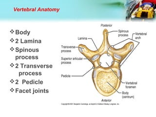
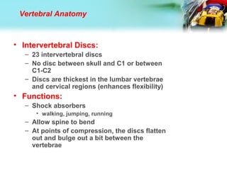
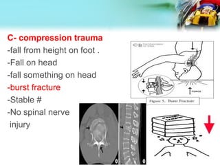
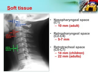
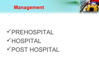
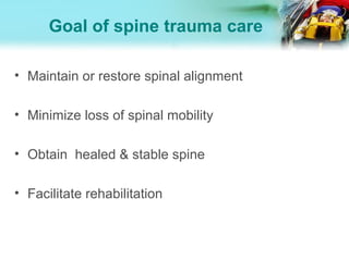
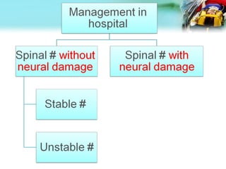
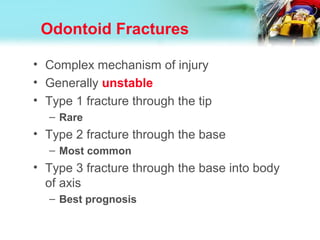
Done by  malika hameed
Supervision by  dr - Anwer Naif
Group D
Spine
fractur
e
Clinical Anatomy
• Vertebral Column:
– Extends from skull
to the pelvis
– 33 total vertebrae:
• Superiorly: 24
individual vertebrae
(separated by
intervertebral discs)
• Inferiorly: 9 fuse to
form 2 composite
bones
– Sacrum (5)
– Coccyx (4)
Clinical Anatomy
• Vertebral Column:
– Functions:
1. Transmits weight
of the trunk to the
lower limbs
2. Surrounds/protect
s spinal cord
3. Attachment point
for the ribs and
muscles of neck
and back
Vertebral Anatomy
Body
2 Lamina
Spinous
process
2 Transverse
process
2 Pedicle
Facet joints
Vertebral Anatomy
Vertebral Anatomy
• Intervertebral Discs:
– 23 intervertebral discs
– No disc between skull and C1 or between
C1-C2
– Discs are thickest in the lumbar vertebrae
and cervical regions (enhances flexibility)
• Functions:
– Shock absorbers
• walking, jumping, running
– Allow spine to bend
– At points of compression, the discs flatten
out and bulge out a bit between the
vertebrae
Vertebral Anatomy
Clinical Anatomy
Clinical Anatomy
• Major Supporting
Ligaments
– Anterior Longitudinal
Ligament
– runs vertically along
anterior surface of
vertebral bodies
• Neck - Sacrum
• Attaches strongly to both
vertebrae and
intervertebral discs (very
wide)
• Prevents back
hyperextension
Clinical Anatomy
• Major Supporting
Ligaments
– *Posterior Longitudinal
Ligament
– - runs vertically along
posterior surfaces of
vertebral bodies
• Narrower, weaker
• Attaches to
intervertebral discs
• Prevents hyperflexion
• Major Supporting Ligaments
– Ligamentum Flavum - strong
ligament that connects the
laminae of the vertebrae
• Protects the neural elements
and the spinal cord
• Stabilizes the spine to prevent
excessive vertebral body
motion
• Strongest of the spinal
ligaments
• Forms the posterior wall of the
spinal canal with the laminae
• Stretches with forward
bending / recoils in erect
position
Clinical Anatomy
Clinical Anatomy
sporting Ligaments
– Intertransverse Ligament
- located between the
transverse processes
• Cervical region:
consist of a few
irregular, scattered
fibers
• Thoracic region:
rounded cords
connected with deep
muscles of the back
• Lumbar region: thin
and membranous
Clinical Anatomy
Supporting
Ligaments
– Interspinal Ligament
- connect spinous
processes (spans the
entire process)
• Meets the
ligamentum
flavum in front
and the
supraspinal
ligament behind
Clinical Anatomy
Supporting Ligaments
– Supraspinal Ligament
-connects together the
apexes of the spinous
processes
• Extends from 7th
cervical vertebra to
sacrum
• Strong fibrous cord
• At points of attachment
(tips of the spinous
processes) fibrocartilage
is developed in the
ligament
Supraspinal
Ligament
Clinical Anatomy
vertebral column
Significance
Unstable
if middle column + either Anterior or Posterior column
is damaged
Rupture of interspinous ligament is :
- associated with avulsion of spinous process
- Unstable spine
- Further flexion increases neurological injury
• Cervical 40%
• Thoracic 10%
• Lumbar 3%
• Dorso lumbar
35%
Incidence :-
-More in males 20-40 (more
exposure to mechanism of
injury) .
Most frequently injured spinal regions
C5-C7 (Mid cervical)
C1-C2 (Upper cervical)
T12-L2 (Dorso-lumbar)
Mechanism of injury
:
Penetrating trauma as
knives bullets.
Or blunt trauma as car
accident
I. Direct trauma
:
A. Hyper-flexion trauma:
-Fall in bent position.
-Deceleration.
II-indirect Trauma
-Wedge fracture
-Stable
-No spinal cord
injury
B- Hyper-extension injuries
-sudden acceleration
Blow on forehead-
INCLUDE
hangman’s #
# of arch of atlas and axis
ant. Longitudinal tear
-stable except hangman fracture-
-Spinal cord may be damage
C- compression trauma
-fall from height on foot .
-Fall on head
-fall something on head
-burst fracture
-Stable #
-No spinal nerve
injury
D-Shearing (specially rotation)
-rotation cause ligaments Damage
, usually associated with flexion.
As:
-Fall from height with the body twisted
Or
Weight fall asymmetrically onto the back
**Wedge shape # with shearing
of posterior ligament.
spinal usually affect
unstable #
Denis classification
1.Compression fracture (wedge)
2.Flexion compression (Burst)
3.Flexion distraction
4.(Seat belt fracture, Chance fr.)
5.Fracture Dislocation
Classification
• I- according to morphology
• II- according to stability
I- according to morphology
1. Wedge compression fracture:
Hyper-flexion trauma crushes the vertebral body into the shape of a wedge.
2. Fracture dislocation:
Commonly there is damage to the spinal cord and nerve roots.
3. Dislocation:
Pure dislocation without fracture is common in cervical vertebrae because its
articular processes are rather horizontal.
4. Comminuted (burst) fracture:
This is an uncommon injury due to vertical compression of the straight spine
.
5. Avulsion fractures of transverse and spinous
processes:
Not accompanied by neurological injury and require no special
treatment.
II- according to stability
1-Stable fractures: Have intact posterior
ligaments, e.g., wedge compression,
comminuted and avulsion fracture of the
transverse and spinous processes.
2-Unstable fractures: Have torn posterior
ligaments and are liable to injure the
cord and nerve roots, these include fracture
dislocation and pure dislocations
Spinal cord injury
classification
I- According to injury
1-Primary injuries
Occur at time of trauma.
2-Secondary injuries
Occur later due to:
-Swelling
-Ischemia
-Movement of bony fragments
Type of injury:
Concussed
Contused
Compressed
Lacerated
Severity of injuries depends on:
Amount and type of force
Duration of injury
II – According to neurology DAMAGE
Complete Cord Lesions –
Death --aboveC4
-Quadriplegia  below C4
-Paraplegia  thoracic or lumbar level
Incomplete Cord Lesions -
mixed loss
- Anterior sc syndrome
- Posterior sc syndrome
- Central cord syndrome
- Brown sequard’s syndrome
- Cauda equina syndrome
Incomplete cord lesion:
1-Central cord syndrome
Flaccid weakness of arm and spastic
weakness of the leg
2- anterior cord syndrome
complete loss of muscle strength below the
level of injury
loss pain and temperature
3-brown sequard syndrome
Motor deficit on side of lesion while sensory
deficit on the other side
4-Posterior cord syndrome
Loose of light touch and
proprioception
Diagnosis :
History
• Mechanism of injury
• Position of the patient when found
• Transient motor or sensory loss
• Paradoxical breathing
• Seat belt
Physical examination
• A- General examination:
- -Vital sign
- -Look for missed injury
- -look for associated fracture
B-Local examination:
Inspection:
Abrasion - hematoma- deformity
Soft tissue swelling and bruising
Palpation :
Point of spinal tenderness
Gap or Step-off
Spasm of associated muscles
DON’T MOVE
C- neurological
examination
1-Dermatome and
myotome.
2- superficial and deep
reflex .
Is the patient awake or
“unexaminable”?
• What’s the difference ?
– Awake
• ask/answer question
• pain/tenderness
• motor/sensory exam
– Not awake
• you can ask (but they won’t answer)
• can’t assess tenderness
• no motor/sensory exam
OW!
------
“Unexaminable”
≠
“No exam”
Neurogenic Shock
• Temporary loss of autonomic function of the
cord at the level of injury
– results from cervical or high thoracic injury
• Presentation
– Flaccid paralysis distal to injury site
– Loss of autonomic function
• hypotension
• vasodilatation
• loss of bladder and bowel control
• loss of thermoregulation
• warm, pink, dry below injury site
• bradycardia
42
Neurogenic Hypovolemic
Etiology Loss of sympathetic
outflow
Loss of blood volume
Blood
pressure
Hypotension Hypotension
Heart rate Bradycardia Tachycardia
Skin
temperature
Warm Cold
Urine
output
Normal Low
Comparison of neurogenic and
hypovolemic shock
Cervical Spine Imaging Options
– Plain films
• AP, lateral and open mouth view
– Optional: Oblique and Swimmer’s
– CT
• Better for occult fractures
– MRI
• Very good for spinal cord, soft tissue and
ligamentous injuries
– Flexion-Extension Plain Films
• to determine stability
Radiolographic evaluation
X-ray Guidelines (cervical)
ABCDS
• Adequacy, Alignment
• Bone abnormality, Base of skull
• Cartilage
• Disc space
• Soft tissue
Adequacy
• Must visualize entire C-spine
• A film that does not show the
upper border of T1 is
inadequate
• Caudal traction on the arms
may help
• If can not, get swimmer’s view
or CT
Swimmer’s view
Alignment
• The anterior vertebral line,
posterior vertebral line, and
spinolaminar line should
have a smooth curve with
no steps or discontinuities
• Malalignment of the
posterior vertebral bodies is
more significant than that
anteriorly, which may be
due to rotation
• A step-off of >3.5mm is
significant anywhere
Lateral Cervical Spine X-Ray
• Anterior subluxation of
one vertebra on another
indicates facet
dislocation
–< 50% of the width of a
vertebral body 
unilateral facet
dislocation
–> 50%  bilateral facet
dislocation
Bones
Disc
• Disc Spaces
– Should be
uniform
• Assess spaces
between the
spinous
processes
Soft tissue
• Nasopharyngeal space
(C1)
– 10 mm (adult)
• Retropharyngeal space
(C2-C4)
– 5-7 mm
• Retrotracheal space
(C5-C7)
– 14 mm (children)
– 22 mm (adults)
AP C-spine Films
• Spinous processes
should line up
• Disc space should be
uniform
• Vertebral body height
should be uniform.
Check for oblique
fractures.
Open mouth view
• Adequacy: all of: all of
the dens andthe dens and
lateral borders oflateral borders of
C1 & C2C1 & C2
• Alignment: lateral: lateral
masses of C1 andmasses of C1 and
C2C2
• Bone: Inspect dens
for lucent fracture
lines
CT Scan
• Thin cut CT scan should
be used to evaluate
abnormal, suspicious or
poorly visualized areas
on plain film
• The combination of plain
film and directed CT scan
provides a false negative
rate of less than 0.1%
MRI
• Ideally all patients
with abnormal
neurological
examination
should be
evaluated with MRI
scan
Management
• Primary Goal
– Prevent secondary injury
• Immobilization of the spine begins in the initial
assessment
– Treat the spine as a long bone
• Secure joint above and below
– Caution with “partial” spine splinting
Management
PREHOSPITAL
HOSPITAL
POST HOSPITAL
Goal of spine trauma care
• Protect further injury during evaluation and
management
• Identify spine injury or document absence of
spine injury
• Optimize conditions for maximal neurologic
recovery
Goal of spine trauma care
• Maintain or restore spinal alignment
• Minimize loss of spinal mobility
• Obtain healed & stable spine
• Facilitate rehabilitation
Pre-hospital management
1- ensure the ventilation and circulation
:2- CAREFUL transportation OF PATIENT if
we Suspect Spinal Injury
When should we Suspect Spinal Injury
-High speed crash
-Unconscious
-Multiple injuries
-Neurological deficit
-Spinal pain/tenderness
• PROTECTION  PRIORITY
• Detection  Secondary
• Up to 15% of spinal injuries have a second
(possibly non adjacent) fracture elsewhere in the
spine
• Ideally, whole spine should be immobilized in
neutral position on a firm surface
Clinical assessment
• Advance Trauma Life Support (ATLS)
guidelines
• Primary and secondary surveys
• Adequate airway and ventilation are the
most important factors
• Supplemental oxygenation
• Early intubation is critical to limit secondary
injury from hypoxia
Principle of treatment
• Spinal alignment
– deformity/subluxation/dislocation reduction
• Spinal column stability
– unstable  stabilization
• Neurological status
– neurological deficit  decompression
Stable # :
-no reduction
-immobilization with cervical
collar or
plaster jacket for 4-6 weeks .
Unstable #
Unstable cervical #
1- reduction
-close reduction : by skull traction –skin traction
or manipulation
Open reduction
2- fixation:
By cervical collar or halo vest(6-12 weeks)
or internal fixation
or post fusion.
Unstable lumbar #
1- reduction:
By gradual postural reduction  by use of pillow or
sand bags under appropriate spinal level for 12 weeks
or
Open reduction
2- immoilization:
By plaster jacket or
By internal fixation
Local management
-Restore alignment
-Decompress
neural damage
-Stabilize the spine
by fixation and
fusion
-Allow early
mobilization
-Restore alignment by traction
Decompress not retard - recovery
or decrease further injury
-No need for stabilization  most
injury stable
-Mobilization achieved by active
physiotherapy in confined bed pt
General management
1- care of skin:
-Trophic ulcer
-Skin keep dry by regular washing and powder
2- care of bladder:
Decompress by catheter-
Antibiotics if infection occurred
3- care of bowel:
By evacuation by enema and giving low residual diet
4- care of muscles and joints:
By passive movement twice daily
complication
• 1- Spinal cord and/or root injury including roots
of cauda spina
• 2- Shock may be:
• a- Spinal shock
• - Sympathatic affection leading to loss of
vascular tone & bradycardia
• - loss of muscle tone ) venous pooling &
hypovolemia
• b- Hemorrhage lead to true hypovolemia
FRECTURE OF
CERVICAL
Jefferson Fracture
• Burst fracture of C1 ring
• Unstable fracture
• Increased lateral ADI on
lateral film if ruptured
transverse ligament and
displacement of C1 lateral
masses on open mouth view
• Need CT scan
the key radiographic view is
the AP open mouth
Jefferson Fracture
Burst Fracture
• Fracture of C3-C7 from
axial loading
• Spinal cord injury is
common from posterior
displacement of fragments
into the spinal canal
• Unstable
Clay Shoveler’s Fracture
• Flexion fracture of
spinous process
• C7>C6>T1
• Stable fracture
Flexion Teardrop Fracture
• Flexion injury causing a
fracture of the
• anteroinferior portion
of the vertebral body
• Unstable because
usually associated with
posterior ligamentous
injury
Bilateral Facet Dislocation
• Flexion injury
• Subluxation of dislocated
vertebra of greater than
½ the AP diameter of the
vertebral body below it
• High incidence of spinal
cord injury
• Extremely unstable
Hangman’s Fracture
• Extension injury
• Bilateral fractures of
C2 pedicles
(white arrow)
• Anterior dislocation of
C2 vertebral body
(red arrow)
• Unstable
Odontoid Fractures
• Complex mechanism of injury
• Generally unstable
• Type 1 fracture through the tip
– Rare
• Type 2 fracture through the base
– Most common
• Type 3 fracture through the base into body
of axis
– Best prognosis
Odontoid fracture Type I
fracture in superior tip of
the odontoid.
potentially unstable.
It is a relatively rare
fracture.
Odontoid Fracture Type II
Odontoid Fracture Type III
• This is an example of dens type III fracture.
The first image is an odontoid view, which
shows the fracture line extending beyond the
base of the dens.
• The second image is a CT that confirms the
fracture in the body of C2
Question
Which of the following fractures is caused by
hyperextension?
Jefferson fracture.
Hangman's fracture.
Teardrop fracture.
THANK YOU
FOR YOUR ATTENTION

‫Spinal injury

  • 1.
    Done by malika hameed Supervision by dr - Anwer Naif Group D Spine fractur e
  • 2.
    Clinical Anatomy • VertebralColumn: – Extends from skull to the pelvis – 33 total vertebrae: • Superiorly: 24 individual vertebrae (separated by intervertebral discs) • Inferiorly: 9 fuse to form 2 composite bones – Sacrum (5) – Coccyx (4)
  • 3.
    Clinical Anatomy • VertebralColumn: – Functions: 1. Transmits weight of the trunk to the lower limbs 2. Surrounds/protect s spinal cord 3. Attachment point for the ribs and muscles of neck and back
  • 4.
    Vertebral Anatomy Body 2 Lamina Spinous process 2Transverse process 2 Pedicle Facet joints
  • 5.
  • 7.
  • 8.
    • Intervertebral Discs: –23 intervertebral discs – No disc between skull and C1 or between C1-C2 – Discs are thickest in the lumbar vertebrae and cervical regions (enhances flexibility) • Functions: – Shock absorbers • walking, jumping, running – Allow spine to bend – At points of compression, the discs flatten out and bulge out a bit between the vertebrae Vertebral Anatomy
  • 9.
  • 10.
    Clinical Anatomy • MajorSupporting Ligaments – Anterior Longitudinal Ligament – runs vertically along anterior surface of vertebral bodies • Neck - Sacrum • Attaches strongly to both vertebrae and intervertebral discs (very wide) • Prevents back hyperextension
  • 11.
    Clinical Anatomy • MajorSupporting Ligaments – *Posterior Longitudinal Ligament – - runs vertically along posterior surfaces of vertebral bodies • Narrower, weaker • Attaches to intervertebral discs • Prevents hyperflexion
  • 12.
    • Major SupportingLigaments – Ligamentum Flavum - strong ligament that connects the laminae of the vertebrae • Protects the neural elements and the spinal cord • Stabilizes the spine to prevent excessive vertebral body motion • Strongest of the spinal ligaments • Forms the posterior wall of the spinal canal with the laminae • Stretches with forward bending / recoils in erect position Clinical Anatomy
  • 13.
    Clinical Anatomy sporting Ligaments –Intertransverse Ligament - located between the transverse processes • Cervical region: consist of a few irregular, scattered fibers • Thoracic region: rounded cords connected with deep muscles of the back • Lumbar region: thin and membranous
  • 14.
    Clinical Anatomy Supporting Ligaments – InterspinalLigament - connect spinous processes (spans the entire process) • Meets the ligamentum flavum in front and the supraspinal ligament behind
  • 15.
    Clinical Anatomy Supporting Ligaments –Supraspinal Ligament -connects together the apexes of the spinous processes • Extends from 7th cervical vertebra to sacrum • Strong fibrous cord • At points of attachment (tips of the spinous processes) fibrocartilage is developed in the ligament Supraspinal Ligament
  • 16.
  • 17.
  • 18.
    Significance Unstable if middle column+ either Anterior or Posterior column is damaged Rupture of interspinous ligament is : - associated with avulsion of spinous process - Unstable spine - Further flexion increases neurological injury
  • 19.
    • Cervical 40% •Thoracic 10% • Lumbar 3% • Dorso lumbar 35% Incidence :- -More in males 20-40 (more exposure to mechanism of injury) . Most frequently injured spinal regions C5-C7 (Mid cervical) C1-C2 (Upper cervical) T12-L2 (Dorso-lumbar)
  • 20.
  • 21.
    : Penetrating trauma as knivesbullets. Or blunt trauma as car accident I. Direct trauma
  • 22.
    : A. Hyper-flexion trauma: -Fallin bent position. -Deceleration. II-indirect Trauma -Wedge fracture -Stable -No spinal cord injury
  • 23.
    B- Hyper-extension injuries -suddenacceleration Blow on forehead- INCLUDE hangman’s # # of arch of atlas and axis ant. Longitudinal tear -stable except hangman fracture- -Spinal cord may be damage
  • 24.
    C- compression trauma -fallfrom height on foot . -Fall on head -fall something on head -burst fracture -Stable # -No spinal nerve injury
  • 25.
    D-Shearing (specially rotation) -rotationcause ligaments Damage , usually associated with flexion. As: -Fall from height with the body twisted Or Weight fall asymmetrically onto the back **Wedge shape # with shearing of posterior ligament. spinal usually affect unstable #
  • 26.
    Denis classification 1.Compression fracture(wedge) 2.Flexion compression (Burst) 3.Flexion distraction 4.(Seat belt fracture, Chance fr.) 5.Fracture Dislocation
  • 27.
    Classification • I- accordingto morphology • II- according to stability
  • 28.
    I- according tomorphology 1. Wedge compression fracture: Hyper-flexion trauma crushes the vertebral body into the shape of a wedge. 2. Fracture dislocation: Commonly there is damage to the spinal cord and nerve roots. 3. Dislocation: Pure dislocation without fracture is common in cervical vertebrae because its articular processes are rather horizontal. 4. Comminuted (burst) fracture: This is an uncommon injury due to vertical compression of the straight spine . 5. Avulsion fractures of transverse and spinous processes: Not accompanied by neurological injury and require no special treatment.
  • 29.
    II- according tostability 1-Stable fractures: Have intact posterior ligaments, e.g., wedge compression, comminuted and avulsion fracture of the transverse and spinous processes. 2-Unstable fractures: Have torn posterior ligaments and are liable to injure the cord and nerve roots, these include fracture dislocation and pure dislocations
  • 30.
  • 31.
    classification I- According toinjury 1-Primary injuries Occur at time of trauma. 2-Secondary injuries Occur later due to: -Swelling -Ischemia -Movement of bony fragments
  • 32.
    Type of injury: Concussed Contused Compressed Lacerated Severityof injuries depends on: Amount and type of force Duration of injury
  • 33.
    II – Accordingto neurology DAMAGE Complete Cord Lesions – Death --aboveC4 -Quadriplegia  below C4 -Paraplegia  thoracic or lumbar level Incomplete Cord Lesions - mixed loss - Anterior sc syndrome - Posterior sc syndrome - Central cord syndrome - Brown sequard’s syndrome - Cauda equina syndrome
  • 34.
    Incomplete cord lesion: 1-Centralcord syndrome Flaccid weakness of arm and spastic weakness of the leg 2- anterior cord syndrome complete loss of muscle strength below the level of injury loss pain and temperature 3-brown sequard syndrome Motor deficit on side of lesion while sensory deficit on the other side 4-Posterior cord syndrome Loose of light touch and proprioception
  • 35.
  • 36.
    History • Mechanism ofinjury • Position of the patient when found • Transient motor or sensory loss • Paradoxical breathing • Seat belt
  • 37.
    Physical examination • A-General examination: - -Vital sign - -Look for missed injury - -look for associated fracture
  • 38.
    B-Local examination: Inspection: Abrasion -hematoma- deformity Soft tissue swelling and bruising Palpation : Point of spinal tenderness Gap or Step-off Spasm of associated muscles DON’T MOVE C- neurological examination 1-Dermatome and myotome. 2- superficial and deep reflex .
  • 39.
    Is the patientawake or “unexaminable”? • What’s the difference ? – Awake • ask/answer question • pain/tenderness • motor/sensory exam – Not awake • you can ask (but they won’t answer) • can’t assess tenderness • no motor/sensory exam OW! ------
  • 40.
  • 41.
    Neurogenic Shock • Temporaryloss of autonomic function of the cord at the level of injury – results from cervical or high thoracic injury • Presentation – Flaccid paralysis distal to injury site – Loss of autonomic function • hypotension • vasodilatation • loss of bladder and bowel control • loss of thermoregulation • warm, pink, dry below injury site • bradycardia
  • 42.
    42 Neurogenic Hypovolemic Etiology Lossof sympathetic outflow Loss of blood volume Blood pressure Hypotension Hypotension Heart rate Bradycardia Tachycardia Skin temperature Warm Cold Urine output Normal Low Comparison of neurogenic and hypovolemic shock
  • 43.
    Cervical Spine ImagingOptions – Plain films • AP, lateral and open mouth view – Optional: Oblique and Swimmer’s – CT • Better for occult fractures – MRI • Very good for spinal cord, soft tissue and ligamentous injuries – Flexion-Extension Plain Films • to determine stability
  • 44.
    Radiolographic evaluation X-ray Guidelines(cervical) ABCDS • Adequacy, Alignment • Bone abnormality, Base of skull • Cartilage • Disc space • Soft tissue
  • 45.
    Adequacy • Must visualizeentire C-spine • A film that does not show the upper border of T1 is inadequate • Caudal traction on the arms may help • If can not, get swimmer’s view or CT
  • 46.
  • 47.
    Alignment • The anteriorvertebral line, posterior vertebral line, and spinolaminar line should have a smooth curve with no steps or discontinuities • Malalignment of the posterior vertebral bodies is more significant than that anteriorly, which may be due to rotation • A step-off of >3.5mm is significant anywhere
  • 48.
    Lateral Cervical SpineX-Ray • Anterior subluxation of one vertebra on another indicates facet dislocation –< 50% of the width of a vertebral body  unilateral facet dislocation –> 50%  bilateral facet dislocation
  • 49.
  • 50.
    Disc • Disc Spaces –Should be uniform • Assess spaces between the spinous processes
  • 51.
    Soft tissue • Nasopharyngealspace (C1) – 10 mm (adult) • Retropharyngeal space (C2-C4) – 5-7 mm • Retrotracheal space (C5-C7) – 14 mm (children) – 22 mm (adults)
  • 52.
    AP C-spine Films •Spinous processes should line up • Disc space should be uniform • Vertebral body height should be uniform. Check for oblique fractures.
  • 53.
    Open mouth view •Adequacy: all of: all of the dens andthe dens and lateral borders oflateral borders of C1 & C2C1 & C2 • Alignment: lateral: lateral masses of C1 andmasses of C1 and C2C2 • Bone: Inspect dens for lucent fracture lines
  • 54.
    CT Scan • Thincut CT scan should be used to evaluate abnormal, suspicious or poorly visualized areas on plain film • The combination of plain film and directed CT scan provides a false negative rate of less than 0.1%
  • 55.
    MRI • Ideally allpatients with abnormal neurological examination should be evaluated with MRI scan
  • 56.
    Management • Primary Goal –Prevent secondary injury • Immobilization of the spine begins in the initial assessment – Treat the spine as a long bone • Secure joint above and below – Caution with “partial” spine splinting
  • 57.
  • 58.
    Goal of spinetrauma care • Protect further injury during evaluation and management • Identify spine injury or document absence of spine injury • Optimize conditions for maximal neurologic recovery
  • 59.
    Goal of spinetrauma care • Maintain or restore spinal alignment • Minimize loss of spinal mobility • Obtain healed & stable spine • Facilitate rehabilitation
  • 60.
    Pre-hospital management 1- ensurethe ventilation and circulation :2- CAREFUL transportation OF PATIENT if we Suspect Spinal Injury When should we Suspect Spinal Injury -High speed crash -Unconscious -Multiple injuries -Neurological deficit -Spinal pain/tenderness
  • 61.
    • PROTECTION PRIORITY • Detection  Secondary • Up to 15% of spinal injuries have a second (possibly non adjacent) fracture elsewhere in the spine • Ideally, whole spine should be immobilized in neutral position on a firm surface
  • 67.
    Clinical assessment • AdvanceTrauma Life Support (ATLS) guidelines • Primary and secondary surveys • Adequate airway and ventilation are the most important factors • Supplemental oxygenation • Early intubation is critical to limit secondary injury from hypoxia
  • 69.
    Principle of treatment •Spinal alignment – deformity/subluxation/dislocation reduction • Spinal column stability – unstable  stabilization • Neurological status – neurological deficit  decompression
  • 70.
    Stable # : -noreduction -immobilization with cervical collar or plaster jacket for 4-6 weeks .
  • 71.
    Unstable # Unstable cervical# 1- reduction -close reduction : by skull traction –skin traction or manipulation Open reduction 2- fixation: By cervical collar or halo vest(6-12 weeks) or internal fixation or post fusion.
  • 73.
    Unstable lumbar # 1-reduction: By gradual postural reduction  by use of pillow or sand bags under appropriate spinal level for 12 weeks or Open reduction 2- immoilization: By plaster jacket or By internal fixation
  • 77.
    Local management -Restore alignment -Decompress neuraldamage -Stabilize the spine by fixation and fusion -Allow early mobilization -Restore alignment by traction Decompress not retard - recovery or decrease further injury -No need for stabilization  most injury stable -Mobilization achieved by active physiotherapy in confined bed pt
  • 78.
    General management 1- careof skin: -Trophic ulcer -Skin keep dry by regular washing and powder 2- care of bladder: Decompress by catheter- Antibiotics if infection occurred 3- care of bowel: By evacuation by enema and giving low residual diet 4- care of muscles and joints: By passive movement twice daily
  • 79.
    complication • 1- Spinalcord and/or root injury including roots of cauda spina • 2- Shock may be: • a- Spinal shock • - Sympathatic affection leading to loss of vascular tone & bradycardia • - loss of muscle tone ) venous pooling & hypovolemia • b- Hemorrhage lead to true hypovolemia
  • 80.
  • 81.
    Jefferson Fracture • Burstfracture of C1 ring • Unstable fracture • Increased lateral ADI on lateral film if ruptured transverse ligament and displacement of C1 lateral masses on open mouth view • Need CT scan
  • 82.
    the key radiographicview is the AP open mouth Jefferson Fracture
  • 83.
    Burst Fracture • Fractureof C3-C7 from axial loading • Spinal cord injury is common from posterior displacement of fragments into the spinal canal • Unstable
  • 84.
    Clay Shoveler’s Fracture •Flexion fracture of spinous process • C7>C6>T1 • Stable fracture
  • 85.
    Flexion Teardrop Fracture •Flexion injury causing a fracture of the • anteroinferior portion of the vertebral body • Unstable because usually associated with posterior ligamentous injury
  • 86.
    Bilateral Facet Dislocation •Flexion injury • Subluxation of dislocated vertebra of greater than ½ the AP diameter of the vertebral body below it • High incidence of spinal cord injury • Extremely unstable
  • 87.
    Hangman’s Fracture • Extensioninjury • Bilateral fractures of C2 pedicles (white arrow) • Anterior dislocation of C2 vertebral body (red arrow) • Unstable
  • 89.
    Odontoid Fractures • Complexmechanism of injury • Generally unstable • Type 1 fracture through the tip – Rare • Type 2 fracture through the base – Most common • Type 3 fracture through the base into body of axis – Best prognosis
  • 90.
    Odontoid fracture TypeI fracture in superior tip of the odontoid. potentially unstable. It is a relatively rare fracture.
  • 91.
  • 92.
  • 94.
    • This isan example of dens type III fracture. The first image is an odontoid view, which shows the fracture line extending beyond the base of the dens. • The second image is a CT that confirms the fracture in the body of C2
  • 95.
    Question Which of thefollowing fractures is caused by hyperextension? Jefferson fracture. Hangman's fracture. Teardrop fracture.
  • 96.