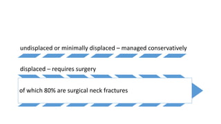
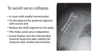
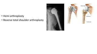
2. Introduction
• Proximal humerus #s account for up to 45% of all humeral fractures.
• The proximal humerus comprises four main osseous segments,
namely the humeral head,
the lesser tuberosity,
the greater tuberosity &
the humeral shaft.
3. The humeral head is connected to the shaft through the anatomical
neck.
Anatomical neck # s are uncommon, but they have a poor prognosis
because of the interference with the vascular supply to the humeral
head.
The surgical neck of the humerus distal to the tuberosities is a common
site for fractures.
4. Incidence
4% to 6% of all fractures
75% of humerus fractures in patients older than age 40
3rd most common type of fragility fracture
5. Incidence varies with age
In young – high energy trauma – mostly a
part of polytrauma
After age 50, Mainly due to osteoporosis
Low energy trauma is sufficient
6. undisplaced or minimally displaced – managed conservatively
displaced – requires surgery
of which 80% are surgical neck fractures
7. Etiology
• The most common mechanism - fall on the outstretched hand from a
standing height
• Seizure activity
• Electrical shock
• Athletic events
• Direct blow to the proximal humerus
9. • The axillary artery is known as “tethered trifurcation” at the level of
the surgical neck.
• Tethered Trifurcation - In the third part of axillary artery where the
vessel is tethered to the humerus by the anterior and posterior
humeral circumflex branches
10. On examination
• Presents immediately after injury
• Shoulder pain
• Swelling and
• Restricted shoulder movements and
• Arm is supported by the other hand.
• Ecchymosis will not appear for 24-48 hours after injury
• If appears within few hours - extensive soft tissue disruption.
11. Careful neurovascular examination is essential as
neurovascular injury can occur in fractures with a
medially displaced shaft of the humerus. Axillary
nerve injury can be diagnosed by the absence of
sensation on the lateral aspect of the proximal
arm (referred as the ‘regiment badge area’).
12. Evaluation for associated injuries
• Ipsilateral scapular, acromion, clavicle
and ribs must be thoroughly examined.
• Auscultation of the lungs to evaluate
pneumothorax and hemopneumothorax
13. Nerve injury
• Axillary nerve and brachial plexus.
• Anterior fracture dislocation with displaced GT fracture – most
commonly associated with axillary nerve injury
• Loss of sensation over the lateral deltoid should alert the examiner to
possible axillary nerve injury.
• Isometric contraction of the deltoid also should be tested.
14. Radiographic Examination
• Neer’s trauma series
• Consists of
• True AP view of the glenohumeral joint
• y-view and
• axillary view
• Velpeau view (Modifications of the
axillary view) or CT scan - to evaluate the
relationship of humeral head to glenoid
• It still is estimated that the initial
treating physician misses 50% of all
fracture dislocations
15. CT Scan
• CT is indicated
• Quantitating the amount of tuberosity displacement
• Extent of articular involvement in head-splitting fractures
• Extent of comminution of associated glenoid fractures.
• 3D reconstruction could be helpful in complex fractures
17. Kocher in 1986
• Classified based on the anatomical level of the fracture
• At anatomical neck
• At metaphyseal region and
• At surgical neck.
• Though it is simplest it does not apply to many complex fracture
patterns commonly encountered
18. Codman in 1934 (4 segment concept)
• He recognized that fractures of proximal humerus
typically produced a combination of four possible
fragments which includes
• Articular surface
• Humeral shaft
• Greater tuberosity and
• Lesser tuberosity
• He hypothesized that fracture lines followed remnant
of old epiphyseal plate - epiphyseal scar
• He concluded that all fractures were some
combination of these different fracture fragments
19.
20. Neer’s 4 part classification in 1970
• Refinement of Codman’s four-segment classification
• Incorporates the concepts of displacement and vascular isolation of
articular segment
• 3 of these segments correspond to ossification centers giving rise to
proximal humerus (one for humeral head and one for each
tuberosity).
• Fusion of these ossification centers at the physis creates a weakened
area that is susceptible to fracture
21. • Regardless of the number of fracture lines present
• Proximal humerus fracture is considered to be undisplaced by Neer’s
criteria when plain radiographs reveal less than 1 cm of displacement
and 45° of angulation of any one fragment with respect to all others.
22.
23. Two part fracture
• may involve
• Anatomic neck
• Surgical neck
• Greater tuberosity or Lesser tuberosity
• When one fragment is displaced at least one centimeter or angulated
45° or more with respect to any of the remaining three fragments
24. Three-part fracture
• Result from displaced fracture of Surgical neck
• In combination with either a displaced greater tuberosity or lesser
tuberosity fracture.
25. Four-part fracture
• Result from displaced fractures of the surgical neck and both
tuberosities
• Any fracture pattern may occur in combination with a glenohumeral
dislocation
26. AO classification
• Emphasizes the vascular supply to the articular segments.
• The system is divided into three categories
• according to the severity of injury and the likelihood of avascular necrosis.
• Type A - least severe with no vascular isolation of the articular segment and
the risk of avascular necrosis is small.
• Type B - more severe fracture with partial isolation of the articular segment
with a low risk of avascular necrosis.
• Type C – most severe with total vascular isolation of the articular segment and
a high risk of avascular necrosis.
28. Non operative
• 85% of proximal humeral fractures are undisplaced or minimally
displaced.
• By immobilizing the arm in a sling for comfort and instituting early
range of motion exercises when pain permits
• Patients with medical illnessess that preclude them from surgery
should also be treated conservatively.
29. Mobilisation
• After 1 week of immobilization - pendulum exercises and gentle
isometric strengthening of biceps and triceps to compress fracture
fragments
• After 3 to 4 weeks - supine passive flexion and passive external rotation
exercises may be added
• 4 to 5 weeks - Overhead pulley exercises
• 6 to 8 weeks - stretching and strengthening
31. Indication for surgery include
• Displacement
• Polytrauma
• Association with other upper extremity fractures
• Vascular injury and
• Open fracture
• Absolute indication for fixations are
1. The fracture dislocation of proximal humerus
2. Head splitting fractures
3. Fractures with neurovascular injuries
32. • Communited fractures and
• Completely displaced surgical neck fractures
• have poor results with nonoperative treatment because they are unstable and
will redisplace after reduction
33. Operative methods
• ORIF + locking plate is the most common type of intervention
• closed reduction with percutaneous pinning
• Hemiarthroplasty
• Proximal humeral nailing and
• Reverse total shoulder arthroplasty
34. Percutaneous pin fixation
• Closed treatment in 2 part
fracture - satisfactory or excellent
outcome if closed reduction can
be achieved and maintained.
• It is useful in selected patients
with unstable fractures.
35. Closed reduction with percutaneous pinning
• Indicated in patients
• With good bone quality
• Noncomminuted or
minimally comminuted
fractures that can be
reduced adequately by
closed means
• Contraindicated in
• Severe comminution
• Osteopenia - are
absolute
contraindications.
36. Advantages Disadvantages
• Avoidance of
devascularization of
fracture fragments
• Minimization of the risk of
injury to the blood supply
of the humeral head
• Reduced operative
morbidity by avoidance of
an open procedure
• Potential for pin migration
• loss of reduction
• Pin-site infection.
37. • Closed reduction under fluoroscopy.
• If reduction is adequate
• Assistant maintains the position with a posteriorly directed force on the
humeral shaft
• Surgeon places two to three terminally threaded or smooth pins from the
shaft into the head.
38. Two part fractures
• Can be managed with intra medullary nailing
• Incision from anterolateral corner of acromion
• Splitting the deltoid in line with its fibers in the
raphe between the anterior and middle thirds
of the deltoid
• To protect the axillary nerve, avoid splitting the
deltoid more than 5 cm distal to the acromion.
• Incise the rotator cuff in line with its fibers.
• Use full-thickness sutures to protect the cuff
from damage during reaming of the humeral
canal.
• Use a threaded pin as a “joystick” in the
posterior humeral head to derotate the head
into a reduced position
39. • Advance guide wire posterior to
biceps tendon
• advance the proximal reamer
• Serial reaming done
• pass the nail down the humeral
canal, ensure that the nail is below
the articular surface of the
humeral head.
• Proximal locking with locking bolts
• Rotator cuff repaired
40. Greater tuberosity fractures
• Often associated with anterior
dislocation
• Reduces to a good position when the
shoulder is relocated.
• If it does not reduce, the fragment
can be re-attached through a small
incision with interosseous sutures or
in young hard bone - cancellous
screws.
41. Anatomical neck fractures
• Very rare
• In young patients the fracture should be fixed with a screw.
• In older patients prosthetic replacement (hemiarthroplasty) is
preferable because of the high risk of avascular necrosis of the
humeral head.
42. Three part fractures
• These usually involve displacement of surgical neck and greater
tuberosity
• Extremely difficult to reduce closed.
• In active individuals this injury is best managed by open reduction
and internal fixation.
43. Four part fractures
• The surgical neck and both tuberosities are displaced.
• Severe injuries with a high risk of complication
• Injuries of the chest wall and avascular necrosis of the humeral head.
44. Fixation or Replacement ????
• In young patients an attempt should be made at fixation.
• In older patients, closed treatment and attempts at open reduction
and fixation can result in continuing pain and stiffness and additional
surgical treatment can compromise the blood supply still further.
• If the fracture pattern is such that
• Blood-supply is likely to be compromised
• Reconstruction and internal fixation will be extremely difficult
• Treatment of choice is prosthetic replacement of the proximal humerus
45. Open reduction and Internal fixation
• Indicated in fractures with
• Inadequate closed reduction
• Severe comminution or
• Poor bone quality
46. • Through deltopectoral approach to the proximal humerus.
• Release the anterior portion of the deltoid to expose the fracture site.
• If necessary, threaded pin as a joystick in the posterior humeral head
or sutures placed through the rotator cuff tendon to derotate the
head into a reduced position.
• Reduce the fracture and provisionally fix it with K wires
• Confirm reduction with fluoroscopy
• If medial comminution is present, check to ensure that a varus
malreduction has not occurred
47. • Place the plate onto the greater tuberosity,
• Posterior to the biceps tendon
• Provisionally fix it in place with Kirschner wires
• Confirm correct plate position with fluoroscopy.
• If placed too far proximally may cause impingement
• If placed too close to the biceps tendon may damage the anterior humeral
circumflex artery.
48. • Place two locking screws through the plate holes into humeral head
segment and one or two screws into the shaft.
• Confirm subchondral placement of the proximal screws and the
quality of the reduction with fluoroscopy
• When accurate reduction is confirmed, insert remaining screws under
direct fluoroscopic guidance
49. To avoid varus collapse
• In cases with medial comminution
• Fix the plate to the proximal segment
with screws and
• Reduce the shaft segment to the plate.
• This helps avoid varus malposition.
• Screw fixation into the inferomedial
humeral head also adds stability for
fractures with medial comminution
51. • Court-Brown et al. reported good or excellent results, however, in
81% of impacted valgus fractures in elderly patients treated
nonoperatively, and in a comparison of operative and nonoperative
treatment of displaced two-part fractures, these authors found
similar results in the two treatment groups.
52. SUMMARY
• The proximal humerus is the most common site for humeral fractures
(45%).
● Depending upon the number of fractured fragments and their
displacement, fractures of the proximal humerus are classified as one-
part to four-part fractures.
● The options of surgical management range from
• K-wire fixation, tension band cerclage wires and
• intramedullary nails to various kinds of plates.
• Fractures in elderly patients need rigid fixation for early shoulder
rehabilitation.
● Four-part fractures and fracture dislocations affect the blood supply of
the humerus and can cause avascular necrosis. These fractures are
better managed by hemiarthroplasty.