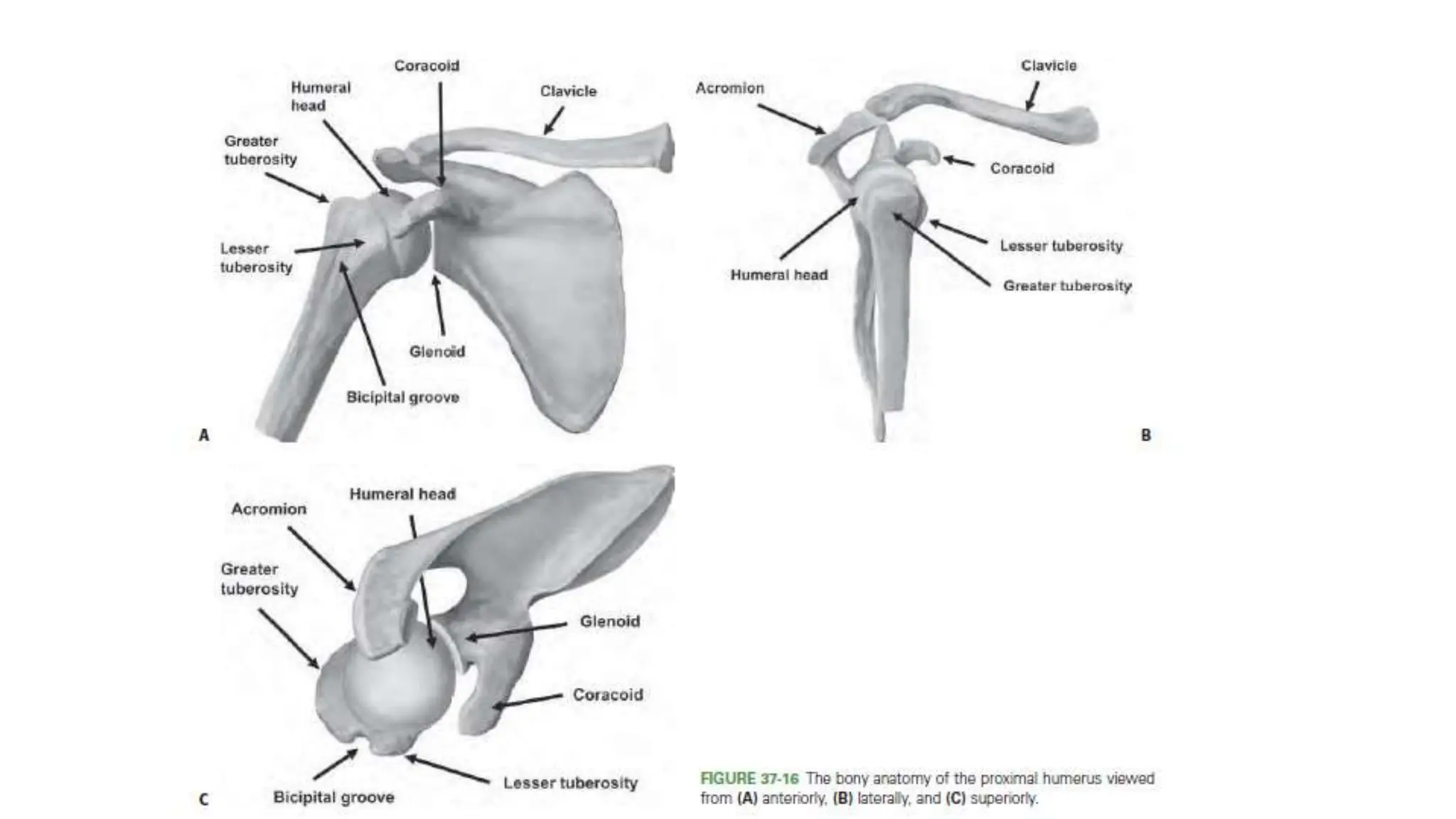
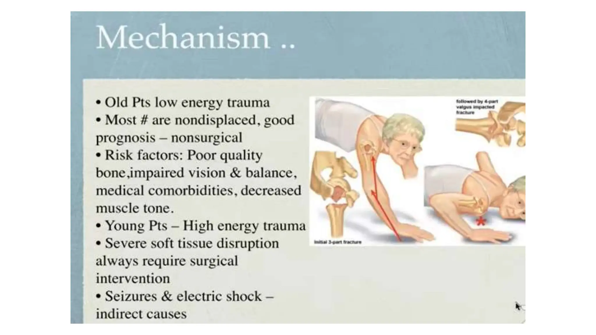
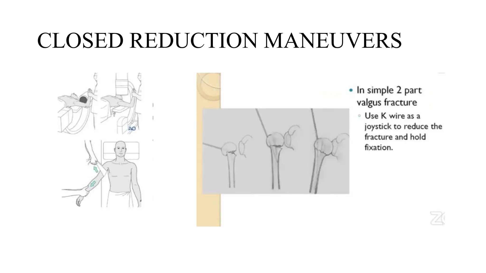
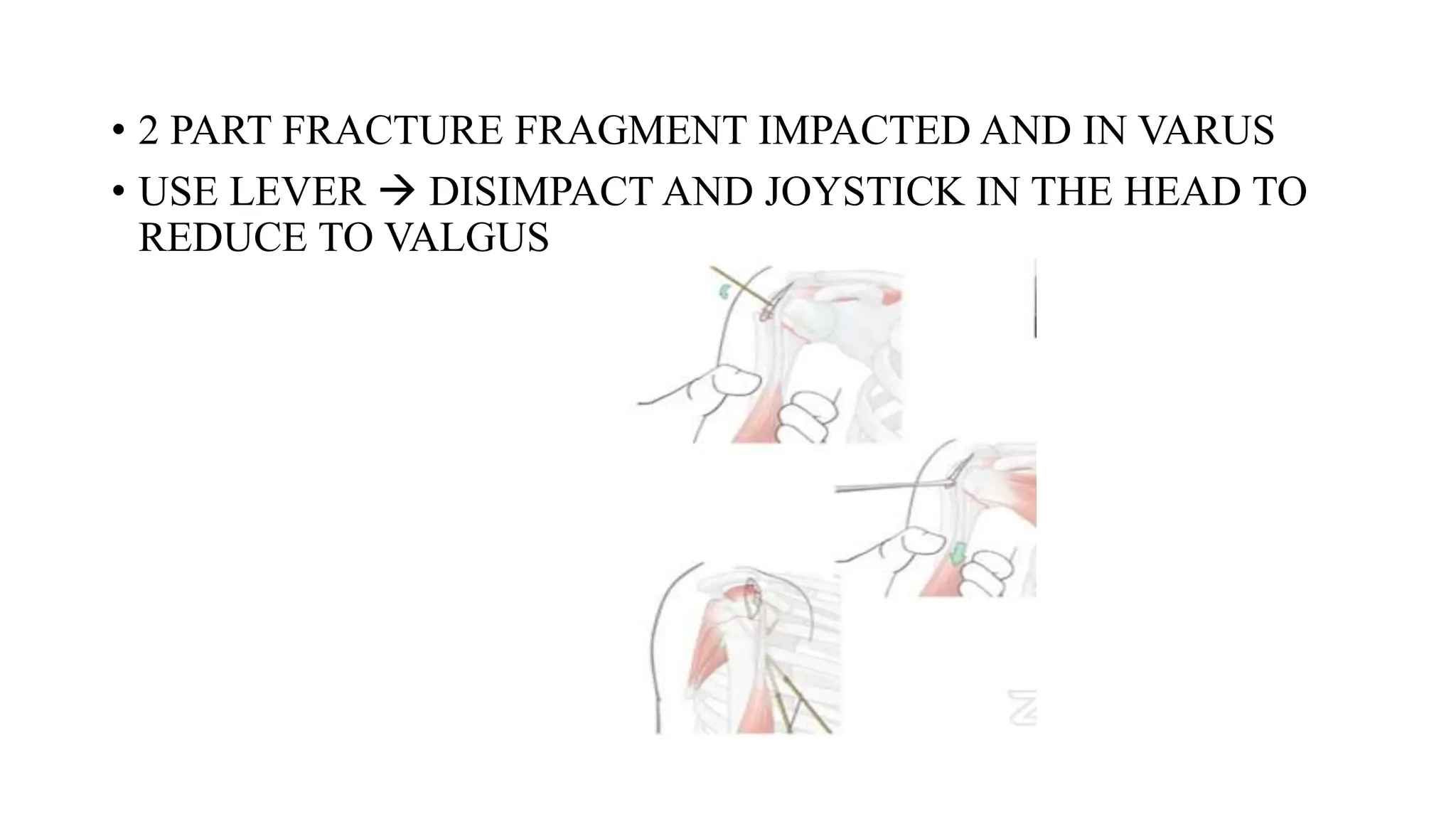
PROXIMAL HUMERUS FRACTURE
TREATMENT
• MODERATOR : DR. T. VINOTH M.S.ORTHO,
CONSULTANT DEPT OF ORTHOPAEDICS PIMS
• PRESENTOR : DR. ARAVINDAN DNB ORTHO
PIMS
• Use adequate radiograms to understand the traumatic lesion, be careful
denying older patients effective treatment, use a safe and simple
surgical approach, know the options for internal fixation, recognize the
value of prosthetic replacement, avoid technical pitfalls, and
thoughtfully supervise the postoperative patient care.
R.H. Cofield (1988)
• Cofield’s summary of treatment of proximal humeral fractures is an
indication of the difficulty of treating these injuries—from first
evaluation to final outcome. Much controversy and confusion still
exist, and no single treatment protocol or algorithm has been proved to
be universally effective.
• Areas still in question include radiographic diagnosis, operative or
nonoperative treatment, consideration of patient age in treatment
decision making, surgical approach, fracture fixation or
hemiarthroplasty, type of internal fixation, and rehabilitation protocol.
• Nonoperative treatment may be preferable for two-, three-, and four-
part proximal humeral fractures in elderly patients, but pain and loss of
function have been reported in high percentages of patients after this
treatment approach.
Good or excellent results in 81% of impacted valgus fractures in elderly
patients treated nonoperatively, and in a comparison of operative and
nonoperative treatment of displaced two-part fractures, these authors
found similar results in the two treatment groups.
• 231 patients, the authors were unable to show superiority of operative
or nonoperative treatment using the Oxford Shoulder Score as the
primary outcome.
• Found large variations in the percentage of proximal humeral fractures
treated operatively, ranging from 6.4% to 60%; in eight regions of the
United States, at least 40% were treated operatively, whereas in 35
regions, fewer than 20% were treated operatively. The fact that 10
different fixation techniques were evaluated for a single fracture type
(fractures of the surgical neck of the humerus) is further indication of
the complexity of treating proximal humeral fractures.
INTRODUCTION
• 80% OF ALL HUMERAL FRACTURES
• 7% OF ALL FRACTURES
• PT > 65 YRS  SECOND MOST COMMON # OF UPPER
EXTREMITY
• 65% OCCUR IN PT > 60YRS
• F/M = 3:1
• INCIDENCE INCREASES WITH AGE
ARTICULAR SEGMENT DIAMETER 46MM(37-55)
INCLINATION OF HUMERAL HEAD TO SHAFT 130 DEG
RETROVERSION OF THE HEAD 18 TO 40
• CLASS 3 LEVER
• TO IMPROVE
EFFICACY 
LARGE MUSCLES
• IMPORTANCE OF
TUBEROSITIES
BLOOD SUPPLY
METAPHYSEAL “TORUS “ RING
CONCEPT
CLINICAL EXAMINATION
• HISTORY
• SHOULDER  PAIN AND RESTRICTION
• ECCHYMOSIS 24-48 HRS
• ASSOCIATED INJURIES IN HIGH ENERGY TRAUMA 
SCAPULA ,CLAVICLE CERVICAL RIB
• AXILLARY NERVE
• ARTERIAL INJURIES -- RARE
• Glenoid fractures
If the amount of displacement of the humeral head or tuberosity fragments is unclear
on radiographs, an axial CT scan with 2-mm sections is indicated
CT SCAN  DETAILED UNDERSTANDING OF FRACTURE
CONFIGURATION
DEGREE OF OSTEOPENIA
LOCATION OF BONE IMPACTION
EXTENT OF FRACTURE COMMUNITION
MRI  RARELY INDICATED IN TRAUMA SETTING
CONFIRMING A NON DISPLACED #
CLASSIFICATIONS
• KOCHER
• CODMAN
• NEER
• AO
NEER CLASSIFICATION
• MOST COMMONLY USED
• 1970 MODIFIED 2002
• BASED  ANATOMIC RELATIONSHIP OF 4 PARTS
GT
LT
ARTICULAR SURFACE COMPONENT
HUMERAL SHAFT
MODERATE INTEROBSERVER RELABILITY
PART IS CONSIDERED DISPLACED IF >1CM SEPARATION AND >45
DEG ANGULATION
MODALITIES OF TREATMENT
• CONSERVATIVE
• OPERATIVE
Philos plating
Nailing
K wire
Hemiarthroplasty
Reverse shoulder arthroplasty
Conservative
INIDICATIONS
Stable nondisplaced or minimally displaced
Valgus impacted
Patient not fit for surgery
Elderly patients with low functional demand
Significant comorbidity
TREATMENT DECISION
• The first step in treatment decision-making is to determine if
displacement (66%) and angulation (varus is poorly tolerated) are
acceptable for a particular patient;
• The second is to determine if the humeral head and shaft move as a
unit.
If both of these conditions are present, the fracture is stable and in an
acceptable position
• Sling is used for comfort
• Physical therapy with pendulum exercises 1st week
• If the humeral head and shaft do not move as a unit, physical therapy
can be delayed for 2 to 4 weeks in patients who are poor surgical
candidates because of age, low functional demands, or comorbidities
that preclude participation in rehabilitation
• A randomized controlled trial involving 74 patients with impacted
proximal humeral fractures found that early (within 72 hours of injury)
passive mobilization was safe and more effective in restoring function
than conventional immobilization (3 weeks) followed by physical
therapy.
• Another study, however, pointed out that fracture settling continues to
occur with conservative treatment
• Weekly radiographs  1st month of treatment
• biweekly radiographs until 6 weeks after injury or initial callus
formation is visible
• Final radiographs are taken at 3 months to confirm union
• Pain usually subsides within 2 weeks after injury passive
range-of-motion exercises of the shoulder
Non operative complications
• Restriction of movements
• Necrosis of the head of humerus
• Pain subacromial impingement
• pseudoarthrosis
OPERATIVE TREATMENT
• It is imperative to recreate the normal proximal humeral anatomy with
respect to tuberosity reduction and the headneck relationship.
Indications
• Displaced two-part surgical neck fractures,
• Displaced (>5 mm) greater tuberosity fractures,
• Displaced three-part fractures, and
• Displaced four-part fractures in young patients.
• The type of fixation (transosseous suture fixation, percutaneous
pinning, intramedullary nailing, or plate fixation) used depends on the
• Patient’s age,
• Activity level, and
• Bone quality; the fracture type and associated fractures; and the
• Surgeon’s technical ability
Age
• In their series of 154 fractures with proximal humeral fractures treated
with plating,
• Boesmueller et al. found that the risk of screw cut-out was four times
higher in patients over the age of 60 years and the overall risk for
complications was three times higher than in younger patients
HERTEL CRITERIA - RADIOGRAPHIC
• A, Metaphyseal extension of
humeral head greater than 9
mm.
• B, Metaphyseal extension of
humeral head less than 8
mm.
• C, Undisplaced medial
hinge.
• D, Medial hinge with greater
than 2-mm displacement.
• Metaphyseal extension of the humeral head of less than 8 mm and
medial hinge disruption of more than 2 mm are predictive of ischemia.
• Combined with anatomic neck fracture pattern has a 97% PPV for
humeral head ischemia.
According to the AO/ASIF classification
system
• Extraarticular type A fractures have an intact
vascular supply
• Type B fractures have a possible injury to the
vascular supply and
• Type C articular fractures have a high
probability of osteonecrosis.
CORTICAL THICKNESS
• Reliable and reproducible factor of BMD
• The combined cortical thickness is the
average of the medial and lateral cortical
thickness at two levels.
• Generally, a cortical thickness of less than
4 mm precludes internal fixation because
adequate screw purchase cannot be
obtained , where, sling immobilization,
transosseous suture, or hemiarthroplasty
may be better options.
Closed reduction and percutaneous fixation
• DECREASED SOFT TISSUE TRAUMA
• REDUCED AVN
• TWO PART , 3 PART AND 4 PART VALGUS IMPACTED FRACTURES
• TECHNICALLY CHALLENGING
• REQUIRES A SATISFACTORY CLOSED REDUCTION,
• ADEQUATE BONE STOCK
• MINIMAL COMMINUTION (PARTICULARLY OF THE TUBEROSITIES)
C/I – METAPHYSEAL COMMINUTION
• AN INTACT MEDIAL CALCAR
• A COMPLIANT PATIENT.
CLOSED REDUCTION MANEUVERS
• 2 PART FRACTURE FRAGMENT IMPACTED AND IN VARUS
• USE LEVER  DISIMPACT AND JOYSTICK IN THE HEAD TO
REDUCE TO VALGUS
Terminally threaded Schanz pins and bicortical
pins inserted from the greater tuberosity to the
medial humeral shaft add stability to the overall
construct
PROBLEMS
INADEQUATE FIXATION
ADDITIONAL IMMOBILIZATION MAY
BE REQUIRED
PIN TRACT INFECTION
PIN LOOSENING AND LOSS OF
REDUCTION
PIN MIGRATION INSIDE
ORIF WITH PHILOS PLATE
• INDICATIONS
Deep dissection
the short head of the biceps and coracobrachialis arise from the coracoid process
and are retracted medially.
The musculocutaneous nerve enters the biceps 5-8cm distal to the coracoid
process; retraction of the conjoint tendon must be done with care.
the fascia on the lateral side of the conjoint tendon is incised to reveal the
subscapularis
External rotation puts the subscapularis fibers on stretch.
the subscapularis may be released from its insertion on the lesser tuberosity
through the tendon or via an osteotomy
the capsule is then incised (as needed) to enter the joint
DELTOID SPLITTING APPROACH
Locked plates
• Most favoured implant for 3 and 4 part #
• Variable angles on either sides
• Multiple nonabsorbable sutures can be used
• Humeral head shoud be supported by the shaft
• Valgus impaction osteotomy will help in cases of medial metaphyseal
comminution
• accurate reduction of varus humeral head
• Restore natural version of head
• Gt below head ,plate below gt
• Restore medial buttress ( head shaft displacement <5mm)
• Manage porosis / head hollowness
• NA suture via bone tendon junction of GT & LT
• Suture tuberosities to plate
• Calcar screw must!
• Screws few mm distal to s/c bone
• 7 locking head screws
• Including 2 locking screws
• Augmented with traction cuff sutures
• Comminuted medial calcar ,an endosteal fibular strut was used
• Subchondral metaphyseal bone  filled with injectable calcium
phosphate cement
problems
• Extensive exposure
• Cephalic vein injury /bleeding
• Prolonged surgical time
• Comminuted fragments come free
• Axillary and musculocutaneous nerve injury
• Bulky deltoid prevents lateral retraction
Proximal humerus im nailing
• Less extensive soft tissue
dissection
• Nail within loading axis of
humerus
• Shorter lever arm of locking
screw
INDICATION
MARKED METAPHYSEAL
COMMINUTION
Spiral fracture
Pathological fractures
Impending pathological fracture
• Older nails for simple 2 part fractures
• 90% had satisfactory results
Overall complications
Screw back out (the addition of polyethylene bushings may increase
stability and prevent)
Proximal screw migration
Loss of proximal fixation
Infection,
nonunion
Impingement and rotator cuff pain
Multiloc nail
• New generation
• 3 dimensional proximal locking pattern
• Straight ( no proximal bent)
• Adaptable to diameter of medullary cavity
• Modular locking options
• Adequate for 2 to 4part fractures
• Contraindication - A comminuted lateral cortex fracture or fractures
involving the tuberosities.
• A recent randomized controlled trial demonstrated that complications
were fewer with a straight nail design compared with a curvilinear
design.
MIPO
• 2 or 3 part fractures
• Proximal humerus with
shaft extension
• 4 part difficult
Transosseous fixation
• Park et al. reported 78% excellent results in patients with two-part
and three-part proximal humeral fractures treated with suture fixation.
• The use of strong nonabsorbable suture provides the advantage of
incorporating the rotator cuff insertion to increase fixation in patients
with poor bone quality.
Dimakopoulos et al. reported good results in 188 displaced proximal
humeral fractures treated with transosseous fixation.
• Advantages are
• Less surgical dissection
• Low rate of AVN
• Fixation sufficient to allow early passive joint motion, and the
• Avoidance of bulky and expensive implants.
Parachute technique
Hemiarthroplasty
• Not reconstructable
• Delayed presentations/neglected fractures
• Osteopenia and poor fixation
• Multiple small fragments
• Greater than 40% impaction
• Failed osteosynthesis
• Non union of major fragments
• Avn with collapse
goals
• Pain relief
• Optimal shoulder function
• Technical aspect
Restore proximal humeral anatomy
Obtain tuberosity healing
Balance soft tissue
Outcomes
• Pain relief : reliable and predicatable
• Function : unpredicatable
Influenced by
Age
Fracture type , time to surgery
Rotator cuff status
Duration and type of rehabilitation
Reverse shoulder Artroplasty
REQUIREMENTS
• Intact axillary nerve
• Full function of deltoid
• Adequate glenoid bone stock
• Soft tissue tension is adjusted by varying the length of
the glenosphere and humeral socket.
• Stability is provided by:
• Deltoid tensioning
• Adjust humeral offset.
• Glenoid tilt
• Head diameter
• Larger diameter is more stable.
• Component positioning
COMPONENT POSITIONING
■ Glenosphere must be positioned as low as possible on the
glenoid.
■ This minimizes risk for scapular notching by humeral
socket.
■ Glenoid baseplate (and glenosphere) should be tilted inferiorly
10 to 15 degrees.
■ This is key to enhancing deltoid tensioning and thus
providing implant stability.
■ Humeral stem is positioned in retroversion.
■ 25 to 40 degrees retroversion preferred
HEMI VS RSA
Areas of research
osteoporosis
CARBONFIBRE REINFORCED PEEK PLATE
Radiolucent plate - Better intraop reduction
Monitor fracture healing
MRI compatible
No cold welding (Easy removal)
Low Wear rate compared to titanium
Less incidence of varus collapse
PROXIMAL HUMERUS FRACTURE TREATMENT.pptx

PROXIMAL HUMERUS FRACTURE TREATMENT.pptx

  • 1.
    PROXIMAL HUMERUS FRACTURE TREATMENT •MODERATOR : DR. T. VINOTH M.S.ORTHO, CONSULTANT DEPT OF ORTHOPAEDICS PIMS • PRESENTOR : DR. ARAVINDAN DNB ORTHO PIMS
  • 2.
    • Use adequateradiograms to understand the traumatic lesion, be careful denying older patients effective treatment, use a safe and simple surgical approach, know the options for internal fixation, recognize the value of prosthetic replacement, avoid technical pitfalls, and thoughtfully supervise the postoperative patient care. R.H. Cofield (1988)
  • 3.
    • Cofield’s summaryof treatment of proximal humeral fractures is an indication of the difficulty of treating these injuries—from first evaluation to final outcome. Much controversy and confusion still exist, and no single treatment protocol or algorithm has been proved to be universally effective. • Areas still in question include radiographic diagnosis, operative or nonoperative treatment, consideration of patient age in treatment decision making, surgical approach, fracture fixation or hemiarthroplasty, type of internal fixation, and rehabilitation protocol.
  • 4.
    • Nonoperative treatmentmay be preferable for two-, three-, and four- part proximal humeral fractures in elderly patients, but pain and loss of function have been reported in high percentages of patients after this treatment approach. Good or excellent results in 81% of impacted valgus fractures in elderly patients treated nonoperatively, and in a comparison of operative and nonoperative treatment of displaced two-part fractures, these authors found similar results in the two treatment groups.
  • 5.
    • 231 patients,the authors were unable to show superiority of operative or nonoperative treatment using the Oxford Shoulder Score as the primary outcome.
  • 6.
    • Found largevariations in the percentage of proximal humeral fractures treated operatively, ranging from 6.4% to 60%; in eight regions of the United States, at least 40% were treated operatively, whereas in 35 regions, fewer than 20% were treated operatively. The fact that 10 different fixation techniques were evaluated for a single fracture type (fractures of the surgical neck of the humerus) is further indication of the complexity of treating proximal humeral fractures.
  • 7.
    INTRODUCTION • 80% OFALL HUMERAL FRACTURES • 7% OF ALL FRACTURES • PT > 65 YRS  SECOND MOST COMMON # OF UPPER EXTREMITY • 65% OCCUR IN PT > 60YRS • F/M = 3:1 • INCIDENCE INCREASES WITH AGE
  • 9.
    ARTICULAR SEGMENT DIAMETER46MM(37-55) INCLINATION OF HUMERAL HEAD TO SHAFT 130 DEG RETROVERSION OF THE HEAD 18 TO 40
  • 10.
    • CLASS 3LEVER • TO IMPROVE EFFICACY  LARGE MUSCLES • IMPORTANCE OF TUBEROSITIES
  • 12.
  • 13.
  • 15.
    CLINICAL EXAMINATION • HISTORY •SHOULDER  PAIN AND RESTRICTION • ECCHYMOSIS 24-48 HRS • ASSOCIATED INJURIES IN HIGH ENERGY TRAUMA  SCAPULA ,CLAVICLE CERVICAL RIB • AXILLARY NERVE • ARTERIAL INJURIES -- RARE
  • 17.
  • 19.
    If the amountof displacement of the humeral head or tuberosity fragments is unclear on radiographs, an axial CT scan with 2-mm sections is indicated CT SCAN  DETAILED UNDERSTANDING OF FRACTURE CONFIGURATION DEGREE OF OSTEOPENIA LOCATION OF BONE IMPACTION EXTENT OF FRACTURE COMMUNITION MRI  RARELY INDICATED IN TRAUMA SETTING CONFIRMING A NON DISPLACED #
  • 20.
  • 21.
    NEER CLASSIFICATION • MOSTCOMMONLY USED • 1970 MODIFIED 2002 • BASED  ANATOMIC RELATIONSHIP OF 4 PARTS GT LT ARTICULAR SURFACE COMPONENT HUMERAL SHAFT MODERATE INTEROBSERVER RELABILITY PART IS CONSIDERED DISPLACED IF >1CM SEPARATION AND >45 DEG ANGULATION
  • 26.
    MODALITIES OF TREATMENT •CONSERVATIVE • OPERATIVE Philos plating Nailing K wire Hemiarthroplasty Reverse shoulder arthroplasty
  • 27.
    Conservative INIDICATIONS Stable nondisplaced orminimally displaced Valgus impacted Patient not fit for surgery Elderly patients with low functional demand Significant comorbidity
  • 28.
    TREATMENT DECISION • Thefirst step in treatment decision-making is to determine if displacement (66%) and angulation (varus is poorly tolerated) are acceptable for a particular patient; • The second is to determine if the humeral head and shaft move as a unit. If both of these conditions are present, the fracture is stable and in an acceptable position
  • 29.
    • Sling isused for comfort • Physical therapy with pendulum exercises 1st week • If the humeral head and shaft do not move as a unit, physical therapy can be delayed for 2 to 4 weeks in patients who are poor surgical candidates because of age, low functional demands, or comorbidities that preclude participation in rehabilitation
  • 30.
    • A randomizedcontrolled trial involving 74 patients with impacted proximal humeral fractures found that early (within 72 hours of injury) passive mobilization was safe and more effective in restoring function than conventional immobilization (3 weeks) followed by physical therapy. • Another study, however, pointed out that fracture settling continues to occur with conservative treatment
  • 31.
    • Weekly radiographs 1st month of treatment • biweekly radiographs until 6 weeks after injury or initial callus formation is visible • Final radiographs are taken at 3 months to confirm union • Pain usually subsides within 2 weeks after injury passive range-of-motion exercises of the shoulder
  • 32.
    Non operative complications •Restriction of movements • Necrosis of the head of humerus • Pain subacromial impingement • pseudoarthrosis
  • 33.
    OPERATIVE TREATMENT • Itis imperative to recreate the normal proximal humeral anatomy with respect to tuberosity reduction and the headneck relationship. Indications • Displaced two-part surgical neck fractures, • Displaced (>5 mm) greater tuberosity fractures, • Displaced three-part fractures, and • Displaced four-part fractures in young patients.
  • 34.
    • The typeof fixation (transosseous suture fixation, percutaneous pinning, intramedullary nailing, or plate fixation) used depends on the • Patient’s age, • Activity level, and • Bone quality; the fracture type and associated fractures; and the • Surgeon’s technical ability
  • 35.
    Age • In theirseries of 154 fractures with proximal humeral fractures treated with plating, • Boesmueller et al. found that the risk of screw cut-out was four times higher in patients over the age of 60 years and the overall risk for complications was three times higher than in younger patients
  • 36.
    HERTEL CRITERIA -RADIOGRAPHIC • A, Metaphyseal extension of humeral head greater than 9 mm. • B, Metaphyseal extension of humeral head less than 8 mm. • C, Undisplaced medial hinge. • D, Medial hinge with greater than 2-mm displacement.
  • 37.
    • Metaphyseal extensionof the humeral head of less than 8 mm and medial hinge disruption of more than 2 mm are predictive of ischemia. • Combined with anatomic neck fracture pattern has a 97% PPV for humeral head ischemia.
  • 38.
    According to theAO/ASIF classification system • Extraarticular type A fractures have an intact vascular supply • Type B fractures have a possible injury to the vascular supply and • Type C articular fractures have a high probability of osteonecrosis.
  • 39.
    CORTICAL THICKNESS • Reliableand reproducible factor of BMD • The combined cortical thickness is the average of the medial and lateral cortical thickness at two levels. • Generally, a cortical thickness of less than 4 mm precludes internal fixation because adequate screw purchase cannot be obtained , where, sling immobilization, transosseous suture, or hemiarthroplasty may be better options.
  • 40.
    Closed reduction andpercutaneous fixation • DECREASED SOFT TISSUE TRAUMA • REDUCED AVN • TWO PART , 3 PART AND 4 PART VALGUS IMPACTED FRACTURES • TECHNICALLY CHALLENGING • REQUIRES A SATISFACTORY CLOSED REDUCTION, • ADEQUATE BONE STOCK • MINIMAL COMMINUTION (PARTICULARLY OF THE TUBEROSITIES) C/I – METAPHYSEAL COMMINUTION • AN INTACT MEDIAL CALCAR • A COMPLIANT PATIENT.
  • 42.
  • 43.
    • 2 PARTFRACTURE FRAGMENT IMPACTED AND IN VARUS • USE LEVER  DISIMPACT AND JOYSTICK IN THE HEAD TO REDUCE TO VALGUS
  • 46.
    Terminally threaded Schanzpins and bicortical pins inserted from the greater tuberosity to the medial humeral shaft add stability to the overall construct PROBLEMS INADEQUATE FIXATION ADDITIONAL IMMOBILIZATION MAY BE REQUIRED PIN TRACT INFECTION PIN LOOSENING AND LOSS OF REDUCTION PIN MIGRATION INSIDE
  • 48.
    ORIF WITH PHILOSPLATE • INDICATIONS
  • 51.
    Deep dissection the shorthead of the biceps and coracobrachialis arise from the coracoid process and are retracted medially. The musculocutaneous nerve enters the biceps 5-8cm distal to the coracoid process; retraction of the conjoint tendon must be done with care. the fascia on the lateral side of the conjoint tendon is incised to reveal the subscapularis External rotation puts the subscapularis fibers on stretch. the subscapularis may be released from its insertion on the lesser tuberosity through the tendon or via an osteotomy the capsule is then incised (as needed) to enter the joint
  • 52.
  • 53.
    Locked plates • Mostfavoured implant for 3 and 4 part # • Variable angles on either sides • Multiple nonabsorbable sutures can be used
  • 58.
    • Humeral headshoud be supported by the shaft • Valgus impaction osteotomy will help in cases of medial metaphyseal comminution • accurate reduction of varus humeral head • Restore natural version of head • Gt below head ,plate below gt • Restore medial buttress ( head shaft displacement <5mm) • Manage porosis / head hollowness
  • 59.
    • NA suturevia bone tendon junction of GT & LT • Suture tuberosities to plate • Calcar screw must! • Screws few mm distal to s/c bone
  • 60.
    • 7 lockinghead screws • Including 2 locking screws • Augmented with traction cuff sutures • Comminuted medial calcar ,an endosteal fibular strut was used • Subchondral metaphyseal bone  filled with injectable calcium phosphate cement
  • 63.
    problems • Extensive exposure •Cephalic vein injury /bleeding • Prolonged surgical time • Comminuted fragments come free • Axillary and musculocutaneous nerve injury • Bulky deltoid prevents lateral retraction
  • 66.
    Proximal humerus imnailing • Less extensive soft tissue dissection • Nail within loading axis of humerus • Shorter lever arm of locking screw INDICATION MARKED METAPHYSEAL COMMINUTION Spiral fracture Pathological fractures Impending pathological fracture
  • 67.
    • Older nailsfor simple 2 part fractures • 90% had satisfactory results Overall complications Screw back out (the addition of polyethylene bushings may increase stability and prevent) Proximal screw migration Loss of proximal fixation Infection, nonunion Impingement and rotator cuff pain
  • 68.
    Multiloc nail • Newgeneration • 3 dimensional proximal locking pattern • Straight ( no proximal bent) • Adaptable to diameter of medullary cavity • Modular locking options • Adequate for 2 to 4part fractures
  • 69.
    • Contraindication -A comminuted lateral cortex fracture or fractures involving the tuberosities. • A recent randomized controlled trial demonstrated that complications were fewer with a straight nail design compared with a curvilinear design.
  • 70.
    MIPO • 2 or3 part fractures • Proximal humerus with shaft extension • 4 part difficult
  • 72.
  • 73.
    • Park etal. reported 78% excellent results in patients with two-part and three-part proximal humeral fractures treated with suture fixation. • The use of strong nonabsorbable suture provides the advantage of incorporating the rotator cuff insertion to increase fixation in patients with poor bone quality.
  • 74.
    Dimakopoulos et al.reported good results in 188 displaced proximal humeral fractures treated with transosseous fixation. • Advantages are • Less surgical dissection • Low rate of AVN • Fixation sufficient to allow early passive joint motion, and the • Avoidance of bulky and expensive implants.
  • 75.
  • 76.
    Hemiarthroplasty • Not reconstructable •Delayed presentations/neglected fractures • Osteopenia and poor fixation • Multiple small fragments • Greater than 40% impaction • Failed osteosynthesis • Non union of major fragments • Avn with collapse
  • 77.
    goals • Pain relief •Optimal shoulder function • Technical aspect Restore proximal humeral anatomy Obtain tuberosity healing Balance soft tissue
  • 81.
    Outcomes • Pain relief: reliable and predicatable • Function : unpredicatable Influenced by Age Fracture type , time to surgery Rotator cuff status Duration and type of rehabilitation
  • 83.
    Reverse shoulder Artroplasty REQUIREMENTS •Intact axillary nerve • Full function of deltoid • Adequate glenoid bone stock • Soft tissue tension is adjusted by varying the length of the glenosphere and humeral socket. • Stability is provided by: • Deltoid tensioning • Adjust humeral offset. • Glenoid tilt • Head diameter • Larger diameter is more stable. • Component positioning
  • 84.
    COMPONENT POSITIONING ■ Glenospheremust be positioned as low as possible on the glenoid. ■ This minimizes risk for scapular notching by humeral socket. ■ Glenoid baseplate (and glenosphere) should be tilted inferiorly 10 to 15 degrees. ■ This is key to enhancing deltoid tensioning and thus providing implant stability. ■ Humeral stem is positioned in retroversion. ■ 25 to 40 degrees retroversion preferred
  • 85.
  • 86.
  • 87.
  • 88.
  • 90.
    Radiolucent plate -Better intraop reduction Monitor fracture healing MRI compatible No cold welding (Easy removal) Low Wear rate compared to titanium Less incidence of varus collapse