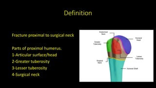
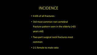
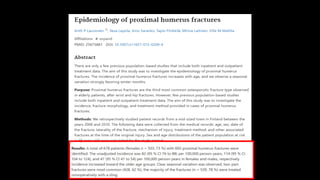
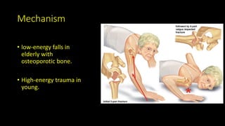
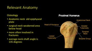
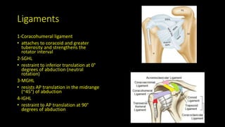
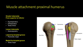
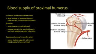
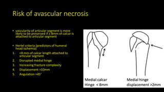
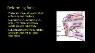
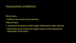
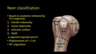
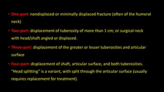
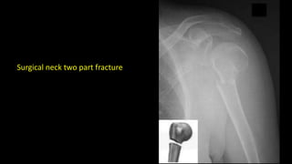
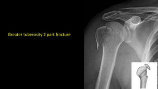
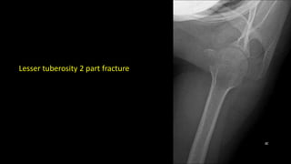
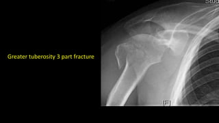
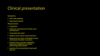
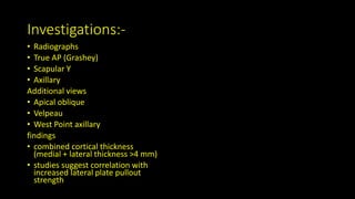
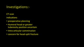
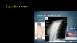
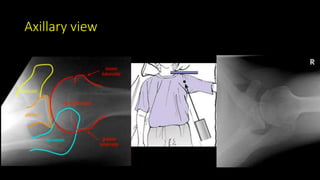
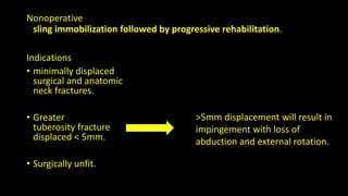
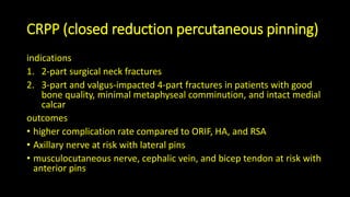
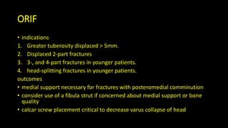
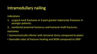
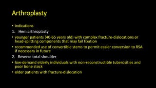
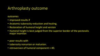
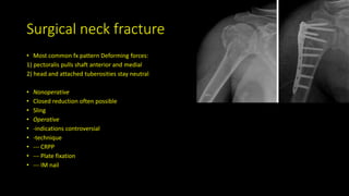
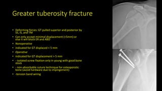
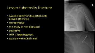
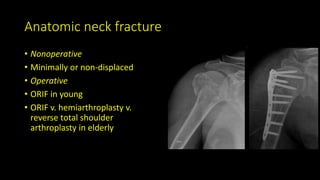
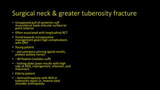
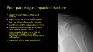
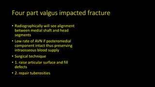
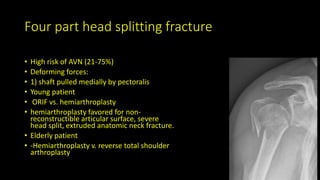
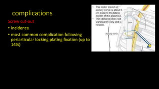
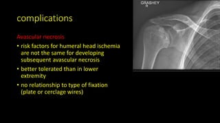
Proximal Humerus Fracture
Frcs orthopaedic trauma fisrt aid
Definition
Fracture proximal to surgical neck
Parts of proximal humerus.
1-Articular surface/head
2-Greater tuberosity
3-Lesser tuberosity
4-Surgical neck
INCIDENCE
• 4-6% of all fractures
• 3rd most common non-vertebral
fracture pattern seen in the elderly (>65
years old)
• Two-part surgical neck fractures most
common
• 2:1 female to male ratio
Risk Factors
• Osteoporosis
• Diabetes
• Epilepsy
• Female
Mechanism
• low-energy falls in
elderly with
osteoporotic bone.
• High-energy trauma in
young.
Relevant Anatomy
Osteology
• Anatomic neck old epiphyseal
plate
• surgical neck weakened area
below head
• more often involved in
fractures
• average neck-shaft angle is
135 degrees
Ligaments
1-Coracohumeral ligament
• attaches to coracoid and greater
tuberosity and strengthens the
rotator interval
2-SGHL
• restraint to inferior translation at 0°
degrees of abduction (neutral
rotation)
3-MGHL
• resists AP translation in the midrange
(~45°) of abduction
4-IGHL
• restraint to AP translation at 90°
degrees of abduction
Muscle attachment proximal humerus
Greater tuberosity
From superior to inferior
• Supraspinatus.
• Infraspinatus.
• Teres minor.
Lesser tuberosity
• Subscapularis.
Lateral lip bicipital groove
• Pectoralis major
Medial lip bicipital groove
• Teres major
Teres major
Blood supply of proximal humerus
1-Anterior humeral circumflex artery
• large number of anastomosis with
other vessels in the proximal humerus
Branches
• anterolateral ascending branch
• arcuate artery is the terminal branch
and main supply to greater tuberosity.
2-posterior humeral circumflex artery
• recent studies suggest it is the main
blood supply to humeral head
Risk of avascular necrosis
• vascularity of articular segment is more
likely to be preserved if ≥ 8mm of calcar is
attached to articular segment
• Hertel criteria (predictors of humeral
head ischemia)
1. <8 mm of calcar length attached to
articular segment
2. Disrupted medial hinge
3. Increasing fracture complexity
4. Displacement >10mm
5. Angulation >45°
Deforming force
• Pectoralis major displaces shaft
anteriorly and medially
• Supraspinatus, Infraspinatus,
and Teres minor externally
rotate greater tuberosity
• Subscapularis internally rotates
articular segment or lesser
tuberosity
Associated conditions
Nerve injury
• Axillary nerve injury most common
Arterial injury
• uncommon (incidence 5-6%), higher likelihood in older patients
• most often occur at level of surgical neck or with subcoracoid
dislocation of the head
Neer classification
• Based on anatomic relationship
of 4 segments
1. Greater tuberosity
2. Lesser tuberosity
3. Articular surface
4. Shaft
a segment separate part if
• Displacement of > 1 cm
• 45° angulation
• One-part: nondisplaced or minimally displaced fracture (often of the humeral
neck)
• Two-part: displacement of tuberosity of more than 1 cm; or surgical neck
with head/shaft angled or displaced.
• Three-part: displacement of the greater or lesser tuberosities and articular
surface
• Four-part: displacement of shaft, articular surface, and both tuberosities.
“Head splitting” is a variant, with split through the articular surface (usually
requires replacement for treatment).
Surgical neck two part fracture
Greater tuberosity 2 part fracture
Lesser tuberosity 2 part fracture
Greater tuberosity 3 part fracture
Lesser tuberosity 3 part fracture
Greater tuberosity 4 part fracture
Lesser tuberosity 4 part fracture
Clinical presentation
Symptoms
• pain and swelling
• decreased motion
Physical exam
• inspection
• extensive ecchymosis of chest, arm,
and forearm
• neurovascular exam
• axillary nerve injury most common
• determine function of deltoid muscle
and lateral shoulder sensation
• arterial injury may be masked by
extensive collateral circulation
preserving distal pulses
• examine for concomitant chest wall
injuries
Investigations:-
• Radiographs
• True AP (Grashey)
• Scapular Y
• Axillary
Additional views
• Apical oblique
• Velpeau
• West Point axillary
findings
• combined cortical thickness
(medial + lateral thickness >4 mm)
• studies suggest correlation with
increased lateral plate pullout
strength
Investigations:-
CT scan
indications
• preoperative planning
• Humeral head or greater
tuberosity position uncertain
• Intra-articular comminution
• concern for head-split fracture
Ap grashey view
Scapular Y view
Axillary view
Nonoperative
sling immobilization followed by progressive rehabilitation.
Indications
• minimally displaced
surgical and anatomic
neck fractures.
• Greater
tuberosity fracture
displaced < 5mm.
• Surgically unfit.
>5mm displacement will result in
impingement with loss of
abduction and external rotation.
CRPP (closed reduction percutaneous pinning)
indications
1. 2-part surgical neck fractures
2. 3-part and valgus-impacted 4-part fractures in patients with good
bone quality, minimal metaphyseal comminution, and intact medial
calcar
outcomes
• higher complication rate compared to ORIF, HA, and RSA
• Axillary nerve at risk with lateral pins
• musculocutaneous nerve, cephalic vein, and bicep tendon at risk with
anterior pins
ORIF
• indications
1. Greater tuberosity displaced > 5mm.
2. Displaced 2-part fractures
3. 3-, and 4-part fractures in younger patients.
4. head-splitting fractures in younger patients.
outcomes
• medial support necessary for fractures with posteromedial comminution
• consider use of a fibula strut if concerned about medial support or bone
quality
• calcar screw placement critical to decrease varus collapse of head
Intramedullary nailing
indications
1. surgical neck fractures or 3-part greater tuberosity fractures in
younger patients.
2. combined proximal humerus and humeral shaft fractures.
outcomes
• biomechanically inferior with torsional stress compared to plates
• favorable rates of fracture healing and ROM compared to ORIF
Arthroplasty
• indications
1. Hemiarthroplasty
• younger patients (40-65 years old) with complex fracture-dislocations or
head-splitting components that may fail fixation
• recommended use of convertible stems to permit easier conversion to RSA
if necessary in future
2. Reverse total shoulder
• low-demand elderly individuals with non-reconstructible tuberosities and
poor bone stock
• older patients with fracture-dislocation
Arthroplasty outcome
outcomes
• improved results if
• Anatomic tuberosity reduction and healing.
• Restoration of humeral height and version
• Humeral height is best judged from the superior border of the pectoralis
major insertion
• poor results with
• tuberosity nonunion or malunion.
• retroversion of humeral component > 40.
Surgical neck fracture
• Most common fx pattern Deforming forces:
1) pectoralis pulls shaft anterior and medial
2) head and attached tuberosities stay neutral
• Nonoperative
• Closed reduction often possible
• Sling
• Operative
• -indications controversial
• -technique
• --- CRPP
• --- Plate fixation
• --- IM nail
Greater tuberosity fracture
• Deforming forces: GT pulled superior and posterior by
SS, IS, and TM
• Can only accept minimal displacement (<5mm) or
else it will block ER and ABD
• Nonoperative
• indicated for GT displaced < 5 mm
• Operative
• indicated for GT displacement > 5 mm
• - isolated screw fixation only in young with good bone
stock
• - non-absorbable suture technique for osteoporotic
bone (avoid hardware due to impingement)
• -tension band wiring
Lesser tuberosity fracture
• Assume posterior dislocation until
proven otherwise
• Nonoperative
• Minimally or non-displaced
• Operative
• ORIF if large fragment
• excision with RCR if small
Anatomic neck fracture
• Nonoperative
• Minimally or non-displaced
• Operative
• ORIF in young
• ORIF v. hemiarthroplasty v.
reverse total shoulder
arthroplasty in elderly
Surgical neck & greater tuberosity fracture
• Unopposed pull of posterior cuff
musculature leads articular surface to
point anterior
• Often associated with longitudinal RCT
• Trend towards nonoperative
management given high complications
with ORIF
• Young patient
• - percutaneous pinning (good results,
protect axillary nerve)
• - IM fixation (violates cuff)
• - locking plate (poor results with high
rate of AVN, impingement, infection, and
malunion)
• Elderly patient
• - Hemiarthroplasty with RCR or
tuberosity repair vs. reverse total
shoulder arthroplasty
Four part valgus impacted fracture
• Specific type of displaced four-part
fracture.
• valgus impaction of the head fragment.
• low rate of rate of avascular necrosis.
• axial trauma to the abducted upper limb,
• direct impaction between the humeral
head and the glenoid cavity.
• posteromedial displacement (dorsal
tilting of the head) because of its
physiological anatomical conformation in
retroversion.
• low rate of rate of avascular necrosis
Four part valgus impacted fracture
• Radiographically will see alignment
between medial shaft and head
segments
• Low rate of AVN if posteromedial
component intact thus preserving
intraosseous blood supply
• Surgical technique
• 1. raise articular surface and fill
defects
• 2. repair tuberosities
Four part head splitting fracture
• High risk of AVN (21-75%)
• Deforming forces:
• 1) shaft pulled medially by pectoralis
• Young patient
• ORIF vs. hemiarthroplasty
• hemiarthroplasty favored for non-
reconstructible articular surface, severe
head split, extruded anatomic neck fracture.
• Elderly patient
• -Hemiarthroplasty v. reverse total shoulder
arthroplasty
complications
Screw cut-out
• incidence
• most common complication following
periarticular locking plating fixation (up to
14%)
complications
Avascular necrosis
• risk factors for humeral head ischemia
are not the same for developing
subsequent avascular necrosis
• better tolerated than in lower
extremity
• no relationship to type of fixation
(plate or cerclage wires)
Complications
Nerve injury
Axillary nerve (up to 58% )
• increased risk with lateral (deltoid-splitting) approach
• axillary nerve is usually found ~5-7cm distal to the tip of the
acromion
• at risk with lateral pins in CRPP
Suprascapular nerve (up to 48%)
Musculocutaneous nerve
• at risk with anterior pins in CRP
complications
Malunion
• usually varus apex-anterior or malunion of GT
• results inferior if converting from varus malunited fracture to TSA
• use reverse shoulder arthroplasty instead
Nonunion
• most common after two-part surgical neck fracture
• treatment of chronic nonunion/malunion in the elderly should include arthroplasty
• lesser tuberosity nonunion leads to weakness with lift-off testing
• greater tuberosity nonunion after arthroplasty leads to lack of active shoulder elevation
• greatest risk factors for nonunion are age and smoking
complications
Rotator cuff injuries and dysfunction
Long head of biceps tendon injuries
• also at risk with anterior pin in CRPP
Missed posterior dislocation
• consider in all patients with lesser tuberosity fracture
Adhesive capsulitis and scar tissue
Posttraumatic arthritis
Infection

Proximal humerus fracture .pptx

  • 1.
    Proximal Humerus Fracture Frcsorthopaedic trauma fisrt aid
  • 2.
    Definition Fracture proximal tosurgical neck Parts of proximal humerus. 1-Articular surface/head 2-Greater tuberosity 3-Lesser tuberosity 4-Surgical neck
  • 3.
    INCIDENCE • 4-6% ofall fractures • 3rd most common non-vertebral fracture pattern seen in the elderly (>65 years old) • Two-part surgical neck fractures most common • 2:1 female to male ratio
  • 5.
    Risk Factors • Osteoporosis •Diabetes • Epilepsy • Female
  • 7.
    Mechanism • low-energy fallsin elderly with osteoporotic bone. • High-energy trauma in young.
  • 8.
    Relevant Anatomy Osteology • Anatomicneck old epiphyseal plate • surgical neck weakened area below head • more often involved in fractures • average neck-shaft angle is 135 degrees
  • 10.
    Ligaments 1-Coracohumeral ligament • attachesto coracoid and greater tuberosity and strengthens the rotator interval 2-SGHL • restraint to inferior translation at 0° degrees of abduction (neutral rotation) 3-MGHL • resists AP translation in the midrange (~45°) of abduction 4-IGHL • restraint to AP translation at 90° degrees of abduction
  • 11.
    Muscle attachment proximalhumerus Greater tuberosity From superior to inferior • Supraspinatus. • Infraspinatus. • Teres minor. Lesser tuberosity • Subscapularis. Lateral lip bicipital groove • Pectoralis major Medial lip bicipital groove • Teres major Teres major
  • 12.
    Blood supply ofproximal humerus 1-Anterior humeral circumflex artery • large number of anastomosis with other vessels in the proximal humerus Branches • anterolateral ascending branch • arcuate artery is the terminal branch and main supply to greater tuberosity. 2-posterior humeral circumflex artery • recent studies suggest it is the main blood supply to humeral head
  • 13.
    Risk of avascularnecrosis • vascularity of articular segment is more likely to be preserved if ≥ 8mm of calcar is attached to articular segment • Hertel criteria (predictors of humeral head ischemia) 1. <8 mm of calcar length attached to articular segment 2. Disrupted medial hinge 3. Increasing fracture complexity 4. Displacement >10mm 5. Angulation >45°
  • 14.
    Deforming force • Pectoralismajor displaces shaft anteriorly and medially • Supraspinatus, Infraspinatus, and Teres minor externally rotate greater tuberosity • Subscapularis internally rotates articular segment or lesser tuberosity
  • 15.
    Associated conditions Nerve injury •Axillary nerve injury most common Arterial injury • uncommon (incidence 5-6%), higher likelihood in older patients • most often occur at level of surgical neck or with subcoracoid dislocation of the head
  • 16.
    Neer classification • Basedon anatomic relationship of 4 segments 1. Greater tuberosity 2. Lesser tuberosity 3. Articular surface 4. Shaft a segment separate part if • Displacement of > 1 cm • 45° angulation
  • 17.
    • One-part: nondisplacedor minimally displaced fracture (often of the humeral neck) • Two-part: displacement of tuberosity of more than 1 cm; or surgical neck with head/shaft angled or displaced. • Three-part: displacement of the greater or lesser tuberosities and articular surface • Four-part: displacement of shaft, articular surface, and both tuberosities. “Head splitting” is a variant, with split through the articular surface (usually requires replacement for treatment).
  • 18.
    Surgical neck twopart fracture
  • 19.
    Greater tuberosity 2part fracture
  • 20.
    Lesser tuberosity 2part fracture
  • 21.
    Greater tuberosity 3part fracture
  • 22.
    Lesser tuberosity 3part fracture
  • 23.
    Greater tuberosity 4part fracture
  • 24.
    Lesser tuberosity 4part fracture
  • 25.
    Clinical presentation Symptoms • painand swelling • decreased motion Physical exam • inspection • extensive ecchymosis of chest, arm, and forearm • neurovascular exam • axillary nerve injury most common • determine function of deltoid muscle and lateral shoulder sensation • arterial injury may be masked by extensive collateral circulation preserving distal pulses • examine for concomitant chest wall injuries
  • 26.
    Investigations:- • Radiographs • TrueAP (Grashey) • Scapular Y • Axillary Additional views • Apical oblique • Velpeau • West Point axillary findings • combined cortical thickness (medial + lateral thickness >4 mm) • studies suggest correlation with increased lateral plate pullout strength
  • 27.
    Investigations:- CT scan indications • preoperativeplanning • Humeral head or greater tuberosity position uncertain • Intra-articular comminution • concern for head-split fracture
  • 28.
  • 29.
  • 30.
  • 31.
    Nonoperative sling immobilization followedby progressive rehabilitation. Indications • minimally displaced surgical and anatomic neck fractures. • Greater tuberosity fracture displaced < 5mm. • Surgically unfit. >5mm displacement will result in impingement with loss of abduction and external rotation.
  • 32.
    CRPP (closed reductionpercutaneous pinning) indications 1. 2-part surgical neck fractures 2. 3-part and valgus-impacted 4-part fractures in patients with good bone quality, minimal metaphyseal comminution, and intact medial calcar outcomes • higher complication rate compared to ORIF, HA, and RSA • Axillary nerve at risk with lateral pins • musculocutaneous nerve, cephalic vein, and bicep tendon at risk with anterior pins
  • 33.
    ORIF • indications 1. Greatertuberosity displaced > 5mm. 2. Displaced 2-part fractures 3. 3-, and 4-part fractures in younger patients. 4. head-splitting fractures in younger patients. outcomes • medial support necessary for fractures with posteromedial comminution • consider use of a fibula strut if concerned about medial support or bone quality • calcar screw placement critical to decrease varus collapse of head
  • 34.
    Intramedullary nailing indications 1. surgicalneck fractures or 3-part greater tuberosity fractures in younger patients. 2. combined proximal humerus and humeral shaft fractures. outcomes • biomechanically inferior with torsional stress compared to plates • favorable rates of fracture healing and ROM compared to ORIF
  • 35.
    Arthroplasty • indications 1. Hemiarthroplasty •younger patients (40-65 years old) with complex fracture-dislocations or head-splitting components that may fail fixation • recommended use of convertible stems to permit easier conversion to RSA if necessary in future 2. Reverse total shoulder • low-demand elderly individuals with non-reconstructible tuberosities and poor bone stock • older patients with fracture-dislocation
  • 36.
    Arthroplasty outcome outcomes • improvedresults if • Anatomic tuberosity reduction and healing. • Restoration of humeral height and version • Humeral height is best judged from the superior border of the pectoralis major insertion • poor results with • tuberosity nonunion or malunion. • retroversion of humeral component > 40.
  • 37.
    Surgical neck fracture •Most common fx pattern Deforming forces: 1) pectoralis pulls shaft anterior and medial 2) head and attached tuberosities stay neutral • Nonoperative • Closed reduction often possible • Sling • Operative • -indications controversial • -technique • --- CRPP • --- Plate fixation • --- IM nail
  • 38.
    Greater tuberosity fracture •Deforming forces: GT pulled superior and posterior by SS, IS, and TM • Can only accept minimal displacement (<5mm) or else it will block ER and ABD • Nonoperative • indicated for GT displaced < 5 mm • Operative • indicated for GT displacement > 5 mm • - isolated screw fixation only in young with good bone stock • - non-absorbable suture technique for osteoporotic bone (avoid hardware due to impingement) • -tension band wiring
  • 39.
    Lesser tuberosity fracture •Assume posterior dislocation until proven otherwise • Nonoperative • Minimally or non-displaced • Operative • ORIF if large fragment • excision with RCR if small
  • 40.
    Anatomic neck fracture •Nonoperative • Minimally or non-displaced • Operative • ORIF in young • ORIF v. hemiarthroplasty v. reverse total shoulder arthroplasty in elderly
  • 41.
    Surgical neck &greater tuberosity fracture • Unopposed pull of posterior cuff musculature leads articular surface to point anterior • Often associated with longitudinal RCT • Trend towards nonoperative management given high complications with ORIF • Young patient • - percutaneous pinning (good results, protect axillary nerve) • - IM fixation (violates cuff) • - locking plate (poor results with high rate of AVN, impingement, infection, and malunion) • Elderly patient • - Hemiarthroplasty with RCR or tuberosity repair vs. reverse total shoulder arthroplasty
  • 42.
    Four part valgusimpacted fracture • Specific type of displaced four-part fracture. • valgus impaction of the head fragment. • low rate of rate of avascular necrosis. • axial trauma to the abducted upper limb, • direct impaction between the humeral head and the glenoid cavity. • posteromedial displacement (dorsal tilting of the head) because of its physiological anatomical conformation in retroversion. • low rate of rate of avascular necrosis
  • 43.
    Four part valgusimpacted fracture • Radiographically will see alignment between medial shaft and head segments • Low rate of AVN if posteromedial component intact thus preserving intraosseous blood supply • Surgical technique • 1. raise articular surface and fill defects • 2. repair tuberosities
  • 44.
    Four part headsplitting fracture • High risk of AVN (21-75%) • Deforming forces: • 1) shaft pulled medially by pectoralis • Young patient • ORIF vs. hemiarthroplasty • hemiarthroplasty favored for non- reconstructible articular surface, severe head split, extruded anatomic neck fracture. • Elderly patient • -Hemiarthroplasty v. reverse total shoulder arthroplasty
  • 45.
    complications Screw cut-out • incidence •most common complication following periarticular locking plating fixation (up to 14%)
  • 47.
    complications Avascular necrosis • riskfactors for humeral head ischemia are not the same for developing subsequent avascular necrosis • better tolerated than in lower extremity • no relationship to type of fixation (plate or cerclage wires)
  • 48.
    Complications Nerve injury Axillary nerve(up to 58% ) • increased risk with lateral (deltoid-splitting) approach • axillary nerve is usually found ~5-7cm distal to the tip of the acromion • at risk with lateral pins in CRPP Suprascapular nerve (up to 48%) Musculocutaneous nerve • at risk with anterior pins in CRP
  • 49.
    complications Malunion • usually varusapex-anterior or malunion of GT • results inferior if converting from varus malunited fracture to TSA • use reverse shoulder arthroplasty instead Nonunion • most common after two-part surgical neck fracture • treatment of chronic nonunion/malunion in the elderly should include arthroplasty • lesser tuberosity nonunion leads to weakness with lift-off testing • greater tuberosity nonunion after arthroplasty leads to lack of active shoulder elevation • greatest risk factors for nonunion are age and smoking
  • 50.
    complications Rotator cuff injuriesand dysfunction Long head of biceps tendon injuries • also at risk with anterior pin in CRPP Missed posterior dislocation • consider in all patients with lesser tuberosity fracture Adhesive capsulitis and scar tissue Posttraumatic arthritis Infection