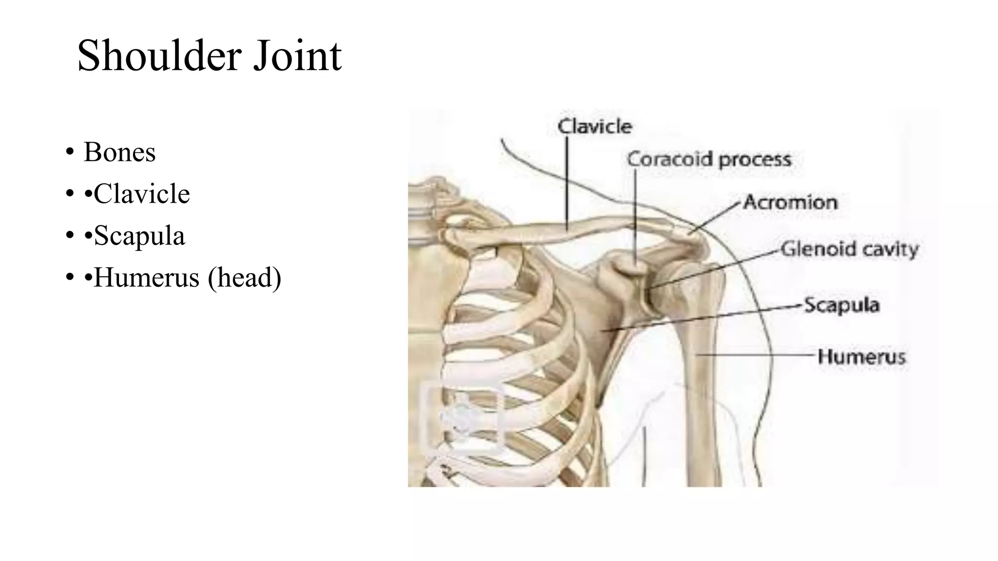
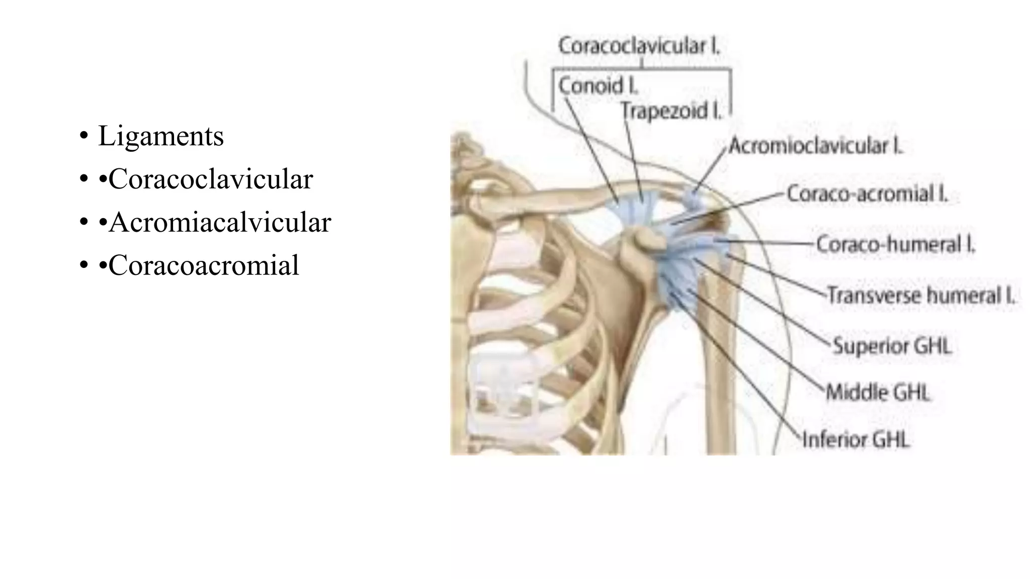
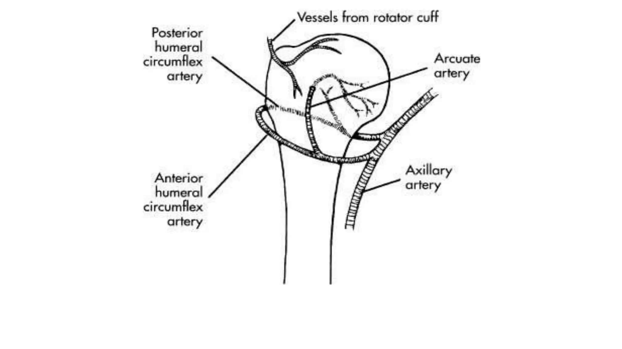
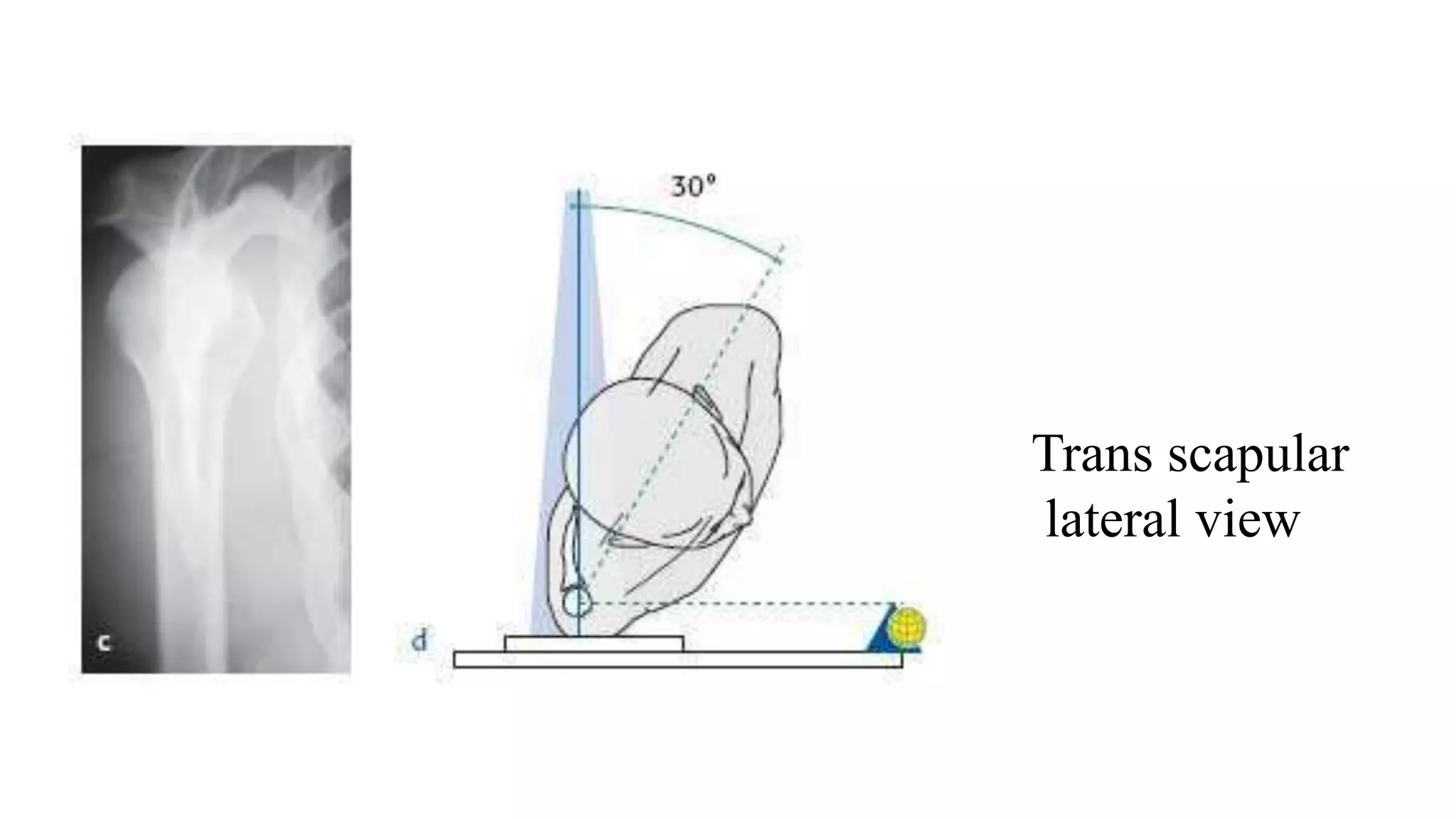
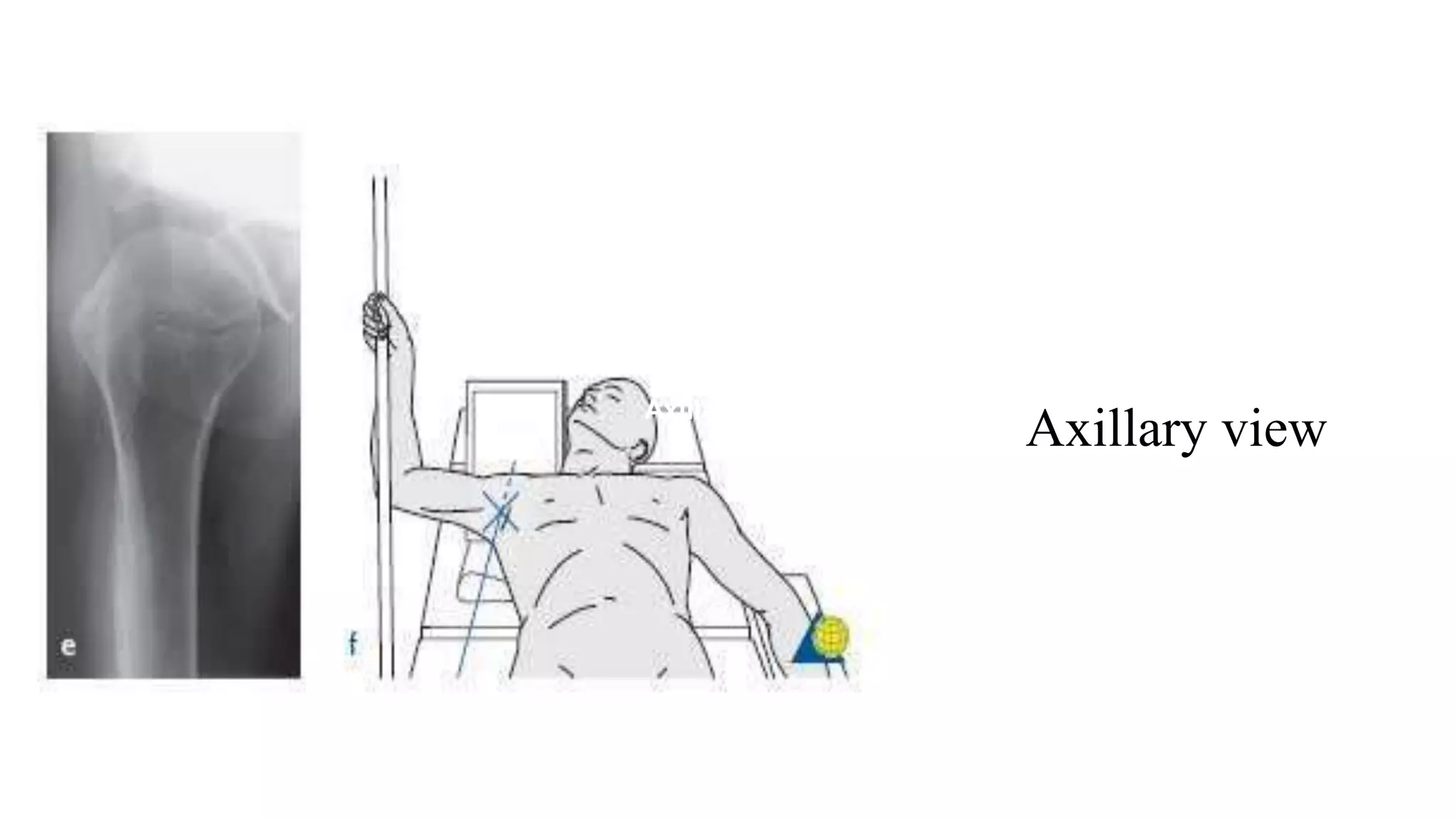
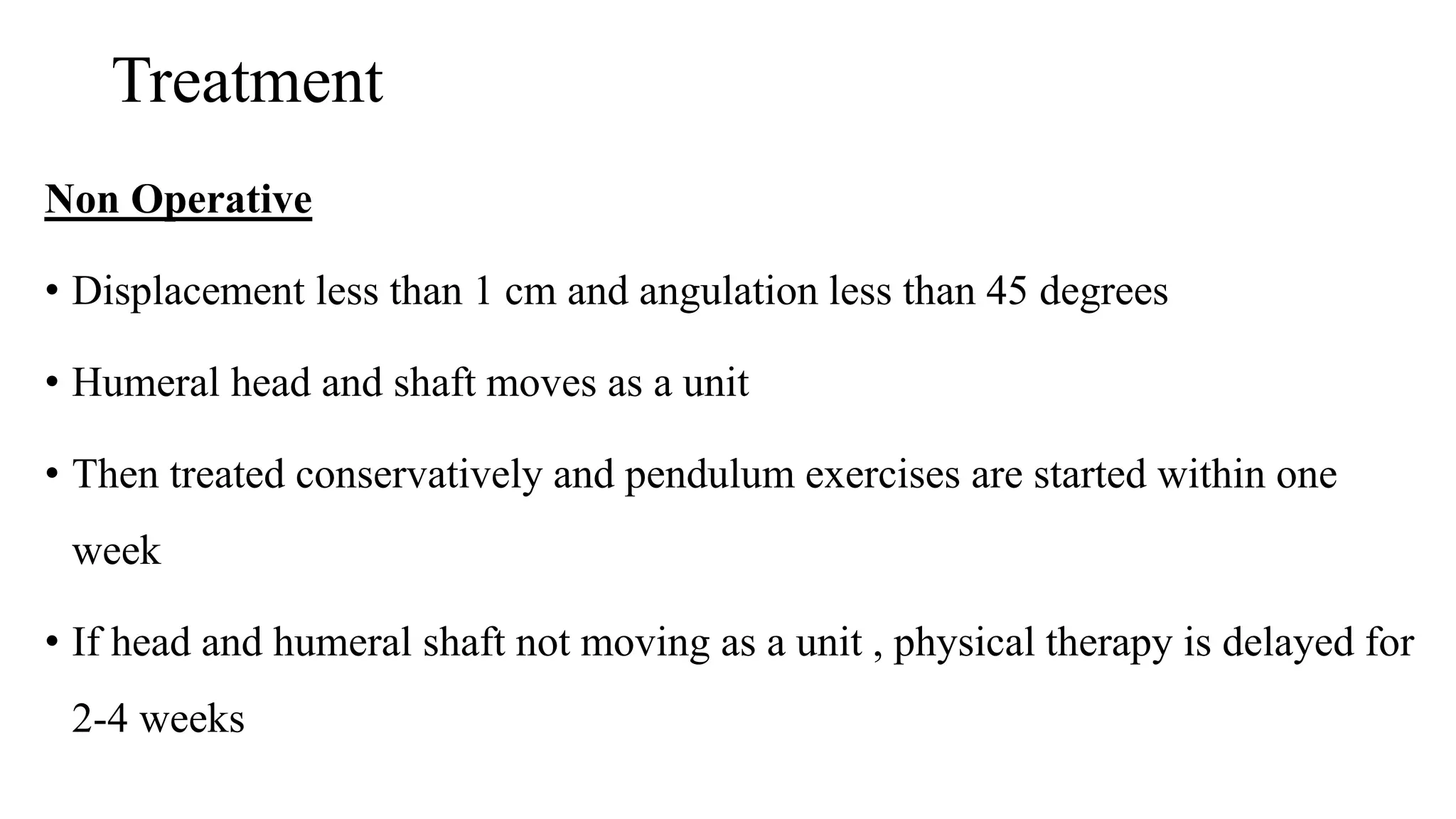
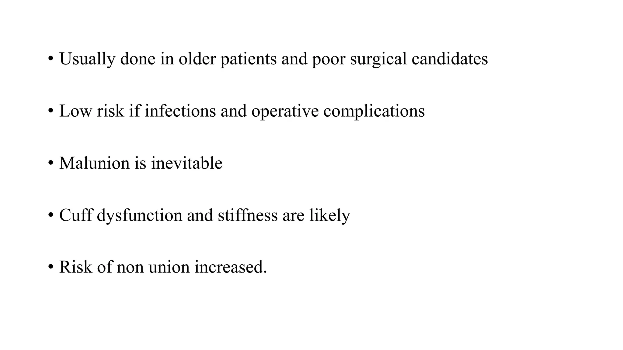
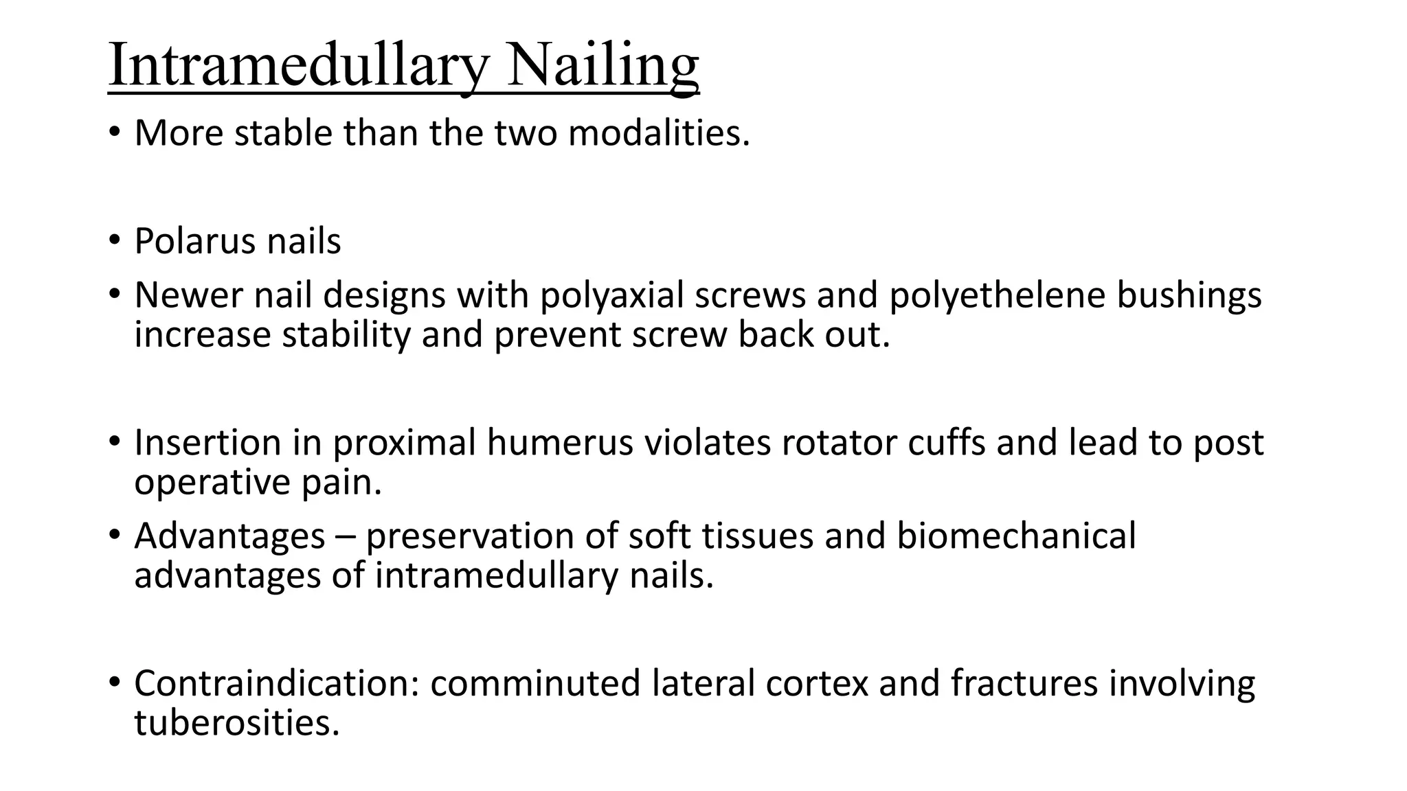
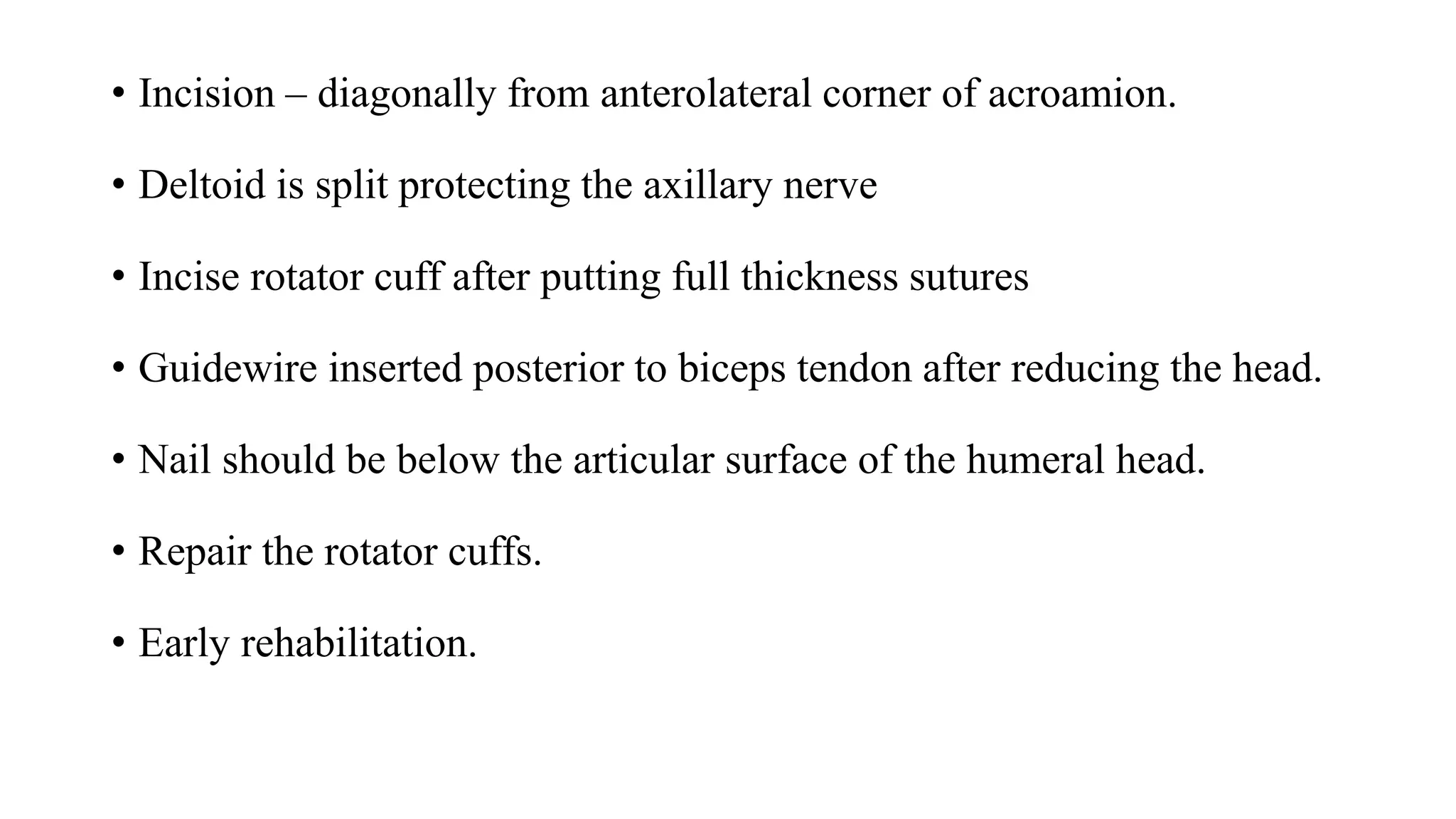
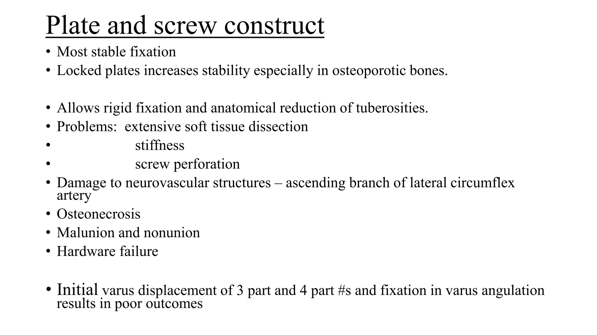
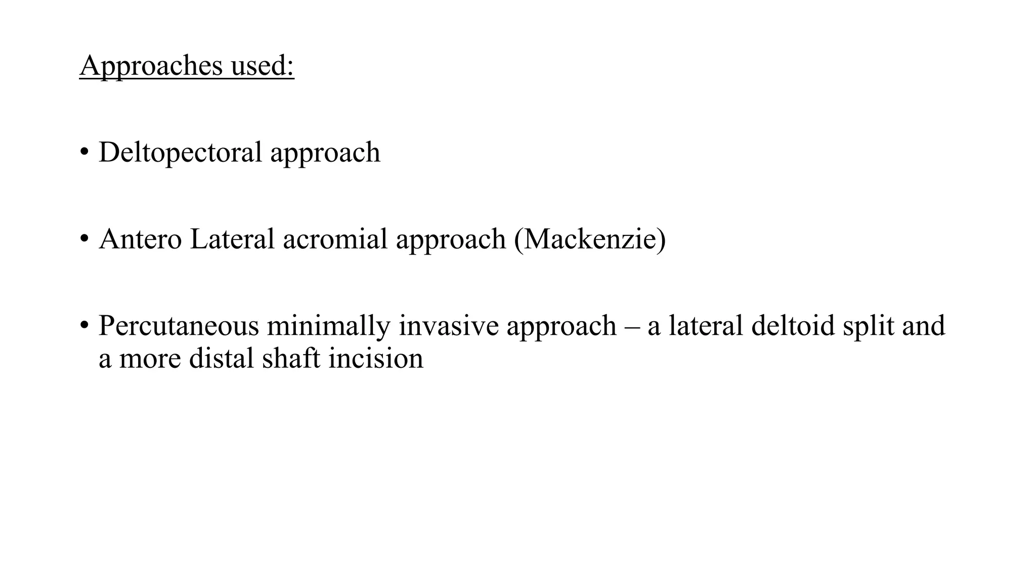
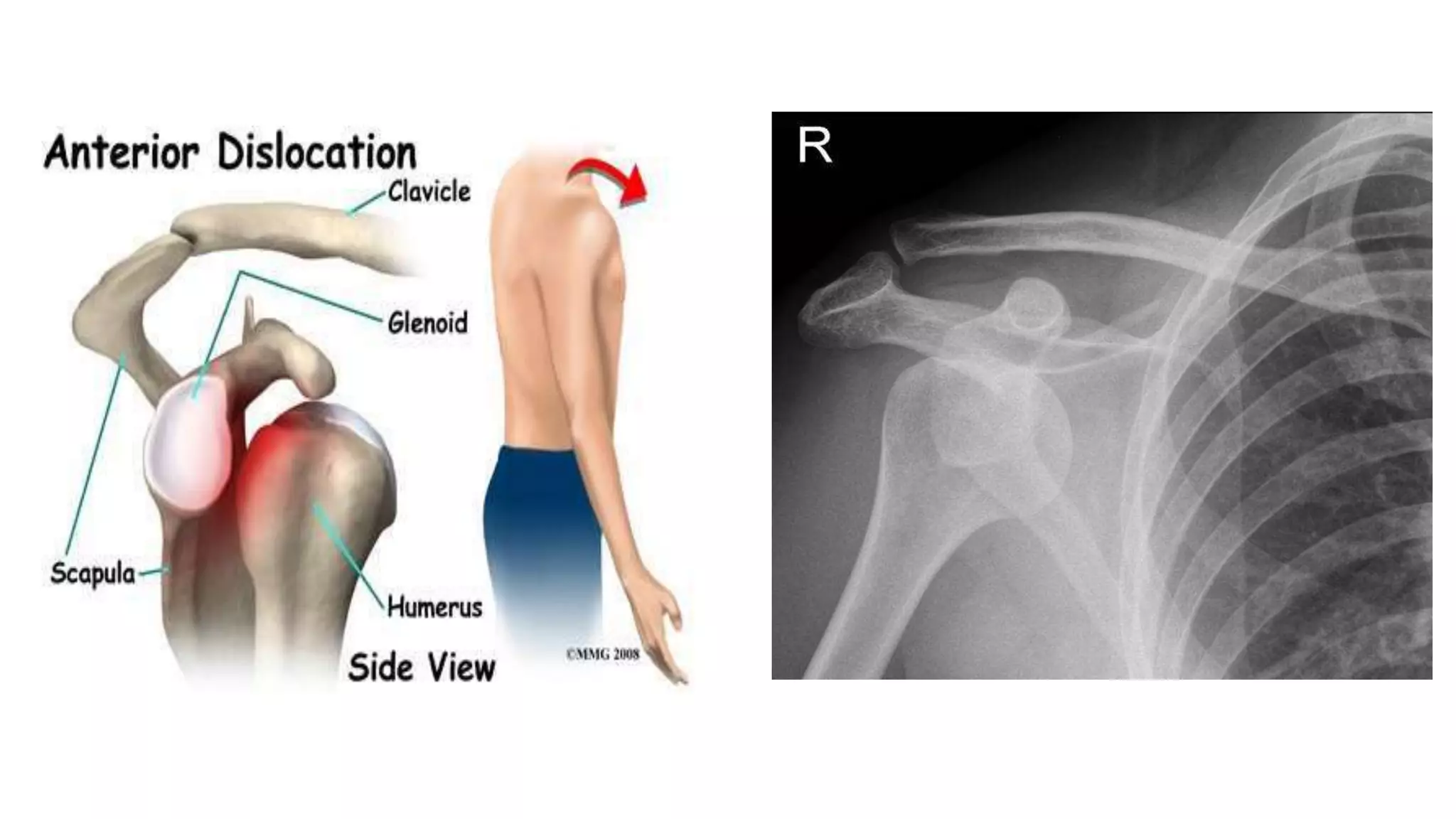
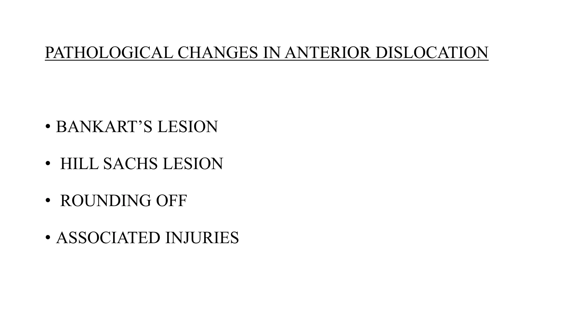
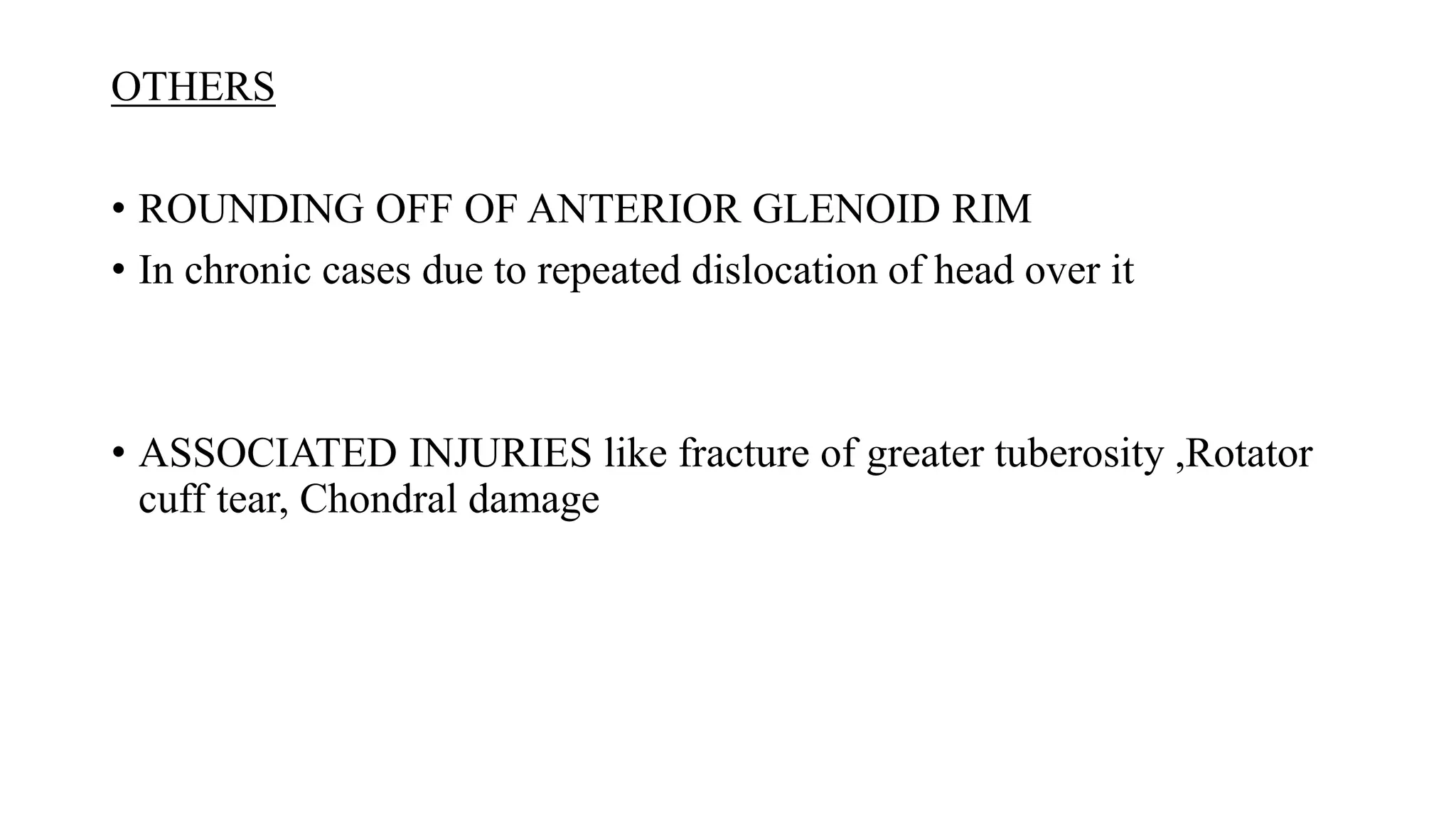
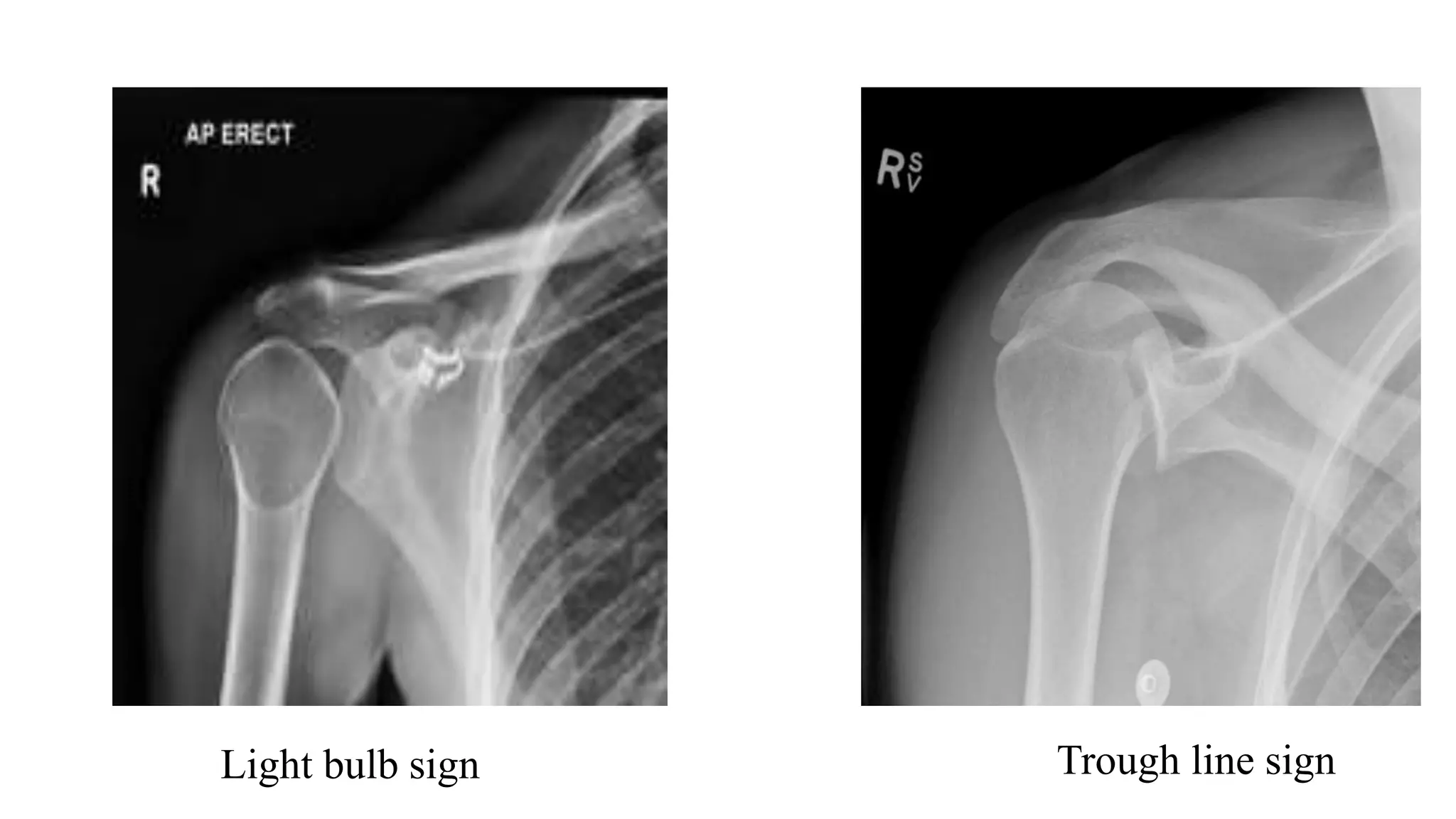
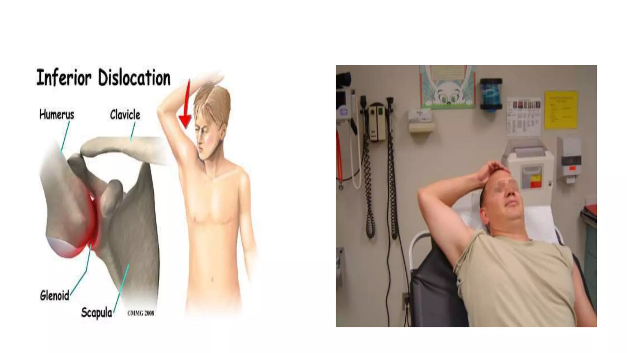
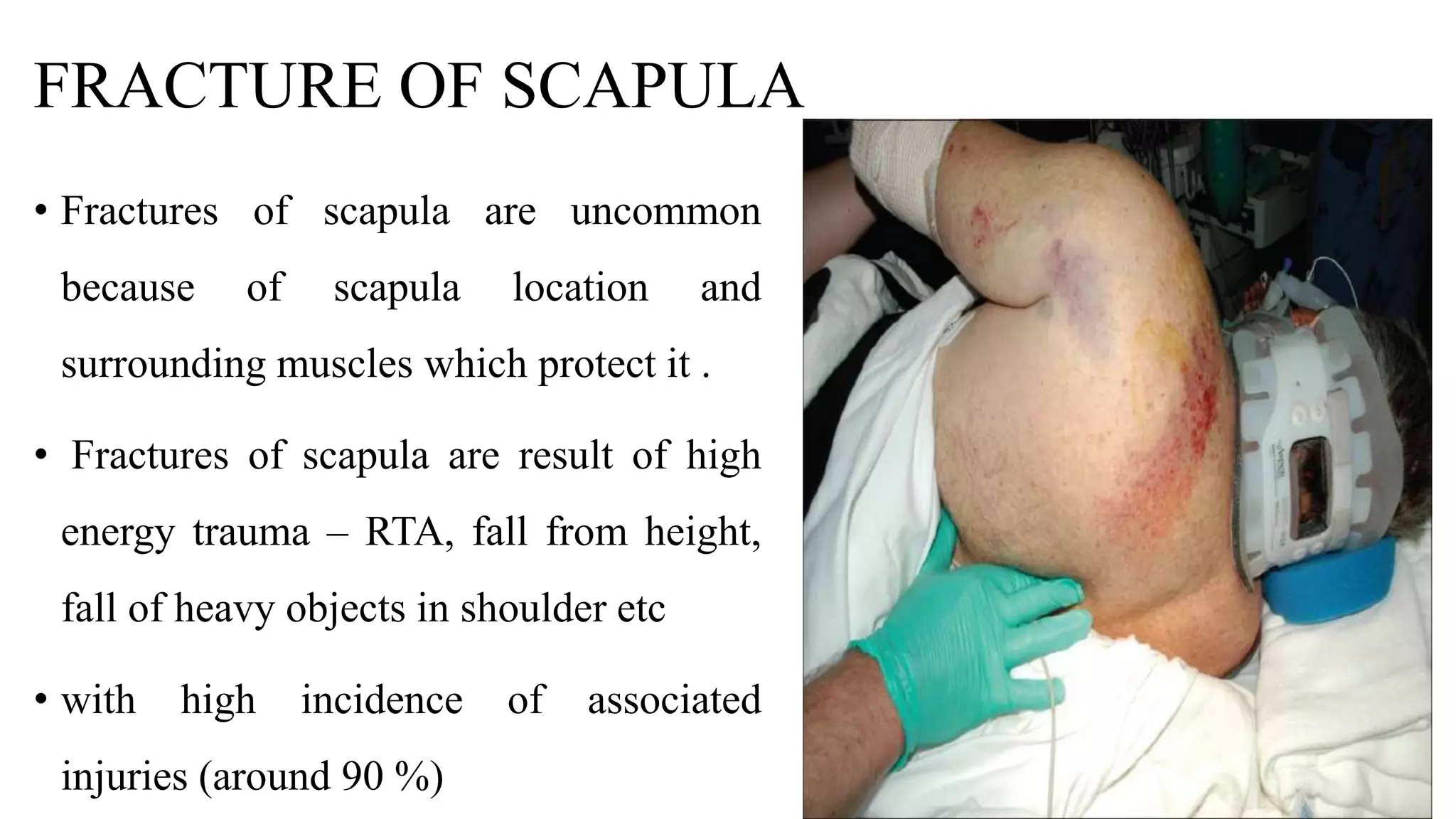
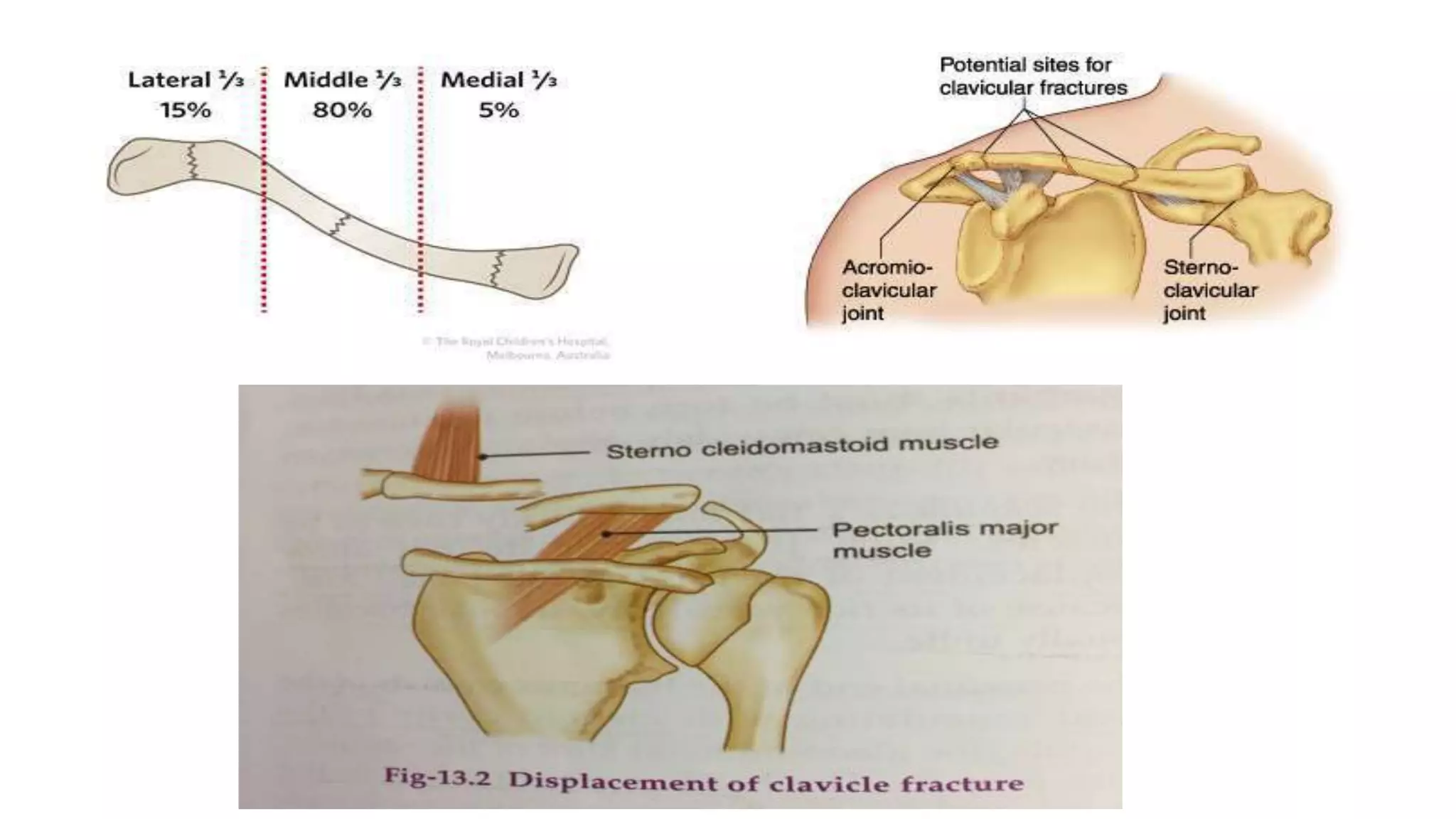
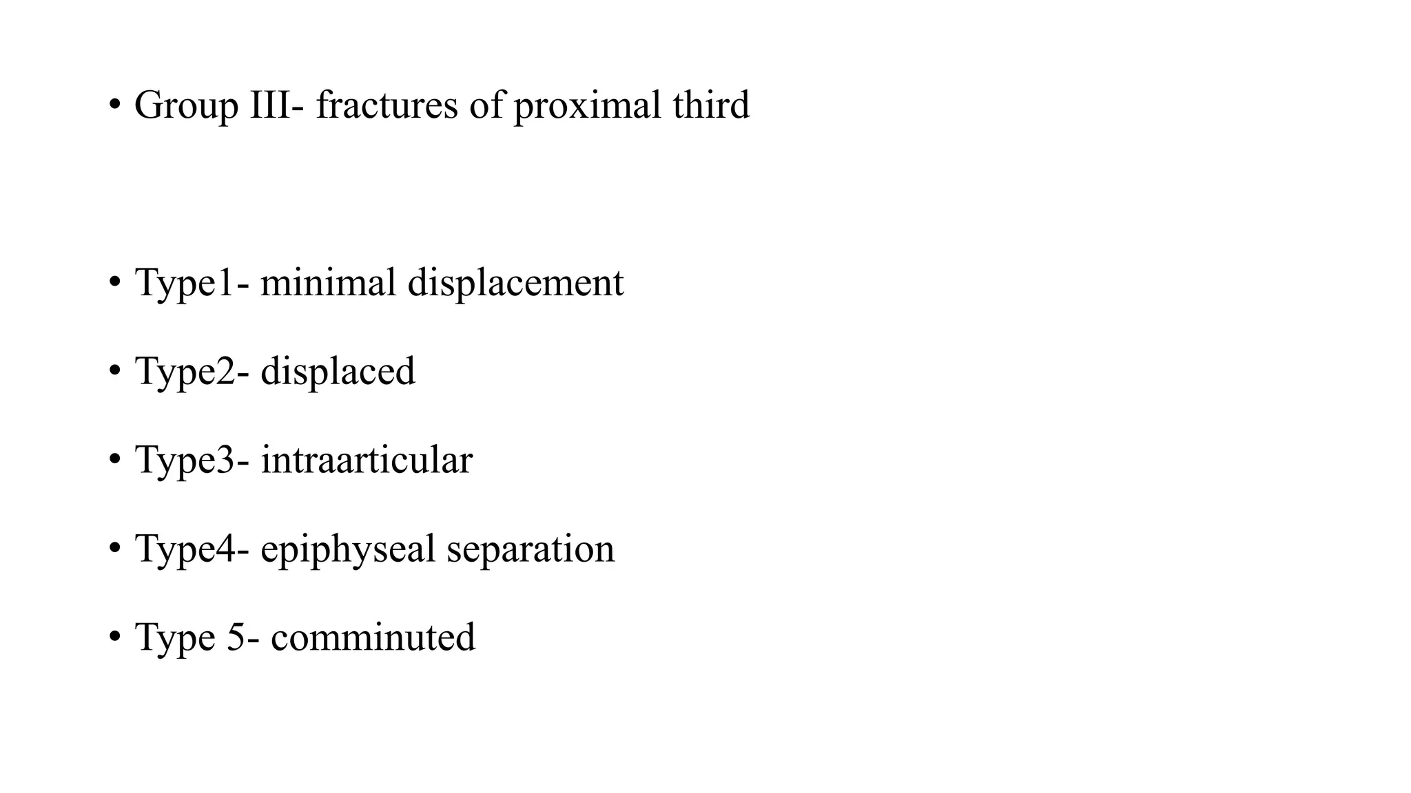
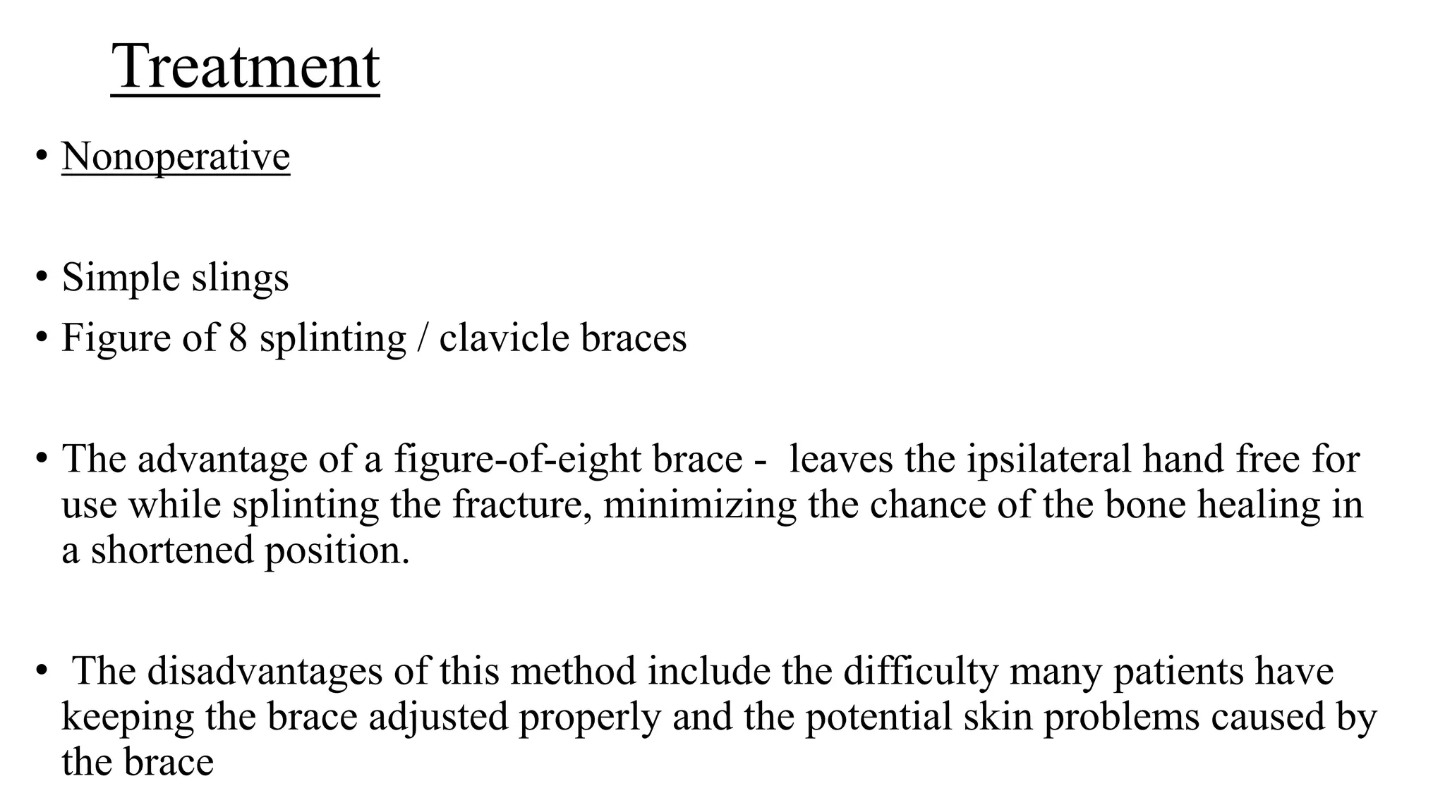
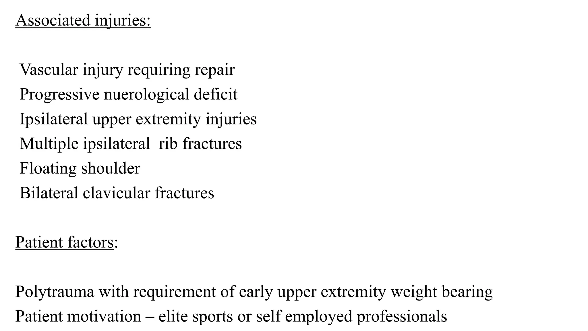
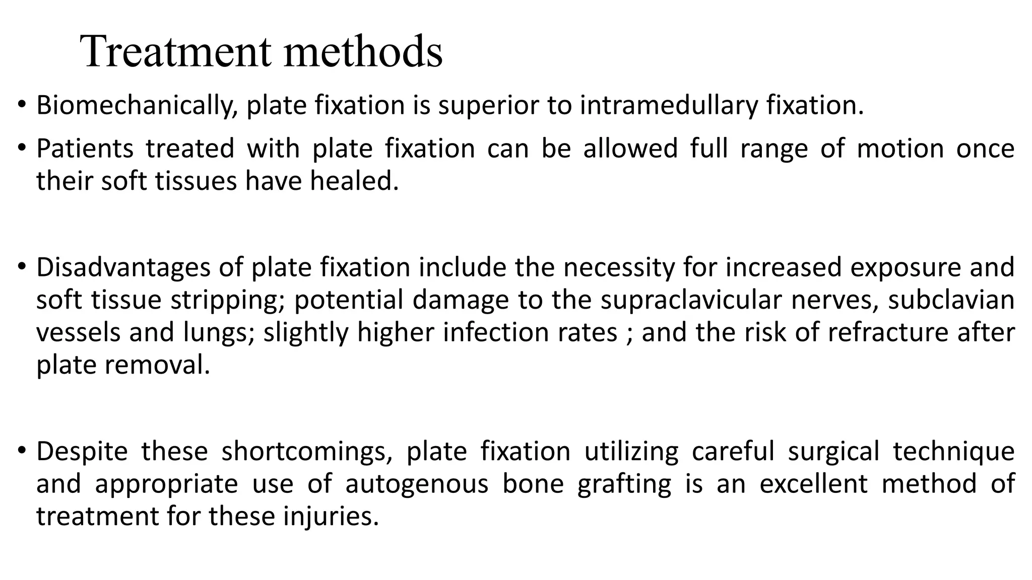
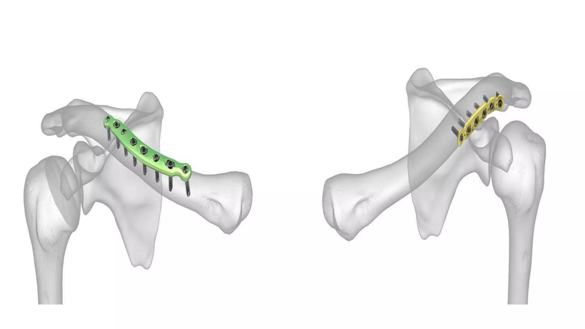
THE FRACTURES AROUND
THE SHOULDER
Dr Bibin K Baby
Shoulder Joint
• Bones
• •Clavicle
• •Scapula
• •Humerus (head)
• Ligaments
• •Coracoclavicular
• •Acromiacalvicular
• •Coracoacromial
Proximal Humerus Fractures
• 2-4 % of upper extremity #
• 5% of all #.
• second most common fracture of the upper extremity.
• F:M – 3:1
• Incidence increases with age.
• The fact that 10 different fixation techniques were evaluated for a
single fracture type, indicates the complexity of treating proximal
humerus fractures.
Neers Classification
• 4 part anatomy-
humeral head , greater tuberosity, lesser tuberosity and humeral shaft
• Displacement greater than 1 cm or angulation greater than 45 degrees
Classification
Trauma-series x-rays
Glenoid
AP view
Trans scapular
lateral view
Axillary view
Axillary view
Treatment
Non Operative
• Displacement less than 1 cm and angulation less than 45 degrees
• Humeral head and shaft moves as a unit
• Then treated conservatively and pendulum exercises are started within one
week
• If head and humeral shaft not moving as a unit , physical therapy is delayed for
2-4 weeks
• Usually done in older patients and poor surgical candidates
• Low risk if infections and operative complications
• Malunion is inevitable
• Cuff dysfunction and stiffness are likely
• Risk of non union increased.
Operative Treatment
• Restoration of proximal humerus anatomy and early achievement of
functional range of motion
• Transosseous suture fixation, percutaneous pinning, intramedullary
nailing and plate fixation
• Depends on age of patient, activity level, bone quality, fracture type
and surgeons technical ability.
Before surgery is considered ,determine the
adequency of blood supply and bone quality
• Hertal radiographic criteria for perfusion-
• Metaphyseal extension of humeral head of less than 8 mm and
medial hinge disruption of more than 2 mm are predictive of
ischemia.
• The cortical thickness of humeral diaphysis of less than 4 mm
precludes internal fixation because adequate screw purchase cannot
be obtained.
Transosseous Suture Fixation
• Mainly for 2 part and 3 part
• Provides the advantage of incorporating the rotator cuff insertion to
increase fixation strength especially in poor bone quality cases.
• Less surgical soft tissue dissection, low rate of osteonecrosis,
avoidance of bulky and expensive implants
• Concern- loss of reduction due to nonrigid construct and problems in
early mobilization of shoulder joint.
Percutaneous pinning
• Avoids damage to soft tissue envelope and blood supply to the head
• 2 part, 3 part and valgus impacted 4 part #s
• Requirements: satisfactory closed reduction, adequate bone stock,
minimal comminution, intact medial calcar and a compliant patient.
• Problems : loss of reduction, pin tract infections and axillary nerve
injuries.
• Contraindicated in fractures with metaphyseal comminution.
Intramedullary Nailing
• More stable than the two modalities.
• Polarus nails
• Newer nail designs with polyaxial screws and polyethelene bushings
increase stability and prevent screw back out.
• Insertion in proximal humerus violates rotator cuffs and lead to post
operative pain.
• Advantages – preservation of soft tissues and biomechanical
advantages of intramedullary nails.
• Contraindication: comminuted lateral cortex and fractures involving
tuberosities.
• Incision – diagonally from anterolateral corner of acroamion.
• Deltoid is split protecting the axillary nerve
• Incise rotator cuff after putting full thickness sutures
• Guidewire inserted posterior to biceps tendon after reducing the head.
• Nail should be below the articular surface of the humeral head.
• Repair the rotator cuffs.
• Early rehabilitation.
Plate and screw construct
• Most stable fixation
• Locked plates increases stability especially in osteoporotic bones.
• Allows rigid fixation and anatomical reduction of tuberosities.
• Problems: extensive soft tissue dissection
• stiffness
• screw perforation
• Damage to neurovascular structures – ascending branch of lateral circumflex
artery
• Osteonecrosis
• Malunion and nonunion
• Hardware failure
• Initial varus displacement of 3 part and 4 part #s and fixation in varus angulation
results in poor outcomes
Approaches used:
• Deltopectoral approach
• Antero Lateral acromial approach (Mackenzie)
• Percutaneous minimally invasive approach – a lateral deltoid split and
a more distal shaft incision
• For 3 part and 4 part suture are placed into the rotator cuff tendons to aid
reduction.
• Provisionally fix with k wires
• Place the plate on to the greater tuberosity and posterior to the biceps
tendon
• Place two locking screws in head segment and one or 2 screws in shaft
segment and confirm reduction with fluoroscopy
• For fractures with medial comminution first fix the plate onto the proximal
segment and then reduce the shaft segment to the plate.
• Helps in reducing varus malposition.
• Screw fixation into the inferomedial humeral head also adds stability to
fractures with medial comminution.
Special cosiderations
• For tuberosity fractures a displacement of 5 mm is considered an indication
for operative treatment
• Fixed usually with transosseous sutures and screws for larger fragments.
• For extremely osteoporotic surgical neck fractures :
Banco et al parachute technique
• Valgus impaction osteotomy and tension band fixation incorporating
transosseous sutures.
• For osteopenic 3 part fractures : hemiarthroplasty can be considered
• But plate fixation is still the preferred mode of treatment.
• 4 part fractures: poor outcome with nonoperative treatment and also
serious complications following surgical treatment .
• But osteonecrosis alone will not lead to poor outcome once the anatomical
relationship of head ,tuberosities and the shaft are reextrablished.
• Rigid fixation with locking plates are currently the choice of treatment.
• Hemiarthroplasty is a viable option in elderly low demand patients.
•
SHOULDER DISLOCATIONS
• anterior > 95%
• posterior 2-4%
• inferior (luxatio erecta) < 1%
MECHANISM OF INJURY
• COMMONEST :Fall on an outstretched hand with shoulder abducted
and externally rotated
POSTERIOR DISLOCATION:
• by direct blow from the front of the shoulder
or
• from epileptiform convulsions or electric Shock.
Clinical Manifestation
• Deformity in the shape of the arm and loss of contour of the shoulder
• If axillary nerve hurts, paresthesia can be detect in lateral and proximal of
the arm.(regimen badge sign)
• We can palpate the head of the humerus
• ROM is limited and painful
PATHOLOGICAL CHANGES IN ANTERIOR DISLOCATION
• BANKART’S LESION
• HILL SACHS LESION
• ROUNDING OFF
• ASSOCIATED INJURIES
BANKARTS LESION
Seen in anterior dislocation.
Stripping of glenoid labrum along with
periosteum .
Antero inferior Surface of glenoid and
scapular neck.
Avulsion of anteroinferior Glenoid rim
causes Bony Bankart Lesion.
HILL SACHS LESION
Depression on humeral head in its postero
lateral Quadrant
Due to impingement by the anterior edge of
glenoid on the head as it dislocates
HILL SACH LESION
BANKART LESION
OTHERS
• ROUNDING OFF OF ANTERIOR GLENOID RIM
• In chronic cases due to repeated dislocation of head over it
• ASSOCIATED INJURIES like fracture of greater tuberosity ,Rotator
cuff tear, Chondral damage
Posterior Shoulder Dislocation
• Radiological signs on AP view:
• Internal rotation of the humerus
• Light bulb sign
• The ‘rim sign’ – Widening of the glenohumeral space
• Trough line sign - the anterior aspect of the humeral head becomes
impacted against the posterior glenoid rim
Light bulb sign Trough line sign
• Inferior shoulder dislocation
• Radiographic features
• The humeral head is displaced directly below the glenoid fossa.
• Arm often in marked abduction (luxatio erecta).
Treatment
• Closed reduction
• Adequate sedation and muscle relaxation should be achieved.
• Simple traction counter traction ( orginally described by Hippocrates)
• Stimson technique: patient in prone , weight is hung from wrist to
provide traction . This steady traction lads to fatigue and relaxation of
shoulder musculature resulting in joint reduction.
Milch technique: patients mostly in supine or sitting , arm is
slowly abducted while stabilizing the humeral head with
opposite hand .
The shoulder is externally rotated , causing head to
spontaneously reduce approximately at 90 degrees of abduction
and ER.
• In posterior dislocation the reduction maneuver is forward flexion with
arm in adduction and internal rotation. An assistant places gentle cross
body traction and digital pressure is placed on the posterior humeral
head.
• The shoulder is immobilized for 3-4 weeks
Indications for open reduction and procedures
• Irreducible or unstable first time acute reduction
• Recurrent instability failing nonoperative treatment
• Open injuries
• Chronic dislocations with poor function and pain
• Dislocations with proximal humerus fractures
• Reduction is accompanied by bony and soft tissue procedures
depending on the associated pathologies.
FRACTURE OF SCAPULA
• Fractures of scapula are uncommon
because of scapula location and
surrounding muscles which protect it .
• Fractures of scapula are result of high
energy trauma – RTA, fall from height,
fall of heavy objects in shoulder etc
• with high incidence of associated
injuries (around 90 %)
Associated injuries with scapula fractures
• Isolated scapula fractures are rare
• Fractures of ribs – most frequent association
• Injuries to thoracic cavities and lungs – pneumothorax, hemothorax,
emphysema and lung contusion
• Injuries to shoulder girdle- clavicle, proximal humerus
• Head injuries- cerebral contusion , intracerebral bleeding
• Other injuries- pelvic fractures, thoracic and lumbar vertebral fractures
, neurovascular injuries
• Mortality – 2 % to 14 %
Imaging in Scapular Fractures
• AP radiograph of the entire shoulder girdle
• Neer I projection- true AP xray of scapula
• Neer II projection- also called the Y view -the lateral scapular
projection
• Chest radiograph
• Axillary views
• CT scans – for revealing exact fracture pattern and involvement of
articular surfaces . 3 Dimensional CT reconstructions are indicated.
• AO classification
• Scapula -14
• 14 A- process #s
• 14B- body #s
• 14F- glenoid fossa #s
• 14 A1- coracoid process
• 14 A2- acromion
• 14 A3- spine of scapula
• B1- # exists body at 2 or less points
• B2- # exists body at 3 or more points
• F0- # through extraarticular subchondral bone of glenoid fossa
( neck #s)
F1- simple fractures -1) anterior rim #s
2) posterior rim #s
3) transverse/short oblique fractures
F2- multifragmentary( 3 or more # lines)
glenoid fossa #
central fracture dislocations
Treatment options
• Majority of scapular fractures especially body and neck fractures are treated
nonoperatively.
• Immoblize the shoulder for 2-3 weeks and active assisted range of
movements are started thereafter, followed by strengthening exercises once
union is radiologically /clinically achieved.
• For all undisplaced fractures of scapula nonoperative treatment is the
choice.
• It should also be used in intra or extra articular displaced fractures when
patients general or local condition does not allow operation.
Operative treatment of scapular fractures
Glenoid fractures-
• To restore the integrity of glenohumeral joint.
• Main indication is displacement – gap more then 3- 4 mm
• Simultaneous involvement of 20%- 30 % articular surface
• Persisting subluxation of humeral head.
Displaced process fractures-
• To achieve healing in anatomical positions
• Healing in displacement causes impingement syndrome and rotator
cuff dysfunction.
• Displacement more than 1 cm is the major indication.
• Displaced extraarticular fractures – body and neck #s
• For restoration of glenopolar angle
• And restoration of length and integrity of lateral border
• Glenopolar angle- this angle is formed by a line drawn from inferior
pole of glenoid fossa to superior pole and
• A second line from the superior pole down through the inferiormost
angle of scapular body.
• Normal is 30 – 45 degrees
Glenopolar angle
Current indications for operative treatment
• 100 % translation of fragments of the lateral border
• 30-40 degrees angulation of main fragments of lateral border
• A 2cm lateral border offset (lateralization)
• Glenopolar angle less than 20 degrees
• Scapular body fractures with injury to the clavicle or clavicle -
acromion complex
• But final decision to be made after considering other injuries like chest
injuries, patient age, skin condition around the shoulder.
Surgical approaches
• Judet approach-
• Excellent exposure to entire scapula – universal approach
• Phase1- boomerang skin incision along the scapular spine and the medial
border of the scapula . Skin flap raised and posterior border of deltoid
identified.
• Phase2- posterior deltoid is detached from the scapular spine and turned
back laterally and distally.
• Phase3- infraspinatus is moblised and retracted proximally.
• Other limited approaches includes- posterosuperior approach, mini-invasive
posterior approach, posterolateral approach, superior approach.
• Operative techniques
• Implants : 3.5 to 2.7 mm reconstruction plates, 3.5 semitubular plate,
3.5 to 2.7 T plates or L plates
• Anatomically contoured plates or locking plates
• Cannulated screws and mini screws for process fractures of glenoid
fossa fractures
• Reduction and fixation: scapula has uneven bone mass
• Areas with sufficient anchorage for implants- lateral border of body,
scapular spine, scapular neck with glenoid
• Less favorable areas- spinomedial and inferior angles
• First step – stabilize the fractures of lateral border
• Scapular neck fractures combination of implants used.
• While fixing glenoid fragment- avoid intraarticular penetration
• Glenoid rim fractures are treated with lag screws and washers or small
plates depending on size of fragment .
• Correct rehabilitation is very important for final outcome.
• Passive range of motion exercises begin in first post op day and continue to
6 weeks
• Active range of motion exercises by 4-5 weeks and resistance exercises by
8 weeks .
Complications
• Injury to suprascapular nerve
• Malreduction
• Intraarticular perforation of screws
• Joint stiffness
• Infections
• Failure of implants
• Impingements
• Heterotopic ossification
CLAVICULAR FRACTURES
• Accounts for 2.6% of all fractures
• Common in young active individuals
• Majority of fractures are in the midshaft
• Usually nonoperative treatment was preferred for most of the clavicular
fractures
• But recent studies showed decreased union rates and high incidence of
functional impairment and morbidities especially in comminuted and
completely displaced clavicular fractures.
• Now the trend is moving towards operative management in special indications
Investigations
• Simple AP chest radiograph
• CT scan – not indicated for simple midshaft fractures
• done in complex fractures involving shoulder girdle
• Also done in medial intraarticular fractures
• Lateral clavicular fractures – zanca view for proper visualization of AC
joint.( beam angles at 15 degree cephalic tilt)
Classification
• Group I- fractures of middle third – most common
• GroupII- fractures of the distal third
• Type1- minimal displacement – coracoclavicular ligaments intact,
attached to medial segment
• Type2- coracoclavicular ligaments detached from medial segment
• Type2a- both conoid and trapezoid attached to distal segment
• Type2b- conoid ligament torn
• Type 3 – intraarticular extension into acromioclavicular joint.
• Group III- fractures of proximal third
• Type1- minimal displacement
• Type2- displaced
• Type3- intraarticular
• Type4- epiphyseal separation
• Type 5- comminuted
Treatment
• Nonoperative
• Simple slings
• Figure of 8 splinting / clavicle braces
• The advantage of a figure-of-eight brace - leaves the ipsilateral hand free for
use while splinting the fracture, minimizing the chance of the bone healing in
a shortened position.
• The disadvantages of this method include the difficulty many patients have
keeping the brace adjusted properly and the potential skin problems caused by
the brace
Operative treatment
• Indications for primary fixation in midshaft fractures
• Fracture specific:
• Displacement > 2 cm
• Shortening > 2 cm
• Increased comminution (> 3 fragments )
• Segmental #s
• Open #s
• Impending open fractures with soft tissue compromise
• Scapular malposition and winging at initial presentation
Associated injuries:
Vascular injury requiring repair
Progressive nuerological deficit
Ipsilateral upper extremity injuries
Multiple ipsilateral rib fractures
Floating shoulder
Bilateral clavicular fractures
Patient factors:
Polytrauma with requirement of early upper extremity weight bearing
Patient motivation – elite sports or self employed professionals
Treatment methods
• Biomechanically, plate fixation is superior to intramedullary fixation.
• Patients treated with plate fixation can be allowed full range of motion once
their soft tissues have healed.
• Disadvantages of plate fixation include the necessity for increased exposure and
soft tissue stripping; potential damage to the supraclavicular nerves, subclavian
vessels and lungs; slightly higher infection rates ; and the risk of refracture after
plate removal.
• Despite these shortcomings, plate fixation utilizing careful surgical technique
and appropriate use of autogenous bone grafting is an excellent method of
treatment for these injuries.
• A 3.5-mm AO dynamic compression plate (DCP) or a low-contact
• Dynamic compression plate with at least three screws (six cortices) in
both the medial and lateral fragment and an interfragmentary lag screw
whenever the fracture pattern allows it.
• 3.5-mm AO reconstruction plates provide acceptable fixation and rigidity
and allows easy contouring.
• One-third of tubular plates have a high rate of fatigue failure when used
for clavicle fractures and should be avoided.
• The plates should be precontoured and placed superiorly (most common)
or Anteroinferior (provides safe screw trajectory).
• Autogenous bone graft should be used in comminuted fractures with bone
loss.
Intramedullary Fixation
• Intramedullary fixation can be accomplished with smooth or threaded Kwires,
rush nails, or Rockwood pins .
• Recently titanium elastic intramedullary nails have been used.
• The advantages of using intramedullary devices are: less surgical dissection and
soft tissue stripping is needed, and the hardware is less prominent.
• Disadvantages include possible pin migration, pin breakage , intrathoracic
migration and poor rotational control during elevation of the extremity above
shoulder level.
• External fixation of the clavicle
• its indications are few
• It may be indicated for severe open fractures with poor quality of
overlying skin.
• External fixation may also be indicated for treatment of clavicle fractures
in the face of infection or infected nonunions following plate removal.
• Even in these cases, plate fixation should be considered first and used
whenever possible.
Lateral third fractures
• Type I fractures are stable because of the intact surrounding ligaments
and can be treated effectively with sling immobilization and progressive
use of the shoulder as pain allows.
• Most type I fractures heal within 4 to 6 weeks with little to no residual
shoulder dysfunction
• Several surgical treatment options are possible
• Transacromial K-wires with or without a tension band
• Coracoclavicular screw
• Plate fixation –Hook Plate
• Coracoclavicular banding or taping with or without acromioclavicular fixation
utilizing dacron or other synthetic materials
• The choice of one technique over another should be based on the size of the distal
fragment, patient and fracture anatomy, and surgeon’s preference.
• If there is a noncomminuted, 2- to 3-cm distal piece, then a small-fragment AO T-
plate or Two K-wires with a tension band placed outside the acromioclavicular
joint are good choices for fixation.
• On the other hand, comminuted and/or small distal fragments require
• Transacromial wire fixation
• coracoclavicular screw fixation
• or
• coracoclavicular ligament repair
• Type III fractures (those with intraarticular extension) of the distal clavicle can
be treated nonoperatively with a sling for support and gradual return to normal
use of the extremity as pain allows.
• If the fracture is unstable, however, then treatment should be similar to that for
type II fractures.
• In severely comminuted fractures, primary excision can be performed with
repair or reconstruction of the coracoclavicular ligaments using the Weaver-
Dunn procedure as necessary to stabilize the clavicle
Shoulder fractures around the shoulder

Shoulder fractures around the shoulder

  • 1.
    THE FRACTURES AROUND THESHOULDER Dr Bibin K Baby
  • 2.
    Shoulder Joint • Bones ••Clavicle • •Scapula • •Humerus (head)
  • 3.
    • Ligaments • •Coracoclavicular ••Acromiacalvicular • •Coracoacromial
  • 5.
    Proximal Humerus Fractures •2-4 % of upper extremity # • 5% of all #. • second most common fracture of the upper extremity. • F:M – 3:1 • Incidence increases with age. • The fact that 10 different fixation techniques were evaluated for a single fracture type, indicates the complexity of treating proximal humerus fractures.
  • 6.
    Neers Classification • 4part anatomy- humeral head , greater tuberosity, lesser tuberosity and humeral shaft • Displacement greater than 1 cm or angulation greater than 45 degrees
  • 7.
  • 8.
  • 9.
  • 10.
  • 11.
    Treatment Non Operative • Displacementless than 1 cm and angulation less than 45 degrees • Humeral head and shaft moves as a unit • Then treated conservatively and pendulum exercises are started within one week • If head and humeral shaft not moving as a unit , physical therapy is delayed for 2-4 weeks
  • 12.
    • Usually donein older patients and poor surgical candidates • Low risk if infections and operative complications • Malunion is inevitable • Cuff dysfunction and stiffness are likely • Risk of non union increased.
  • 13.
    Operative Treatment • Restorationof proximal humerus anatomy and early achievement of functional range of motion • Transosseous suture fixation, percutaneous pinning, intramedullary nailing and plate fixation • Depends on age of patient, activity level, bone quality, fracture type and surgeons technical ability.
  • 14.
    Before surgery isconsidered ,determine the adequency of blood supply and bone quality • Hertal radiographic criteria for perfusion- • Metaphyseal extension of humeral head of less than 8 mm and medial hinge disruption of more than 2 mm are predictive of ischemia. • The cortical thickness of humeral diaphysis of less than 4 mm precludes internal fixation because adequate screw purchase cannot be obtained.
  • 15.
    Transosseous Suture Fixation •Mainly for 2 part and 3 part • Provides the advantage of incorporating the rotator cuff insertion to increase fixation strength especially in poor bone quality cases. • Less surgical soft tissue dissection, low rate of osteonecrosis, avoidance of bulky and expensive implants • Concern- loss of reduction due to nonrigid construct and problems in early mobilization of shoulder joint.
  • 16.
    Percutaneous pinning • Avoidsdamage to soft tissue envelope and blood supply to the head • 2 part, 3 part and valgus impacted 4 part #s • Requirements: satisfactory closed reduction, adequate bone stock, minimal comminution, intact medial calcar and a compliant patient. • Problems : loss of reduction, pin tract infections and axillary nerve injuries. • Contraindicated in fractures with metaphyseal comminution.
  • 17.
    Intramedullary Nailing • Morestable than the two modalities. • Polarus nails • Newer nail designs with polyaxial screws and polyethelene bushings increase stability and prevent screw back out. • Insertion in proximal humerus violates rotator cuffs and lead to post operative pain. • Advantages – preservation of soft tissues and biomechanical advantages of intramedullary nails. • Contraindication: comminuted lateral cortex and fractures involving tuberosities.
  • 18.
    • Incision –diagonally from anterolateral corner of acroamion. • Deltoid is split protecting the axillary nerve • Incise rotator cuff after putting full thickness sutures • Guidewire inserted posterior to biceps tendon after reducing the head. • Nail should be below the articular surface of the humeral head. • Repair the rotator cuffs. • Early rehabilitation.
  • 19.
    Plate and screwconstruct • Most stable fixation • Locked plates increases stability especially in osteoporotic bones. • Allows rigid fixation and anatomical reduction of tuberosities. • Problems: extensive soft tissue dissection • stiffness • screw perforation • Damage to neurovascular structures – ascending branch of lateral circumflex artery • Osteonecrosis • Malunion and nonunion • Hardware failure • Initial varus displacement of 3 part and 4 part #s and fixation in varus angulation results in poor outcomes
  • 20.
    Approaches used: • Deltopectoralapproach • Antero Lateral acromial approach (Mackenzie) • Percutaneous minimally invasive approach – a lateral deltoid split and a more distal shaft incision
  • 21.
    • For 3part and 4 part suture are placed into the rotator cuff tendons to aid reduction. • Provisionally fix with k wires • Place the plate on to the greater tuberosity and posterior to the biceps tendon • Place two locking screws in head segment and one or 2 screws in shaft segment and confirm reduction with fluoroscopy • For fractures with medial comminution first fix the plate onto the proximal segment and then reduce the shaft segment to the plate. • Helps in reducing varus malposition. • Screw fixation into the inferomedial humeral head also adds stability to fractures with medial comminution.
  • 22.
    Special cosiderations • Fortuberosity fractures a displacement of 5 mm is considered an indication for operative treatment • Fixed usually with transosseous sutures and screws for larger fragments. • For extremely osteoporotic surgical neck fractures : Banco et al parachute technique • Valgus impaction osteotomy and tension band fixation incorporating transosseous sutures.
  • 23.
    • For osteopenic3 part fractures : hemiarthroplasty can be considered • But plate fixation is still the preferred mode of treatment. • 4 part fractures: poor outcome with nonoperative treatment and also serious complications following surgical treatment . • But osteonecrosis alone will not lead to poor outcome once the anatomical relationship of head ,tuberosities and the shaft are reextrablished. • Rigid fixation with locking plates are currently the choice of treatment. • Hemiarthroplasty is a viable option in elderly low demand patients. •
  • 24.
    SHOULDER DISLOCATIONS • anterior> 95% • posterior 2-4% • inferior (luxatio erecta) < 1%
  • 25.
    MECHANISM OF INJURY •COMMONEST :Fall on an outstretched hand with shoulder abducted and externally rotated POSTERIOR DISLOCATION: • by direct blow from the front of the shoulder or • from epileptiform convulsions or electric Shock.
  • 26.
    Clinical Manifestation • Deformityin the shape of the arm and loss of contour of the shoulder • If axillary nerve hurts, paresthesia can be detect in lateral and proximal of the arm.(regimen badge sign) • We can palpate the head of the humerus • ROM is limited and painful
  • 28.
    PATHOLOGICAL CHANGES INANTERIOR DISLOCATION • BANKART’S LESION • HILL SACHS LESION • ROUNDING OFF • ASSOCIATED INJURIES
  • 29.
    BANKARTS LESION Seen inanterior dislocation. Stripping of glenoid labrum along with periosteum . Antero inferior Surface of glenoid and scapular neck. Avulsion of anteroinferior Glenoid rim causes Bony Bankart Lesion.
  • 30.
    HILL SACHS LESION Depressionon humeral head in its postero lateral Quadrant Due to impingement by the anterior edge of glenoid on the head as it dislocates
  • 31.
  • 32.
    OTHERS • ROUNDING OFFOF ANTERIOR GLENOID RIM • In chronic cases due to repeated dislocation of head over it • ASSOCIATED INJURIES like fracture of greater tuberosity ,Rotator cuff tear, Chondral damage
  • 33.
    Posterior Shoulder Dislocation •Radiological signs on AP view: • Internal rotation of the humerus • Light bulb sign • The ‘rim sign’ – Widening of the glenohumeral space • Trough line sign - the anterior aspect of the humeral head becomes impacted against the posterior glenoid rim
  • 34.
    Light bulb signTrough line sign
  • 35.
    • Inferior shoulderdislocation • Radiographic features • The humeral head is displaced directly below the glenoid fossa. • Arm often in marked abduction (luxatio erecta).
  • 37.
    Treatment • Closed reduction •Adequate sedation and muscle relaxation should be achieved. • Simple traction counter traction ( orginally described by Hippocrates) • Stimson technique: patient in prone , weight is hung from wrist to provide traction . This steady traction lads to fatigue and relaxation of shoulder musculature resulting in joint reduction.
  • 38.
    Milch technique: patientsmostly in supine or sitting , arm is slowly abducted while stabilizing the humeral head with opposite hand . The shoulder is externally rotated , causing head to spontaneously reduce approximately at 90 degrees of abduction and ER.
  • 39.
    • In posteriordislocation the reduction maneuver is forward flexion with arm in adduction and internal rotation. An assistant places gentle cross body traction and digital pressure is placed on the posterior humeral head. • The shoulder is immobilized for 3-4 weeks
  • 40.
    Indications for openreduction and procedures • Irreducible or unstable first time acute reduction • Recurrent instability failing nonoperative treatment • Open injuries • Chronic dislocations with poor function and pain • Dislocations with proximal humerus fractures • Reduction is accompanied by bony and soft tissue procedures depending on the associated pathologies.
  • 41.
    FRACTURE OF SCAPULA •Fractures of scapula are uncommon because of scapula location and surrounding muscles which protect it . • Fractures of scapula are result of high energy trauma – RTA, fall from height, fall of heavy objects in shoulder etc • with high incidence of associated injuries (around 90 %)
  • 42.
    Associated injuries withscapula fractures • Isolated scapula fractures are rare • Fractures of ribs – most frequent association • Injuries to thoracic cavities and lungs – pneumothorax, hemothorax, emphysema and lung contusion • Injuries to shoulder girdle- clavicle, proximal humerus • Head injuries- cerebral contusion , intracerebral bleeding • Other injuries- pelvic fractures, thoracic and lumbar vertebral fractures , neurovascular injuries • Mortality – 2 % to 14 %
  • 43.
    Imaging in ScapularFractures • AP radiograph of the entire shoulder girdle • Neer I projection- true AP xray of scapula • Neer II projection- also called the Y view -the lateral scapular projection • Chest radiograph • Axillary views • CT scans – for revealing exact fracture pattern and involvement of articular surfaces . 3 Dimensional CT reconstructions are indicated.
  • 44.
    • AO classification •Scapula -14 • 14 A- process #s • 14B- body #s • 14F- glenoid fossa #s • 14 A1- coracoid process • 14 A2- acromion • 14 A3- spine of scapula
  • 45.
    • B1- #exists body at 2 or less points • B2- # exists body at 3 or more points • F0- # through extraarticular subchondral bone of glenoid fossa ( neck #s) F1- simple fractures -1) anterior rim #s 2) posterior rim #s 3) transverse/short oblique fractures F2- multifragmentary( 3 or more # lines) glenoid fossa # central fracture dislocations
  • 46.
    Treatment options • Majorityof scapular fractures especially body and neck fractures are treated nonoperatively. • Immoblize the shoulder for 2-3 weeks and active assisted range of movements are started thereafter, followed by strengthening exercises once union is radiologically /clinically achieved. • For all undisplaced fractures of scapula nonoperative treatment is the choice. • It should also be used in intra or extra articular displaced fractures when patients general or local condition does not allow operation.
  • 47.
    Operative treatment ofscapular fractures Glenoid fractures- • To restore the integrity of glenohumeral joint. • Main indication is displacement – gap more then 3- 4 mm • Simultaneous involvement of 20%- 30 % articular surface • Persisting subluxation of humeral head.
  • 48.
    Displaced process fractures- •To achieve healing in anatomical positions • Healing in displacement causes impingement syndrome and rotator cuff dysfunction. • Displacement more than 1 cm is the major indication.
  • 49.
    • Displaced extraarticularfractures – body and neck #s • For restoration of glenopolar angle • And restoration of length and integrity of lateral border • Glenopolar angle- this angle is formed by a line drawn from inferior pole of glenoid fossa to superior pole and • A second line from the superior pole down through the inferiormost angle of scapular body. • Normal is 30 – 45 degrees
  • 50.
  • 51.
    Current indications foroperative treatment • 100 % translation of fragments of the lateral border • 30-40 degrees angulation of main fragments of lateral border • A 2cm lateral border offset (lateralization) • Glenopolar angle less than 20 degrees • Scapular body fractures with injury to the clavicle or clavicle - acromion complex • But final decision to be made after considering other injuries like chest injuries, patient age, skin condition around the shoulder.
  • 52.
    Surgical approaches • Judetapproach- • Excellent exposure to entire scapula – universal approach • Phase1- boomerang skin incision along the scapular spine and the medial border of the scapula . Skin flap raised and posterior border of deltoid identified. • Phase2- posterior deltoid is detached from the scapular spine and turned back laterally and distally. • Phase3- infraspinatus is moblised and retracted proximally. • Other limited approaches includes- posterosuperior approach, mini-invasive posterior approach, posterolateral approach, superior approach.
  • 53.
    • Operative techniques •Implants : 3.5 to 2.7 mm reconstruction plates, 3.5 semitubular plate, 3.5 to 2.7 T plates or L plates • Anatomically contoured plates or locking plates • Cannulated screws and mini screws for process fractures of glenoid fossa fractures • Reduction and fixation: scapula has uneven bone mass • Areas with sufficient anchorage for implants- lateral border of body, scapular spine, scapular neck with glenoid • Less favorable areas- spinomedial and inferior angles
  • 54.
    • First step– stabilize the fractures of lateral border • Scapular neck fractures combination of implants used. • While fixing glenoid fragment- avoid intraarticular penetration • Glenoid rim fractures are treated with lag screws and washers or small plates depending on size of fragment . • Correct rehabilitation is very important for final outcome. • Passive range of motion exercises begin in first post op day and continue to 6 weeks • Active range of motion exercises by 4-5 weeks and resistance exercises by 8 weeks .
  • 55.
    Complications • Injury tosuprascapular nerve • Malreduction • Intraarticular perforation of screws • Joint stiffness • Infections • Failure of implants • Impingements • Heterotopic ossification
  • 56.
    CLAVICULAR FRACTURES • Accountsfor 2.6% of all fractures • Common in young active individuals • Majority of fractures are in the midshaft • Usually nonoperative treatment was preferred for most of the clavicular fractures • But recent studies showed decreased union rates and high incidence of functional impairment and morbidities especially in comminuted and completely displaced clavicular fractures. • Now the trend is moving towards operative management in special indications
  • 58.
    Investigations • Simple APchest radiograph • CT scan – not indicated for simple midshaft fractures • done in complex fractures involving shoulder girdle • Also done in medial intraarticular fractures • Lateral clavicular fractures – zanca view for proper visualization of AC joint.( beam angles at 15 degree cephalic tilt)
  • 59.
    Classification • Group I-fractures of middle third – most common • GroupII- fractures of the distal third • Type1- minimal displacement – coracoclavicular ligaments intact, attached to medial segment • Type2- coracoclavicular ligaments detached from medial segment • Type2a- both conoid and trapezoid attached to distal segment • Type2b- conoid ligament torn • Type 3 – intraarticular extension into acromioclavicular joint.
  • 60.
    • Group III-fractures of proximal third • Type1- minimal displacement • Type2- displaced • Type3- intraarticular • Type4- epiphyseal separation • Type 5- comminuted
  • 61.
    Treatment • Nonoperative • Simpleslings • Figure of 8 splinting / clavicle braces • The advantage of a figure-of-eight brace - leaves the ipsilateral hand free for use while splinting the fracture, minimizing the chance of the bone healing in a shortened position. • The disadvantages of this method include the difficulty many patients have keeping the brace adjusted properly and the potential skin problems caused by the brace
  • 62.
    Operative treatment • Indicationsfor primary fixation in midshaft fractures • Fracture specific: • Displacement > 2 cm • Shortening > 2 cm • Increased comminution (> 3 fragments ) • Segmental #s • Open #s • Impending open fractures with soft tissue compromise • Scapular malposition and winging at initial presentation
  • 63.
    Associated injuries: Vascular injuryrequiring repair Progressive nuerological deficit Ipsilateral upper extremity injuries Multiple ipsilateral rib fractures Floating shoulder Bilateral clavicular fractures Patient factors: Polytrauma with requirement of early upper extremity weight bearing Patient motivation – elite sports or self employed professionals
  • 64.
    Treatment methods • Biomechanically,plate fixation is superior to intramedullary fixation. • Patients treated with plate fixation can be allowed full range of motion once their soft tissues have healed. • Disadvantages of plate fixation include the necessity for increased exposure and soft tissue stripping; potential damage to the supraclavicular nerves, subclavian vessels and lungs; slightly higher infection rates ; and the risk of refracture after plate removal. • Despite these shortcomings, plate fixation utilizing careful surgical technique and appropriate use of autogenous bone grafting is an excellent method of treatment for these injuries.
  • 65.
    • A 3.5-mmAO dynamic compression plate (DCP) or a low-contact • Dynamic compression plate with at least three screws (six cortices) in both the medial and lateral fragment and an interfragmentary lag screw whenever the fracture pattern allows it. • 3.5-mm AO reconstruction plates provide acceptable fixation and rigidity and allows easy contouring. • One-third of tubular plates have a high rate of fatigue failure when used for clavicle fractures and should be avoided. • The plates should be precontoured and placed superiorly (most common) or Anteroinferior (provides safe screw trajectory). • Autogenous bone graft should be used in comminuted fractures with bone loss.
  • 67.
    Intramedullary Fixation • Intramedullaryfixation can be accomplished with smooth or threaded Kwires, rush nails, or Rockwood pins . • Recently titanium elastic intramedullary nails have been used. • The advantages of using intramedullary devices are: less surgical dissection and soft tissue stripping is needed, and the hardware is less prominent. • Disadvantages include possible pin migration, pin breakage , intrathoracic migration and poor rotational control during elevation of the extremity above shoulder level.
  • 68.
    • External fixationof the clavicle • its indications are few • It may be indicated for severe open fractures with poor quality of overlying skin. • External fixation may also be indicated for treatment of clavicle fractures in the face of infection or infected nonunions following plate removal. • Even in these cases, plate fixation should be considered first and used whenever possible.
  • 69.
    Lateral third fractures •Type I fractures are stable because of the intact surrounding ligaments and can be treated effectively with sling immobilization and progressive use of the shoulder as pain allows. • Most type I fractures heal within 4 to 6 weeks with little to no residual shoulder dysfunction
  • 70.
    • Several surgicaltreatment options are possible • Transacromial K-wires with or without a tension band • Coracoclavicular screw • Plate fixation –Hook Plate • Coracoclavicular banding or taping with or without acromioclavicular fixation utilizing dacron or other synthetic materials • The choice of one technique over another should be based on the size of the distal fragment, patient and fracture anatomy, and surgeon’s preference. • If there is a noncomminuted, 2- to 3-cm distal piece, then a small-fragment AO T- plate or Two K-wires with a tension band placed outside the acromioclavicular joint are good choices for fixation.
  • 71.
    • On theother hand, comminuted and/or small distal fragments require • Transacromial wire fixation • coracoclavicular screw fixation • or • coracoclavicular ligament repair
  • 72.
    • Type IIIfractures (those with intraarticular extension) of the distal clavicle can be treated nonoperatively with a sling for support and gradual return to normal use of the extremity as pain allows. • If the fracture is unstable, however, then treatment should be similar to that for type II fractures. • In severely comminuted fractures, primary excision can be performed with repair or reconstruction of the coracoclavicular ligaments using the Weaver- Dunn procedure as necessary to stabilize the clavicle