SlideShare a Scribd company logo
Radiological Imaging of Corpus Callosum lesions.
Dr/ ABD ALLAH NAZEER. MD.
Normal anatomy of the corpus callosum. T1-weighted
(left), T2-weighted (middle), and FLAIR (right) MR
images show the corpus callosum (CC). The splenium
is the most posterior and thickest portion of the CC.
Lesions of the corpus callosum are uncommon and arise from
multiple different etiologies. The lesions can be classified according
to underlying Pathophysiology .
Congenital
agenesis of the corpus callosum
enlarged perivascular spaces
tubonodular pericallosal lipoma: associated with dysgenesis
of the corpus callosum.
Demyelination
acute disseminated encephalomyelitis
Marchiafava-Bignami disease: demyelination and necrosis, can
appear as cystic lesions
multiple sclerosis
neuromyelitis optica
progressive multifocal leukoencephalopathy
Susac syndrome
wallerian degeneratio
Infection
aspergillosis: can involve corpus callosum
transient lesions of the splenium: many underlying etiologies including infectious agents
subacute sclerosing panencephalitis: can involve the corpus callosum in advanced stages
tuberculosis: callosal tuberculomas have been reported, although rare
Leukodystrophy
Krabbe disease
metachromatic leukodystrophy
Susac syndrome
X-Linked adrenoleukodystrophy
Neoplasm
Typically, amongst neoplasms, only aggressive lesions can invade the corpus callosum as
it is composed of very dense white matter tracts which act as a barrier to tumour
spreading.
anaplastic astrocytoma
callosal oligodendroglioma
glioblastoma (butterfly glioma)
gliomatosis cerebri
meningioma: secondary involvement from primary falx lesion
metastasis: rare, mainly from contiguous extension of lesion adjacent to corpus callosum
primary CNS lymphoma
Other
corpus callosum impingement syndrome
transient lesion of the splenium: many underlying aetiologies
post shunt decompression: after placement of ventriculostomy in patients with
chronic hydrocephalus
Trauma
diffuse axonal injury
Vascular
aneurysm: can occasionally rupture into the corpus callosum
arteriovenous malformation: arising around the corpus callosum
cavernous malformation (cavernoma)
gliosis: secondary to small vessel chronic ischemia, subcortical arteriosclerotic
encephalopathy and radiation therapy; histologically corresponds to
subependymal gliosis
hypoxic-ischemic encephalopathy: corpus callosum only involved in severe or
advanced case
infarction: rare because of extensive collateral vascular supply, most often seen
with emboli, major ischaemic stroke, subfalcine herniation with mass effect and
vasculitides.
Transient lesions of the splenium of the corpus
callosum, also known as mild
encephalitis/encephalopathy with a reversible
isolated SCC lesion (MERS), are occasionally encountered
on MRI studies and may be due to a number of underlying
etiologies.
Clinical presentation
Unlike other causes of splenium of corpus callosum (SCC)
lesions, the small transient lesions of the splenium seen in
epilepsy and antiepileptic drug cessation use do not
demonstrate convincing signs or symptoms of hemispheric
disconnection, such as pseudo-neglect, alien hand
syndrome, apraxia of the left hand, agraphia, alexia,
and visual apraxias.
Aetiology
epilepsy
classic presentation is seen in patients with sudden cessation of antiepileptic
drugs
seizures: focal lesions have been described after focal status epilepticus and
unusually after single seizures and were explained as focal brain edema.
electrolyte imbalance
including extrapontine myelinolysis
demyelination
multiple sclerosis (MS)
acute disseminated encephalomyelitis (ADEM)
posterior reversible encephalopathy syndrome (PRES)
Marchiafava-Bignami disease
diffuse axonal injury (DAI)
AIDS dementia complex
infections
viral: influenza, measles, herpes, mumps, adenovirus, varicella zoster, rotavirus
and HIV
bacterial: salmonella, Legionnaires' disease
mycobacterial: tuberculous meningitis
hypoglycemia
hemolytic-uremic syndrome with encephalopathy.
Radiographic features
Transient lesions of the splenium are only really appreciable on MRI where they
have two distinct patterns:
well circumscribed, small, oval lesions in the midline within the substance of
the corpus callosum
more extensive ill-defined irregular lesions extending throughout the splenium
and into the adjacent hemispheres (boomerang sign)
The smaller well-circumscribed lesions are the typical lesion seen in the setting of
seizures/cessation of antiepileptic medication, whereas the larger lesion is more
typical of other etiologies.
MRI
These lesions tend to demonstrate the following signal characteristics:
T1: hypointense
T2: hyperintense
DWI/ADC: restricted diffusion
T1 C+: no enhancement
Studies have shown that patients recover completely on MRI studies within
1 month, mostly within 1 week following the neurologic recovery .
A 1-year-old female patient with complete corpus callosal
agenesis. T1-weigted axial image (left) shows parallel
configuration of both lateral ventricles. T1-weighted sagittal
image (right) reveals complete agenesis of the corpus callosum.
Agenesis of corpus
callosum.
A, Sagittal midline
T1-weighted
image shows
complete absence
of corpus callosum
(arrows).
Axial T2-weighted image
shows colpocephalic
configuration with dilated
occipital horns, right greater
than left (asterisks), and
parallel (racecar) orientation
of lateral ventricles. Third
ventricle (arrow) is high riding
and forms interhemispheric
arachnoid cyst.
Axial T2-weighted
image at higher
level illustrates
crescentic
(teardrop)
morphology of
frontal horns
(arrows).
lipoma of corpus callosum. Coronal T1-
weighted MR image shows large well-defined
homogeneous midline mass lesion in region
of corpus callosum with characteristic bright
signal of lipoma. Note associated dysgenesis
of corpus callosum.
Corpus callosal
Agenesis with Lipoma.
A 70-year-old male patient with a pericallosal lipoma and partial corpus callosal agenesis.
Axial CT image (left) shows a lipoma in frontal interhemispheric fissure. There is a linear
calcification at left side of the lesion. Sagittal T1-weighted MR image (right) reveals partial
agenesis of the corpus callosum and a hyperintense pericallosal lipoma.
A 64-year-old male patient with a pericallosal lipoma and partial corpus
callosal agenesis. T1-weighted axial (left) and sagittal (right) images show
a hyperintense lipoma superior and posterior to the corpus callosum (CC).
The CC shows partial agenesis. The splenial portion is not fully developed.
Callosal infarcts. A, Left anterior cerebral artery infarct (thick arrow) with extension
into genu (thin arrow) is seen on diffusion-weighted (left) and apparent diffusion
coefficient (ADC) (right) images, which confirm reduced diffusivity in these areas.
A 62-year-old male patient with acute splenial infarction. Axial FLAIR image (left)
and DWI (middle) show multiple hyperintense lesions in bilateral basal ganglia,
left thalamus, the splenium of the corpus callosum, and left occipital lobe. The
lesions are more conspicuously demonstrated on DWI than on FLAIR image. ADC
map image (right) reveals restricted water diffusion of the lesions.
A 70-year old female patient with chronic splenial infarction. Axial FLAIR (left) and
sagittal T2-weighted (middle) images show a hyperintense lesion in the splenium
of the corpus callosum. On DWI (right), the lesion is seen as isointensity.
Traumatic brain injury. A, Unenhanced
CT images at level of lateral (A) and
third (B) ventricles show hyperdense
areas of acute hemorrhage in
splenium (S) and genu (G).
T2* gradient recalled-echo MR images at same
levels confirm magnetic susceptibility in these
areas. There are multiple additional foci of
microhemorrhage throughout gray-white matter
junction and basal ganglia (arrows).
9-year-old female patient with Wallerian degeneration due to intracerebral hematomas.
A, B. Axial T2 (A) and T1-weighted (B) images show two hyperintense hematomas in right
occipital lobe (thick arrows) and left temporooccipital lobes (asterisks). There is moderate
amount of surrounding brain edema. Splenium also shows mild swelling and increased
signal intensity (thin arrows). C. Follow-up T2-weighted image obtained 20 months later
reveals atrophic change and increased signal intensity of splenium (thin arrows).
A 29-year-old female patient with diffuse axonal injury lesion. She had a
motor vehicle accident 9 days age. Axial FLAIR (left) and DWI (middle) images
show a hyperintense lesion in the splenial portion of the corpus callosum. The
lesion is more conspicuously demonstrated on DWI than on FLAIR image. ADC
map image (right) reveals restricted water diffusion of the lesion.
A 29-year-old female patient with diffuse axonal injury
lesion. Follow-up MR images were obtained 17 months
later. On axial T2 (left), FLAIR (middle) and DWI (right)
images, the splenial lesion has disappeared.
A 27-year-old male patient with diffuse axonal injury lesion. He had a motor
vehicle accident 2 days age. Axial FLAIR (left) and DWI (middle) images show
a focal hyperintense lesion in the splenial portion of the corpus callosum. The
lesion is more conspicuously demonstrated on DWI than on FLAIR image. ADC
map image (right) reveals restricted water diffusion of the lesion.
A 56-year-old male patient with diffuse axonal injury lesions. He had a motor
vehicle accident 4 months age. Axial FLAIR image (left) shows a hyperintense
lesion in the splenial portion of the corpus callosum. On DWI (middle), the
lesion is seen as hypointensity. GE T2*-weighted image (right) reveals tiny
hemorrhagic foci in the splenial lesion. There are also multiple tiny
hemorrhage in right thalamus and left frontal white matter.
A 2-month-old female patient with hypoxic-ischemic encephalopathy.
She had hypoxic insult 3 days ago. Axial DWI (left) shows diffuse
hyperintense lesions in the bilateral cerebral cortices and white
matter including the genu and splenium of the corpus callosum. ADC
map image (right) reveals restricted water diffusion of the lesions
2-month-old female patient with hypoxic-ischemic encephalopathy. Follow-up MR images
were obtained 9 days later (12th day from hypoxic insult). On axial T2-weighted image
(left) and DWI (middle left), the bilateral basal ganglia and thalami are newly involved and
the corpus callosal (CC) lesions show more prominent hyperintensities compared to on
initial DWI. Both cerebral cortices and white matter lesions are decreased in their signal
intensities. ADC map image (middle right) reveals restricted water diffusion of the CC
lesions. On contrast-enhanced T1-weighted image, there is strong contrast enhancement
along the cortices and subcortical white matter of both cerebral hemispheres.
2-day-old female with hypoglycemic encephalopathy. Glucose level was 2 mg/dL at
presentation. A. On FLAIR axial image, there is no definite lesion. B. Axial DWI shows
hyperintense lesions in both occipital lobes and splenium (arrows). C. ADC map image
reveals restricted water diffusion of lesions (arrows). ADC = apparent diffusion coefficient,
DWI = diffusion-weighted image, FLAIR = fluid-attenuated inversion recovery
8-year-old female patient with CO-intoxication. Axial FLAIR image (A) and DWI (B)
show multiple hyperintense lesions in bilateral globus pallidus (arrowheads) and
cerebral white matter (thin arrows) including splenium of corpus callosum (thick
arrows). DWI = diffusion-weighted image, FLAIR = fluid-attenuated inversion recovery
59-year-old male patient with chronic alcoholism. A, B. FLAIR (A) and T2-weighted (B)
images show hyperintense lesions in body (thin arrows) and splenial portion (thick
arrows) of corpus callosum. C. There is focal contrast enhancement of lesions (arrows)
on post-contrast T1-weighted image. FLAIR = fluid-attenuated inversion recovery
5-day-old male with rotavirus-related white matter injury. A, B. Initial axial DWI (A) and ADC
map (B) demonstrate extensive areas of restricted diffusion in periventricular white matter,
deep white matter, corpus callosum, internal capsule, and posterior thalami. C. Follow-up
FLAIR image obtained five years later shows residual ischemic lesions due to previous white
matter injury in both periventricular white matters (arrows). ADC = apparent diffusion
coefficient, DWI = diffusion-weighted image, FLAIR = fluid-attenuated inversion recovery
Multiple sclerosis who presented with visual complaints. Sagittal MR
image shows multiple hyperintense lesions (arrows) in corpus callosum.
Axial brain MRI (fluid-attenuated inversion recovery) with tumefactive lesions
(A) developed exacerbation 67 days after the start of interferon-β-1b (IFNβ-1b)
therapy; MRI showed huge tumefactive lesion in right hemisphere. Case 5 (B)
developed exacerbation 32 days after the start of IFNβ-1b therapy; MRI showed
tumefactive lesions in corpus callosum extending to the right parietal white matter.
4-year-old girl with pilocytic
astrocytoma. Axial T2-
weighted MR image shows
that lesion is hyperintense.
4-year-old girl with pilocytic
astrocytoma. Sagittal T1-
weighted MR image shows
well-circumscribed
hypointense lesion in body
of corpus callosum.
4-year-old girl with
pilocytic astrocytoma.
Enhanced coronal T1-
weighted MR image shows
marked contrast
enhancement of lesion.
A 59-year-old female patient with a glioblastoma. Axial T2-weighted image (left)
and DWI (middle left) show a large heterointense mass in right temporoparietal
white matter and adjacent splenium of the corpus callosum. There is large
amount of surrounding brain edema. Perfusion MR image (middle right) reveals
increased perfusion in the peripheral solid portion of the mass. On contrast-
enhanced T1-weighted image, there is peripheral rim enhancement of the mass.
Glioblastoma multiforme. A, Unenhanced CT image shows
heterogeneously hypodense mass with surrounding
vasogenic edema. This involves both frontal lobes and
crosses genu (G) of corpus callosum, producing butterfly
appearance. There is compression and distortion of frontal
horns of both lateral ventricles.
Contrast-enhanced T1-weighted MR image
shows involvement of genu (G). Lesion
shows rim enhancement and central
hypointensity, compatible with necrosis.
A 46-year-old female patient with gliomatosis cerebri. Axial FLAIR image (left)
show an ill-defined hyperintense lesion involving bilateral posterior cerebral white
matter and the splenium of the corpus callosum. On enhanced T1-weighted axial
image (middle), there is no definite contrast enhancement of the lesion. Single
voxel 1H MR spectroscopy (right) reveals increased choline peak.
Nonimmunocompromised woman with primary central nervous system lymphoma
who presented with disorientation. MRI show lesion (arrow) of left parieto—
occipital white matter, crossing corpus callosum in classic butterfly pattern.
Primary CNS lymphoma.
A 23-year-old male patient with a pineal germinoma. Axial T2 (left) and T1-
weighted (middle) images show a mass in the pineal area. The mass extends into
the 3rd ventricle anteriorly and to the splenium of the corpus callosum
posteriorly. On enhanced T1-weighted image, the mass is strongly enhanced.
A 62-year-old male patient with epilepsy. Axial DWI (left) shows a focal
hyperintense lesion in the splenium of the corpus callosum. ADC map
image (middle) reveals restricted water diffusion of the lesion. On follow-
up DWI (right) obtained 14 days later, the splenial lesion has disappeared.
A 38-year-old female patient with bacterial meningoencephalitis due to S.
pneumoniae. Axial FLAIR image (left) and DWI (middle) show a hyperintense lesion in
the splenium of the corpus callosum. The lesion is more conspicuously demonstrated
on DWI than on FLAIR image. There is tissue loss in left frontal lobe. ADC map image
(right) reveals markedly restricted water diffusion of the splenial lesion.
A 15-year-old male patient with abc meningoencephalitis. Axial FLAIR image (left)
and DWI (middle) shows a focal hyperintense lesion in midline corpus callosum.
The lesion is more conspicuously demonstrated on DWI than on FLAIR image. ADC
map image (right) reveals markedly restricted water diffusion of the lesion.
Transient splenial intensity changes.
A, Example images in migraine and visual
scotomas (A), adenovirus encephalitis (B),
and metronidazole toxicity (C) show FLAIR
hyperintensity and reduced diffusivity in
splenium (arrows), which resolved on
follow-up imaging.
A 59-year old male patient with chronic alcoholics. FLAIR (left) and
T2-weighted (middle) images show hyperintense lesions in the body
and splenial portion of the CC. There is focal contrast enhancement
of the lesions on post-contrast T1-weighted image (right).
2-year-old man with arteriovenous
malformation who presented with
intraventricular hemorrhage. Sagittal T1-
weighted MR image shows hemorrhage
(arrows) and multiple flow voids in corpus
callosum.
2-year-old man with arteriovenous
malformation who presented with
intraventricular hemorrhage. Axial T2-
weighted MR image shows hyperintense
lesion (arrow) with flow voids.
Unenhanced CT and Gradient MR image in different patient
who had ruptured anterior communicating artery aneurysm.
Right pericallosal artery aneurysm (arrow).
Aneurysm.
Unenhanced CT image shows round well-
circumscribed hyperdense mass (asterisk) centered
in falx cerebri (arrow) and extending across midline.
There is subtle surrounding edema that involves
bilateral temporo-occipital regions
T1-weighted contrast-enhanced MR image
shows relatively homogeneous contrast
enhancement within mass, which deviates
surrounding pial vessels (arrows).
Contrast-enhanced CT (A) and contrast-
enhanced T1-weighted MR (B) images show
heterogeneously enhancing dominant
metastasis in left frontal lobe (asterisk).
Additional enhancing foci are noted in
splenium (S), genu (G), and periventricular
regions (arrows).
Contrast-enhanced CT (A) and contrast-enhanced
T1-weighted MR (B) images show
heterogeneously enhancing dominant metastasis
in left frontal lobe (asterisk). Additional
enhancing foci are noted in splenium (S), genu
(G), and periventricular regions (arrows).
Hereditary leukoencephalopathies. A, FLAIR MR images show signal abnormalities
in bilateral parietal lobes and intervening splenium of corpus callosum (arrows):
metachromatic leukodystrophy (A) and adrenoleukodystrophy (B)
Periventricular leukomalacia. A, T2-weighted MR image
shows periventricular white matter abnormalities, with
increased signal in both frontal and parietal lobes as
well as genu (G) and splenium (S) of corpus callosum.
Susceptibility artifact within choroid plexus of occipital
horns (asterisks) confirms presence of hemorrhage.
Diffusion-weighted MR image identifies
reduced diffusivity within splenium
(arrows) and multiple other areas.
Virchow-Robin spaces in Hunter syndrome
(mucopolysaccharidosis type 2). Axial FLAIR
image shows diffusely enlarged perivascular
spaces, which are isointense to CSF and involve
body of corpus callosum (arrows).
Sagittal T1 FLAIR contrast-enhanced
image shows large Virchow-Robin
spaces within corpus callosum
(arrow) and brain parenchyma.
Marchiafava-Bignami disease.
(Courtesy of Ginat DT,
Massachusetts General Hospital,
Boston, MA)A, FLAIR MR image
shows increased signal in
splenium (arrow).
Marchiafava-Bignami disease.
(Courtesy of Ginat DT, Massachusetts
General Hospital, Boston,
MA)B, Apparent diffusion coefficient
map confirms reduced diffusivity in
this region (arrow).
Susac's syndrome:(a-d) Magnetic resonance imaging FLAIR T2 image showing multiple
well-defined hyperintense lesions in periventricular and callosal area. Magnetic resonance
imaging T1 image showing hypointense lesions in center of corpus callosum. Magnetic
resonance imaging, 4 weeks later showing almost complete disappearance of lesions
Magnetic resonance imaging of the brain: Diffusion-weighted sequences during 2010 episode (a-d) showing
restricting lesions in left posterior limb of internal capsule and splenium of the corpus callosum. Lesions
involving bilateral subcortical white matter and centrum semi ovale (e-h) during July 2013 episode. Resolution
of the changes (i-l) in the repeat imaging done after 1.5 months. Arrows showing abnormalities.
Callosal thickening of neurofibromatosis type 1. A midsagittal T1W
MR image shows diffuse thickening of the corpus callosum (arrows).
Thank You.

More Related Content

What's hot

Radiological anatomy of the brain
Radiological anatomy of the brainRadiological anatomy of the brain
Radiological anatomy of the brain
Mohamed M.A. Zaitoun
 
MRI KNEE JOINT ANATOMY
MRI KNEE JOINT ANATOMYMRI KNEE JOINT ANATOMY
MRI KNEE JOINT ANATOMY
Nikhil Bansal
 
IMAGING OF INTRAVENTRICULAR TUMORS
IMAGING OF INTRAVENTRICULAR TUMORS IMAGING OF INTRAVENTRICULAR TUMORS
IMAGING OF INTRAVENTRICULAR TUMORS
Ameen Rageh
 
Larynx anatomy ct and mri
Larynx anatomy ct and mriLarynx anatomy ct and mri
Larynx anatomy ct and mri
Anish Choudhary
 
Presentation1, mri imaging of the prostate
Presentation1, mri imaging of the prostatePresentation1, mri imaging of the prostate
Presentation1, mri imaging of the prostate
Abdellah Nazeer
 
Normal Anatomy of Buccal mucosa and cancer
 Normal Anatomy of  Buccal mucosa and cancer Normal Anatomy of  Buccal mucosa and cancer
Normal Anatomy of Buccal mucosa and cancer
Kanhu Charan
 
Presentation1.pptx, radiological imaging of spinal dysraphism.
Presentation1.pptx, radiological imaging of spinal dysraphism.Presentation1.pptx, radiological imaging of spinal dysraphism.
Presentation1.pptx, radiological imaging of spinal dysraphism.
Abdellah Nazeer
 
Radiology Spotters
Radiology Spotters Radiology Spotters
Radiology Spotters
Anish Choudhary
 
Diagnostic Imaging of Cerebellopontine Angle Masses
Diagnostic Imaging of Cerebellopontine Angle MassesDiagnostic Imaging of Cerebellopontine Angle Masses
Diagnostic Imaging of Cerebellopontine Angle Masses
Mohamed M.A. Zaitoun
 
CT anatomy of Neck Spaces RV
CT anatomy of Neck Spaces RVCT anatomy of Neck Spaces RV
CT anatomy of Neck Spaces RV
Roshan Valentine
 
Presentation1.pptx, radiological anatomy of the brain and pituitary gland
Presentation1.pptx, radiological anatomy of the brain and pituitary glandPresentation1.pptx, radiological anatomy of the brain and pituitary gland
Presentation1.pptx, radiological anatomy of the brain and pituitary glandAbdellah Nazeer
 
Presentation2.pptx wrist joint.
Presentation2.pptx wrist joint.Presentation2.pptx wrist joint.
Presentation2.pptx wrist joint.Abdellah Nazeer
 
Presentation1.pptx, radiological anatomy of the thigh and leg.
Presentation1.pptx, radiological anatomy of the thigh and leg.Presentation1.pptx, radiological anatomy of the thigh and leg.
Presentation1.pptx, radiological anatomy of the thigh and leg.Abdellah Nazeer
 
Radiology spotters
Radiology spottersRadiology spotters
Radiology spotters
priyanka rana
 
Presentation1, radiological imaging of undescended testis.
Presentation1, radiological imaging of undescended testis.Presentation1, radiological imaging of undescended testis.
Presentation1, radiological imaging of undescended testis.
Abdellah Nazeer
 
Presentation2, radiological imaging of phakomatosis.
Presentation2, radiological imaging of phakomatosis.Presentation2, radiological imaging of phakomatosis.
Presentation2, radiological imaging of phakomatosis.
Abdellah Nazeer
 
Sellar, Suprasellar and Pineal tumor final pk .ppt
Sellar, Suprasellar and Pineal tumor final pk .pptSellar, Suprasellar and Pineal tumor final pk .ppt
Sellar, Suprasellar and Pineal tumor final pk .ppt
Dr pradeep Kumar
 
Radiology Spots PPT- 3 by Dr Chandni Wadhwani
 Radiology Spots PPT- 3 by Dr Chandni Wadhwani Radiology Spots PPT- 3 by Dr Chandni Wadhwani
Radiology Spots PPT- 3 by Dr Chandni Wadhwani
Chandni Wadhwani
 
Imaging in Neurovascular conflicts [Neurovascular compression syndrome ]
Imaging in Neurovascular conflicts [Neurovascular compression syndrome ]Imaging in Neurovascular conflicts [Neurovascular compression syndrome ]
Imaging in Neurovascular conflicts [Neurovascular compression syndrome ]
Nija Panchal
 
Perfusion MRI (DSC and DCE perfusion techniques) for radiology residents
Perfusion MRI (DSC and DCE perfusion techniques) for radiology residentsPerfusion MRI (DSC and DCE perfusion techniques) for radiology residents
Perfusion MRI (DSC and DCE perfusion techniques) for radiology residents
Riham Dessouky
 

What's hot (20)

Radiological anatomy of the brain
Radiological anatomy of the brainRadiological anatomy of the brain
Radiological anatomy of the brain
 
MRI KNEE JOINT ANATOMY
MRI KNEE JOINT ANATOMYMRI KNEE JOINT ANATOMY
MRI KNEE JOINT ANATOMY
 
IMAGING OF INTRAVENTRICULAR TUMORS
IMAGING OF INTRAVENTRICULAR TUMORS IMAGING OF INTRAVENTRICULAR TUMORS
IMAGING OF INTRAVENTRICULAR TUMORS
 
Larynx anatomy ct and mri
Larynx anatomy ct and mriLarynx anatomy ct and mri
Larynx anatomy ct and mri
 
Presentation1, mri imaging of the prostate
Presentation1, mri imaging of the prostatePresentation1, mri imaging of the prostate
Presentation1, mri imaging of the prostate
 
Normal Anatomy of Buccal mucosa and cancer
 Normal Anatomy of  Buccal mucosa and cancer Normal Anatomy of  Buccal mucosa and cancer
Normal Anatomy of Buccal mucosa and cancer
 
Presentation1.pptx, radiological imaging of spinal dysraphism.
Presentation1.pptx, radiological imaging of spinal dysraphism.Presentation1.pptx, radiological imaging of spinal dysraphism.
Presentation1.pptx, radiological imaging of spinal dysraphism.
 
Radiology Spotters
Radiology Spotters Radiology Spotters
Radiology Spotters
 
Diagnostic Imaging of Cerebellopontine Angle Masses
Diagnostic Imaging of Cerebellopontine Angle MassesDiagnostic Imaging of Cerebellopontine Angle Masses
Diagnostic Imaging of Cerebellopontine Angle Masses
 
CT anatomy of Neck Spaces RV
CT anatomy of Neck Spaces RVCT anatomy of Neck Spaces RV
CT anatomy of Neck Spaces RV
 
Presentation1.pptx, radiological anatomy of the brain and pituitary gland
Presentation1.pptx, radiological anatomy of the brain and pituitary glandPresentation1.pptx, radiological anatomy of the brain and pituitary gland
Presentation1.pptx, radiological anatomy of the brain and pituitary gland
 
Presentation2.pptx wrist joint.
Presentation2.pptx wrist joint.Presentation2.pptx wrist joint.
Presentation2.pptx wrist joint.
 
Presentation1.pptx, radiological anatomy of the thigh and leg.
Presentation1.pptx, radiological anatomy of the thigh and leg.Presentation1.pptx, radiological anatomy of the thigh and leg.
Presentation1.pptx, radiological anatomy of the thigh and leg.
 
Radiology spotters
Radiology spottersRadiology spotters
Radiology spotters
 
Presentation1, radiological imaging of undescended testis.
Presentation1, radiological imaging of undescended testis.Presentation1, radiological imaging of undescended testis.
Presentation1, radiological imaging of undescended testis.
 
Presentation2, radiological imaging of phakomatosis.
Presentation2, radiological imaging of phakomatosis.Presentation2, radiological imaging of phakomatosis.
Presentation2, radiological imaging of phakomatosis.
 
Sellar, Suprasellar and Pineal tumor final pk .ppt
Sellar, Suprasellar and Pineal tumor final pk .pptSellar, Suprasellar and Pineal tumor final pk .ppt
Sellar, Suprasellar and Pineal tumor final pk .ppt
 
Radiology Spots PPT- 3 by Dr Chandni Wadhwani
 Radiology Spots PPT- 3 by Dr Chandni Wadhwani Radiology Spots PPT- 3 by Dr Chandni Wadhwani
Radiology Spots PPT- 3 by Dr Chandni Wadhwani
 
Imaging in Neurovascular conflicts [Neurovascular compression syndrome ]
Imaging in Neurovascular conflicts [Neurovascular compression syndrome ]Imaging in Neurovascular conflicts [Neurovascular compression syndrome ]
Imaging in Neurovascular conflicts [Neurovascular compression syndrome ]
 
Perfusion MRI (DSC and DCE perfusion techniques) for radiology residents
Perfusion MRI (DSC and DCE perfusion techniques) for radiology residentsPerfusion MRI (DSC and DCE perfusion techniques) for radiology residents
Perfusion MRI (DSC and DCE perfusion techniques) for radiology residents
 

Similar to Presentation1, radiological imaging of corpus callosum lesios.

Harbor UCLA Neuro-Radiology Case 7
Harbor UCLA Neuro-Radiology Case 7Harbor UCLA Neuro-Radiology Case 7
Harbor UCLA Neuro-Radiology Case 7
Surgical Neurology International
 
Paraplegia and spinal cord syndromes
Paraplegia and spinal cord syndromesParaplegia and spinal cord syndromes
Paraplegia and spinal cord syndromes
ramtinyoung
 
Picture Medicine
Picture MedicinePicture Medicine
Picture Medicine
Dr Ahmed Sayeed
 
Emergency Quiz Cases
Emergency Quiz CasesEmergency Quiz Cases
Emergency Quiz Cases
ejheffernan
 
Presentation1.pptx, radiological imaging of peri natal acute ischemia and hyp...
Presentation1.pptx, radiological imaging of peri natal acute ischemia and hyp...Presentation1.pptx, radiological imaging of peri natal acute ischemia and hyp...
Presentation1.pptx, radiological imaging of peri natal acute ischemia and hyp...Abdellah Nazeer
 
Stroke and stroke mimics
Stroke and stroke mimicsStroke and stroke mimics
Stroke and stroke mimics
Nathaliazuos
 
Presentation1, radiological imaging of shoulder dislocation.
Presentation1, radiological imaging of shoulder dislocation.Presentation1, radiological imaging of shoulder dislocation.
Presentation1, radiological imaging of shoulder dislocation.
Abdellah Nazeer
 
CNS Rapid Review of Radiology
CNS Rapid Review of RadiologyCNS Rapid Review of Radiology
CNS Rapid Review of Radiology
Double M
 
Ecr2017 c 0909
Ecr2017 c 0909Ecr2017 c 0909
Ecr2017 c 0909
MarintamaIndra
 
23205032
2320503223205032
23205032
radgirl
 
Presentation1, radiological imaging of caudal regression syndrome.
Presentation1, radiological imaging of caudal regression syndrome.Presentation1, radiological imaging of caudal regression syndrome.
Presentation1, radiological imaging of caudal regression syndrome.
Abdellah Nazeer
 
Presentation1.pptx, radiological classical signs and appearances in neuroradi...
Presentation1.pptx, radiological classical signs and appearances in neuroradi...Presentation1.pptx, radiological classical signs and appearances in neuroradi...
Presentation1.pptx, radiological classical signs and appearances in neuroradi...
Abdellah Nazeer
 
Cerebral Infarcts . pptx
Cerebral Infarcts          .         pptxCerebral Infarcts          .         pptx
Cerebral Infarcts . pptx
Dr Abna J
 
23204910
2320491023204910
23204910
radgirl
 
Case record...Spinal multiple sclerosis
Case record...Spinal multiple sclerosisCase record...Spinal multiple sclerosis
Case record...Spinal multiple sclerosis
Professor Yasser Metwally
 
Presentation1, radiological imaging of tuberous sclerosis.
Presentation1, radiological imaging of tuberous sclerosis.Presentation1, radiological imaging of tuberous sclerosis.
Presentation1, radiological imaging of tuberous sclerosis.
Abdellah Nazeer
 
23205056
2320505623205056
23205056
radgirl
 
Presentation1.pptx mri of elbow joint
Presentation1.pptx mri of elbow jointPresentation1.pptx mri of elbow joint
Presentation1.pptx mri of elbow jointAbdellah Nazeer
 

Similar to Presentation1, radiological imaging of corpus callosum lesios. (20)

Harbor UCLA Neuro-Radiology Case 7
Harbor UCLA Neuro-Radiology Case 7Harbor UCLA Neuro-Radiology Case 7
Harbor UCLA Neuro-Radiology Case 7
 
Paraplegia and spinal cord syndromes
Paraplegia and spinal cord syndromesParaplegia and spinal cord syndromes
Paraplegia and spinal cord syndromes
 
Picture Medicine
Picture MedicinePicture Medicine
Picture Medicine
 
Emergency Quiz Cases
Emergency Quiz CasesEmergency Quiz Cases
Emergency Quiz Cases
 
Presentation1.pptx, radiological imaging of peri natal acute ischemia and hyp...
Presentation1.pptx, radiological imaging of peri natal acute ischemia and hyp...Presentation1.pptx, radiological imaging of peri natal acute ischemia and hyp...
Presentation1.pptx, radiological imaging of peri natal acute ischemia and hyp...
 
Stroke and stroke mimics
Stroke and stroke mimicsStroke and stroke mimics
Stroke and stroke mimics
 
Presentation1, radiological imaging of shoulder dislocation.
Presentation1, radiological imaging of shoulder dislocation.Presentation1, radiological imaging of shoulder dislocation.
Presentation1, radiological imaging of shoulder dislocation.
 
CNS Rapid Review of Radiology
CNS Rapid Review of RadiologyCNS Rapid Review of Radiology
CNS Rapid Review of Radiology
 
Ecr2017 c 0909
Ecr2017 c 0909Ecr2017 c 0909
Ecr2017 c 0909
 
23205032
2320503223205032
23205032
 
Ecgs at bedside
Ecgs at bedsideEcgs at bedside
Ecgs at bedside
 
Presentation1, radiological imaging of caudal regression syndrome.
Presentation1, radiological imaging of caudal regression syndrome.Presentation1, radiological imaging of caudal regression syndrome.
Presentation1, radiological imaging of caudal regression syndrome.
 
Presentation1.pptx, radiological classical signs and appearances in neuroradi...
Presentation1.pptx, radiological classical signs and appearances in neuroradi...Presentation1.pptx, radiological classical signs and appearances in neuroradi...
Presentation1.pptx, radiological classical signs and appearances in neuroradi...
 
Cerebral Infarcts . pptx
Cerebral Infarcts          .         pptxCerebral Infarcts          .         pptx
Cerebral Infarcts . pptx
 
Moyamoya disease
Moyamoya diseaseMoyamoya disease
Moyamoya disease
 
23204910
2320491023204910
23204910
 
Case record...Spinal multiple sclerosis
Case record...Spinal multiple sclerosisCase record...Spinal multiple sclerosis
Case record...Spinal multiple sclerosis
 
Presentation1, radiological imaging of tuberous sclerosis.
Presentation1, radiological imaging of tuberous sclerosis.Presentation1, radiological imaging of tuberous sclerosis.
Presentation1, radiological imaging of tuberous sclerosis.
 
23205056
2320505623205056
23205056
 
Presentation1.pptx mri of elbow joint
Presentation1.pptx mri of elbow jointPresentation1.pptx mri of elbow joint
Presentation1.pptx mri of elbow joint
 

More from Abdellah Nazeer

Muculoskeletal Pediatic Imaging..pptx
Muculoskeletal Pediatic Imaging..pptxMuculoskeletal Pediatic Imaging..pptx
Muculoskeletal Pediatic Imaging..pptx
Abdellah Nazeer
 
Presentation1, Ultrasound of the bowel loops and the lymph nodes..pptx
Presentation1, Ultrasound of the bowel loops and the lymph nodes..pptxPresentation1, Ultrasound of the bowel loops and the lymph nodes..pptx
Presentation1, Ultrasound of the bowel loops and the lymph nodes..pptx
Abdellah Nazeer
 
Presentation1 Short cases MD..pptx
Presentation1 Short cases MD..pptxPresentation1 Short cases MD..pptx
Presentation1 Short cases MD..pptx
Abdellah Nazeer
 
Presentation1, MD MCQ Cases..pptx
Presentation1, MD MCQ Cases..pptxPresentation1, MD MCQ Cases..pptx
Presentation1, MD MCQ Cases..pptx
Abdellah Nazeer
 
Presentation1, Short Cases Quiz..pptx
Presentation1, Short Cases Quiz..pptxPresentation1, Short Cases Quiz..pptx
Presentation1, Short Cases Quiz..pptx
Abdellah Nazeer
 
Presentation1, radiological imaging of lateral hindfoot impingement.
Presentation1, radiological imaging of lateral hindfoot impingement.Presentation1, radiological imaging of lateral hindfoot impingement.
Presentation1, radiological imaging of lateral hindfoot impingement.
Abdellah Nazeer
 
Presentation2, radiological anatomy of the liver and spleen.
Presentation2, radiological anatomy of the liver and spleen.Presentation2, radiological anatomy of the liver and spleen.
Presentation2, radiological anatomy of the liver and spleen.
Abdellah Nazeer
 
Presentation1, artifacts and pitfalls of the wrist and elbow joints.
Presentation1, artifacts and pitfalls of the wrist and elbow joints.Presentation1, artifacts and pitfalls of the wrist and elbow joints.
Presentation1, artifacts and pitfalls of the wrist and elbow joints.
Abdellah Nazeer
 
Presentation1, artifact and pitfalls of the knee, hip and ankle joints.
Presentation1, artifact and pitfalls of the knee, hip and ankle joints.Presentation1, artifact and pitfalls of the knee, hip and ankle joints.
Presentation1, artifact and pitfalls of the knee, hip and ankle joints.
Abdellah Nazeer
 
Presentation1, radiological imaging of artifact and pitfalls in shoulder join...
Presentation1, radiological imaging of artifact and pitfalls in shoulder join...Presentation1, radiological imaging of artifact and pitfalls in shoulder join...
Presentation1, radiological imaging of artifact and pitfalls in shoulder join...
Abdellah Nazeer
 
Presentation1, radiological imaging of internal abdominal hernia.
Presentation1, radiological imaging of internal abdominal hernia.Presentation1, radiological imaging of internal abdominal hernia.
Presentation1, radiological imaging of internal abdominal hernia.
Abdellah Nazeer
 
Presentation11, radiological imaging of ovarian torsion.
Presentation11, radiological imaging of ovarian torsion.Presentation11, radiological imaging of ovarian torsion.
Presentation11, radiological imaging of ovarian torsion.
Abdellah Nazeer
 
Presentation1, musculoskeletal anatomy.
Presentation1, musculoskeletal anatomy.Presentation1, musculoskeletal anatomy.
Presentation1, musculoskeletal anatomy.
Abdellah Nazeer
 
Presentation1, new mri techniques in the diagnosis and monitoring of multiple...
Presentation1, new mri techniques in the diagnosis and monitoring of multiple...Presentation1, new mri techniques in the diagnosis and monitoring of multiple...
Presentation1, new mri techniques in the diagnosis and monitoring of multiple...
Abdellah Nazeer
 
Presentation1, radiological application of diffusion weighted mri in neck mas...
Presentation1, radiological application of diffusion weighted mri in neck mas...Presentation1, radiological application of diffusion weighted mri in neck mas...
Presentation1, radiological application of diffusion weighted mri in neck mas...
Abdellah Nazeer
 
Presentation1, radiological application of diffusion weighted images in breas...
Presentation1, radiological application of diffusion weighted images in breas...Presentation1, radiological application of diffusion weighted images in breas...
Presentation1, radiological application of diffusion weighted images in breas...
Abdellah Nazeer
 
Presentation1, radiological application of diffusion weighted images in abdom...
Presentation1, radiological application of diffusion weighted images in abdom...Presentation1, radiological application of diffusion weighted images in abdom...
Presentation1, radiological application of diffusion weighted images in abdom...
Abdellah Nazeer
 
Presentation1, radiological application of diffusion weighted imges in neuror...
Presentation1, radiological application of diffusion weighted imges in neuror...Presentation1, radiological application of diffusion weighted imges in neuror...
Presentation1, radiological application of diffusion weighted imges in neuror...
Abdellah Nazeer
 
Presentation1, mr physics.
Presentation1, mr physics.Presentation1, mr physics.
Presentation1, mr physics.
Abdellah Nazeer
 
Presentation1. ct physics.
Presentation1. ct physics.Presentation1. ct physics.
Presentation1. ct physics.
Abdellah Nazeer
 

More from Abdellah Nazeer (20)

Muculoskeletal Pediatic Imaging..pptx
Muculoskeletal Pediatic Imaging..pptxMuculoskeletal Pediatic Imaging..pptx
Muculoskeletal Pediatic Imaging..pptx
 
Presentation1, Ultrasound of the bowel loops and the lymph nodes..pptx
Presentation1, Ultrasound of the bowel loops and the lymph nodes..pptxPresentation1, Ultrasound of the bowel loops and the lymph nodes..pptx
Presentation1, Ultrasound of the bowel loops and the lymph nodes..pptx
 
Presentation1 Short cases MD..pptx
Presentation1 Short cases MD..pptxPresentation1 Short cases MD..pptx
Presentation1 Short cases MD..pptx
 
Presentation1, MD MCQ Cases..pptx
Presentation1, MD MCQ Cases..pptxPresentation1, MD MCQ Cases..pptx
Presentation1, MD MCQ Cases..pptx
 
Presentation1, Short Cases Quiz..pptx
Presentation1, Short Cases Quiz..pptxPresentation1, Short Cases Quiz..pptx
Presentation1, Short Cases Quiz..pptx
 
Presentation1, radiological imaging of lateral hindfoot impingement.
Presentation1, radiological imaging of lateral hindfoot impingement.Presentation1, radiological imaging of lateral hindfoot impingement.
Presentation1, radiological imaging of lateral hindfoot impingement.
 
Presentation2, radiological anatomy of the liver and spleen.
Presentation2, radiological anatomy of the liver and spleen.Presentation2, radiological anatomy of the liver and spleen.
Presentation2, radiological anatomy of the liver and spleen.
 
Presentation1, artifacts and pitfalls of the wrist and elbow joints.
Presentation1, artifacts and pitfalls of the wrist and elbow joints.Presentation1, artifacts and pitfalls of the wrist and elbow joints.
Presentation1, artifacts and pitfalls of the wrist and elbow joints.
 
Presentation1, artifact and pitfalls of the knee, hip and ankle joints.
Presentation1, artifact and pitfalls of the knee, hip and ankle joints.Presentation1, artifact and pitfalls of the knee, hip and ankle joints.
Presentation1, artifact and pitfalls of the knee, hip and ankle joints.
 
Presentation1, radiological imaging of artifact and pitfalls in shoulder join...
Presentation1, radiological imaging of artifact and pitfalls in shoulder join...Presentation1, radiological imaging of artifact and pitfalls in shoulder join...
Presentation1, radiological imaging of artifact and pitfalls in shoulder join...
 
Presentation1, radiological imaging of internal abdominal hernia.
Presentation1, radiological imaging of internal abdominal hernia.Presentation1, radiological imaging of internal abdominal hernia.
Presentation1, radiological imaging of internal abdominal hernia.
 
Presentation11, radiological imaging of ovarian torsion.
Presentation11, radiological imaging of ovarian torsion.Presentation11, radiological imaging of ovarian torsion.
Presentation11, radiological imaging of ovarian torsion.
 
Presentation1, musculoskeletal anatomy.
Presentation1, musculoskeletal anatomy.Presentation1, musculoskeletal anatomy.
Presentation1, musculoskeletal anatomy.
 
Presentation1, new mri techniques in the diagnosis and monitoring of multiple...
Presentation1, new mri techniques in the diagnosis and monitoring of multiple...Presentation1, new mri techniques in the diagnosis and monitoring of multiple...
Presentation1, new mri techniques in the diagnosis and monitoring of multiple...
 
Presentation1, radiological application of diffusion weighted mri in neck mas...
Presentation1, radiological application of diffusion weighted mri in neck mas...Presentation1, radiological application of diffusion weighted mri in neck mas...
Presentation1, radiological application of diffusion weighted mri in neck mas...
 
Presentation1, radiological application of diffusion weighted images in breas...
Presentation1, radiological application of diffusion weighted images in breas...Presentation1, radiological application of diffusion weighted images in breas...
Presentation1, radiological application of diffusion weighted images in breas...
 
Presentation1, radiological application of diffusion weighted images in abdom...
Presentation1, radiological application of diffusion weighted images in abdom...Presentation1, radiological application of diffusion weighted images in abdom...
Presentation1, radiological application of diffusion weighted images in abdom...
 
Presentation1, radiological application of diffusion weighted imges in neuror...
Presentation1, radiological application of diffusion weighted imges in neuror...Presentation1, radiological application of diffusion weighted imges in neuror...
Presentation1, radiological application of diffusion weighted imges in neuror...
 
Presentation1, mr physics.
Presentation1, mr physics.Presentation1, mr physics.
Presentation1, mr physics.
 
Presentation1. ct physics.
Presentation1. ct physics.Presentation1. ct physics.
Presentation1. ct physics.
 

Recently uploaded

Flu Vaccine Alert in Bangalore Karnataka
Flu Vaccine Alert in Bangalore KarnatakaFlu Vaccine Alert in Bangalore Karnataka
Flu Vaccine Alert in Bangalore Karnataka
addon Scans
 
basicmodesofventilation2022-220313203758.pdf
basicmodesofventilation2022-220313203758.pdfbasicmodesofventilation2022-220313203758.pdf
basicmodesofventilation2022-220313203758.pdf
aljamhori teaching hospital
 
ACUTE SCROTUM.....pdf. ACUTE SCROTAL CONDITIOND
ACUTE SCROTUM.....pdf. ACUTE SCROTAL CONDITIONDACUTE SCROTUM.....pdf. ACUTE SCROTAL CONDITIOND
ACUTE SCROTUM.....pdf. ACUTE SCROTAL CONDITIOND
DR SETH JOTHAM
 
ARTIFICIAL INTELLIGENCE IN HEALTHCARE.pdf
ARTIFICIAL INTELLIGENCE IN  HEALTHCARE.pdfARTIFICIAL INTELLIGENCE IN  HEALTHCARE.pdf
ARTIFICIAL INTELLIGENCE IN HEALTHCARE.pdf
Anujkumaranit
 
KDIGO 2024 guidelines for diabetologists
KDIGO 2024 guidelines for diabetologistsKDIGO 2024 guidelines for diabetologists
KDIGO 2024 guidelines for diabetologists
د.محمود نجيب
 
TEST BANK for Operations Management, 14th Edition by William J. Stevenson, Ve...
TEST BANK for Operations Management, 14th Edition by William J. Stevenson, Ve...TEST BANK for Operations Management, 14th Edition by William J. Stevenson, Ve...
TEST BANK for Operations Management, 14th Edition by William J. Stevenson, Ve...
kevinkariuki227
 
micro teaching on communication m.sc nursing.pdf
micro teaching on communication m.sc nursing.pdfmicro teaching on communication m.sc nursing.pdf
micro teaching on communication m.sc nursing.pdf
Anurag Sharma
 
heat stroke and heat exhaustion in children
heat stroke and heat exhaustion in childrenheat stroke and heat exhaustion in children
heat stroke and heat exhaustion in children
SumeraAhmad5
 
How STIs Influence the Development of Pelvic Inflammatory Disease.pptx
How STIs Influence the Development of Pelvic Inflammatory Disease.pptxHow STIs Influence the Development of Pelvic Inflammatory Disease.pptx
How STIs Influence the Development of Pelvic Inflammatory Disease.pptx
FFragrant
 
Couples presenting to the infertility clinic- Do they really have infertility...
Couples presenting to the infertility clinic- Do they really have infertility...Couples presenting to the infertility clinic- Do they really have infertility...
Couples presenting to the infertility clinic- Do they really have infertility...
Sujoy Dasgupta
 
Pulmonary Thromboembolism - etilogy, types, medical- Surgical and nursing man...
Pulmonary Thromboembolism - etilogy, types, medical- Surgical and nursing man...Pulmonary Thromboembolism - etilogy, types, medical- Surgical and nursing man...
Pulmonary Thromboembolism - etilogy, types, medical- Surgical and nursing man...
VarunMahajani
 
BENIGN PROSTATIC HYPERPLASIA.BPH. BPHpdf
BENIGN PROSTATIC HYPERPLASIA.BPH. BPHpdfBENIGN PROSTATIC HYPERPLASIA.BPH. BPHpdf
BENIGN PROSTATIC HYPERPLASIA.BPH. BPHpdf
DR SETH JOTHAM
 
New Directions in Targeted Therapeutic Approaches for Older Adults With Mantl...
New Directions in Targeted Therapeutic Approaches for Older Adults With Mantl...New Directions in Targeted Therapeutic Approaches for Older Adults With Mantl...
New Directions in Targeted Therapeutic Approaches for Older Adults With Mantl...
i3 Health
 
How to Give Better Lectures: Some Tips for Doctors
How to Give Better Lectures: Some Tips for DoctorsHow to Give Better Lectures: Some Tips for Doctors
How to Give Better Lectures: Some Tips for Doctors
LanceCatedral
 
Novas diretrizes da OMS para os cuidados perinatais de mais qualidade
Novas diretrizes da OMS para os cuidados perinatais de mais qualidadeNovas diretrizes da OMS para os cuidados perinatais de mais qualidade
Novas diretrizes da OMS para os cuidados perinatais de mais qualidade
Prof. Marcus Renato de Carvalho
 
POST OPERATIVE OLIGURIA and its management
POST OPERATIVE OLIGURIA and its managementPOST OPERATIVE OLIGURIA and its management
POST OPERATIVE OLIGURIA and its management
touseefaziz1
 
Knee anatomy and clinical tests 2024.pdf
Knee anatomy and clinical tests 2024.pdfKnee anatomy and clinical tests 2024.pdf
Knee anatomy and clinical tests 2024.pdf
vimalpl1234
 
Physiology of Chemical Sensation of smell.pdf
Physiology of Chemical Sensation of smell.pdfPhysiology of Chemical Sensation of smell.pdf
Physiology of Chemical Sensation of smell.pdf
MedicoseAcademics
 
Evaluation of antidepressant activity of clitoris ternatea in animals
Evaluation of antidepressant activity of clitoris ternatea in animalsEvaluation of antidepressant activity of clitoris ternatea in animals
Evaluation of antidepressant activity of clitoris ternatea in animals
Shweta
 
Alcohol_Dr. Jeenal Mistry MD Pharmacology.pdf
Alcohol_Dr. Jeenal Mistry MD Pharmacology.pdfAlcohol_Dr. Jeenal Mistry MD Pharmacology.pdf
Alcohol_Dr. Jeenal Mistry MD Pharmacology.pdf
Dr Jeenal Mistry
 

Recently uploaded (20)

Flu Vaccine Alert in Bangalore Karnataka
Flu Vaccine Alert in Bangalore KarnatakaFlu Vaccine Alert in Bangalore Karnataka
Flu Vaccine Alert in Bangalore Karnataka
 
basicmodesofventilation2022-220313203758.pdf
basicmodesofventilation2022-220313203758.pdfbasicmodesofventilation2022-220313203758.pdf
basicmodesofventilation2022-220313203758.pdf
 
ACUTE SCROTUM.....pdf. ACUTE SCROTAL CONDITIOND
ACUTE SCROTUM.....pdf. ACUTE SCROTAL CONDITIONDACUTE SCROTUM.....pdf. ACUTE SCROTAL CONDITIOND
ACUTE SCROTUM.....pdf. ACUTE SCROTAL CONDITIOND
 
ARTIFICIAL INTELLIGENCE IN HEALTHCARE.pdf
ARTIFICIAL INTELLIGENCE IN  HEALTHCARE.pdfARTIFICIAL INTELLIGENCE IN  HEALTHCARE.pdf
ARTIFICIAL INTELLIGENCE IN HEALTHCARE.pdf
 
KDIGO 2024 guidelines for diabetologists
KDIGO 2024 guidelines for diabetologistsKDIGO 2024 guidelines for diabetologists
KDIGO 2024 guidelines for diabetologists
 
TEST BANK for Operations Management, 14th Edition by William J. Stevenson, Ve...
TEST BANK for Operations Management, 14th Edition by William J. Stevenson, Ve...TEST BANK for Operations Management, 14th Edition by William J. Stevenson, Ve...
TEST BANK for Operations Management, 14th Edition by William J. Stevenson, Ve...
 
micro teaching on communication m.sc nursing.pdf
micro teaching on communication m.sc nursing.pdfmicro teaching on communication m.sc nursing.pdf
micro teaching on communication m.sc nursing.pdf
 
heat stroke and heat exhaustion in children
heat stroke and heat exhaustion in childrenheat stroke and heat exhaustion in children
heat stroke and heat exhaustion in children
 
How STIs Influence the Development of Pelvic Inflammatory Disease.pptx
How STIs Influence the Development of Pelvic Inflammatory Disease.pptxHow STIs Influence the Development of Pelvic Inflammatory Disease.pptx
How STIs Influence the Development of Pelvic Inflammatory Disease.pptx
 
Couples presenting to the infertility clinic- Do they really have infertility...
Couples presenting to the infertility clinic- Do they really have infertility...Couples presenting to the infertility clinic- Do they really have infertility...
Couples presenting to the infertility clinic- Do they really have infertility...
 
Pulmonary Thromboembolism - etilogy, types, medical- Surgical and nursing man...
Pulmonary Thromboembolism - etilogy, types, medical- Surgical and nursing man...Pulmonary Thromboembolism - etilogy, types, medical- Surgical and nursing man...
Pulmonary Thromboembolism - etilogy, types, medical- Surgical and nursing man...
 
BENIGN PROSTATIC HYPERPLASIA.BPH. BPHpdf
BENIGN PROSTATIC HYPERPLASIA.BPH. BPHpdfBENIGN PROSTATIC HYPERPLASIA.BPH. BPHpdf
BENIGN PROSTATIC HYPERPLASIA.BPH. BPHpdf
 
New Directions in Targeted Therapeutic Approaches for Older Adults With Mantl...
New Directions in Targeted Therapeutic Approaches for Older Adults With Mantl...New Directions in Targeted Therapeutic Approaches for Older Adults With Mantl...
New Directions in Targeted Therapeutic Approaches for Older Adults With Mantl...
 
How to Give Better Lectures: Some Tips for Doctors
How to Give Better Lectures: Some Tips for DoctorsHow to Give Better Lectures: Some Tips for Doctors
How to Give Better Lectures: Some Tips for Doctors
 
Novas diretrizes da OMS para os cuidados perinatais de mais qualidade
Novas diretrizes da OMS para os cuidados perinatais de mais qualidadeNovas diretrizes da OMS para os cuidados perinatais de mais qualidade
Novas diretrizes da OMS para os cuidados perinatais de mais qualidade
 
POST OPERATIVE OLIGURIA and its management
POST OPERATIVE OLIGURIA and its managementPOST OPERATIVE OLIGURIA and its management
POST OPERATIVE OLIGURIA and its management
 
Knee anatomy and clinical tests 2024.pdf
Knee anatomy and clinical tests 2024.pdfKnee anatomy and clinical tests 2024.pdf
Knee anatomy and clinical tests 2024.pdf
 
Physiology of Chemical Sensation of smell.pdf
Physiology of Chemical Sensation of smell.pdfPhysiology of Chemical Sensation of smell.pdf
Physiology of Chemical Sensation of smell.pdf
 
Evaluation of antidepressant activity of clitoris ternatea in animals
Evaluation of antidepressant activity of clitoris ternatea in animalsEvaluation of antidepressant activity of clitoris ternatea in animals
Evaluation of antidepressant activity of clitoris ternatea in animals
 
Alcohol_Dr. Jeenal Mistry MD Pharmacology.pdf
Alcohol_Dr. Jeenal Mistry MD Pharmacology.pdfAlcohol_Dr. Jeenal Mistry MD Pharmacology.pdf
Alcohol_Dr. Jeenal Mistry MD Pharmacology.pdf
 

Presentation1, radiological imaging of corpus callosum lesios.

  • 1. Radiological Imaging of Corpus Callosum lesions. Dr/ ABD ALLAH NAZEER. MD.
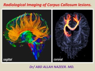
  • 2. Normal anatomy of the corpus callosum. T1-weighted (left), T2-weighted (middle), and FLAIR (right) MR images show the corpus callosum (CC). The splenium is the most posterior and thickest portion of the CC.
  • 3. Lesions of the corpus callosum are uncommon and arise from multiple different etiologies. The lesions can be classified according to underlying Pathophysiology . Congenital agenesis of the corpus callosum enlarged perivascular spaces tubonodular pericallosal lipoma: associated with dysgenesis of the corpus callosum. Demyelination acute disseminated encephalomyelitis Marchiafava-Bignami disease: demyelination and necrosis, can appear as cystic lesions multiple sclerosis neuromyelitis optica progressive multifocal leukoencephalopathy Susac syndrome wallerian degeneratio
  • 4. Infection aspergillosis: can involve corpus callosum transient lesions of the splenium: many underlying etiologies including infectious agents subacute sclerosing panencephalitis: can involve the corpus callosum in advanced stages tuberculosis: callosal tuberculomas have been reported, although rare Leukodystrophy Krabbe disease metachromatic leukodystrophy Susac syndrome X-Linked adrenoleukodystrophy Neoplasm Typically, amongst neoplasms, only aggressive lesions can invade the corpus callosum as it is composed of very dense white matter tracts which act as a barrier to tumour spreading. anaplastic astrocytoma callosal oligodendroglioma glioblastoma (butterfly glioma) gliomatosis cerebri meningioma: secondary involvement from primary falx lesion metastasis: rare, mainly from contiguous extension of lesion adjacent to corpus callosum primary CNS lymphoma
  • 5. Other corpus callosum impingement syndrome transient lesion of the splenium: many underlying aetiologies post shunt decompression: after placement of ventriculostomy in patients with chronic hydrocephalus Trauma diffuse axonal injury Vascular aneurysm: can occasionally rupture into the corpus callosum arteriovenous malformation: arising around the corpus callosum cavernous malformation (cavernoma) gliosis: secondary to small vessel chronic ischemia, subcortical arteriosclerotic encephalopathy and radiation therapy; histologically corresponds to subependymal gliosis hypoxic-ischemic encephalopathy: corpus callosum only involved in severe or advanced case infarction: rare because of extensive collateral vascular supply, most often seen with emboli, major ischaemic stroke, subfalcine herniation with mass effect and vasculitides.
  • 6. Transient lesions of the splenium of the corpus callosum, also known as mild encephalitis/encephalopathy with a reversible isolated SCC lesion (MERS), are occasionally encountered on MRI studies and may be due to a number of underlying etiologies. Clinical presentation Unlike other causes of splenium of corpus callosum (SCC) lesions, the small transient lesions of the splenium seen in epilepsy and antiepileptic drug cessation use do not demonstrate convincing signs or symptoms of hemispheric disconnection, such as pseudo-neglect, alien hand syndrome, apraxia of the left hand, agraphia, alexia, and visual apraxias.
  • 7. Aetiology epilepsy classic presentation is seen in patients with sudden cessation of antiepileptic drugs seizures: focal lesions have been described after focal status epilepticus and unusually after single seizures and were explained as focal brain edema. electrolyte imbalance including extrapontine myelinolysis demyelination multiple sclerosis (MS) acute disseminated encephalomyelitis (ADEM) posterior reversible encephalopathy syndrome (PRES) Marchiafava-Bignami disease diffuse axonal injury (DAI) AIDS dementia complex infections viral: influenza, measles, herpes, mumps, adenovirus, varicella zoster, rotavirus and HIV bacterial: salmonella, Legionnaires' disease mycobacterial: tuberculous meningitis hypoglycemia hemolytic-uremic syndrome with encephalopathy.
  • 8. Radiographic features Transient lesions of the splenium are only really appreciable on MRI where they have two distinct patterns: well circumscribed, small, oval lesions in the midline within the substance of the corpus callosum more extensive ill-defined irregular lesions extending throughout the splenium and into the adjacent hemispheres (boomerang sign) The smaller well-circumscribed lesions are the typical lesion seen in the setting of seizures/cessation of antiepileptic medication, whereas the larger lesion is more typical of other etiologies. MRI These lesions tend to demonstrate the following signal characteristics: T1: hypointense T2: hyperintense DWI/ADC: restricted diffusion T1 C+: no enhancement Studies have shown that patients recover completely on MRI studies within 1 month, mostly within 1 week following the neurologic recovery .
  • 9. A 1-year-old female patient with complete corpus callosal agenesis. T1-weigted axial image (left) shows parallel configuration of both lateral ventricles. T1-weighted sagittal image (right) reveals complete agenesis of the corpus callosum.
  • 10. Agenesis of corpus callosum. A, Sagittal midline T1-weighted image shows complete absence of corpus callosum (arrows). Axial T2-weighted image shows colpocephalic configuration with dilated occipital horns, right greater than left (asterisks), and parallel (racecar) orientation of lateral ventricles. Third ventricle (arrow) is high riding and forms interhemispheric arachnoid cyst. Axial T2-weighted image at higher level illustrates crescentic (teardrop) morphology of frontal horns (arrows).
  • 11. lipoma of corpus callosum. Coronal T1- weighted MR image shows large well-defined homogeneous midline mass lesion in region of corpus callosum with characteristic bright signal of lipoma. Note associated dysgenesis of corpus callosum. Corpus callosal Agenesis with Lipoma.
  • 12. A 70-year-old male patient with a pericallosal lipoma and partial corpus callosal agenesis. Axial CT image (left) shows a lipoma in frontal interhemispheric fissure. There is a linear calcification at left side of the lesion. Sagittal T1-weighted MR image (right) reveals partial agenesis of the corpus callosum and a hyperintense pericallosal lipoma.
  • 13. A 64-year-old male patient with a pericallosal lipoma and partial corpus callosal agenesis. T1-weighted axial (left) and sagittal (right) images show a hyperintense lipoma superior and posterior to the corpus callosum (CC). The CC shows partial agenesis. The splenial portion is not fully developed.
  • 14. Callosal infarcts. A, Left anterior cerebral artery infarct (thick arrow) with extension into genu (thin arrow) is seen on diffusion-weighted (left) and apparent diffusion coefficient (ADC) (right) images, which confirm reduced diffusivity in these areas.
  • 15. A 62-year-old male patient with acute splenial infarction. Axial FLAIR image (left) and DWI (middle) show multiple hyperintense lesions in bilateral basal ganglia, left thalamus, the splenium of the corpus callosum, and left occipital lobe. The lesions are more conspicuously demonstrated on DWI than on FLAIR image. ADC map image (right) reveals restricted water diffusion of the lesions.
  • 16. A 70-year old female patient with chronic splenial infarction. Axial FLAIR (left) and sagittal T2-weighted (middle) images show a hyperintense lesion in the splenium of the corpus callosum. On DWI (right), the lesion is seen as isointensity.
  • 17. Traumatic brain injury. A, Unenhanced CT images at level of lateral (A) and third (B) ventricles show hyperdense areas of acute hemorrhage in splenium (S) and genu (G). T2* gradient recalled-echo MR images at same levels confirm magnetic susceptibility in these areas. There are multiple additional foci of microhemorrhage throughout gray-white matter junction and basal ganglia (arrows).
  • 18. 9-year-old female patient with Wallerian degeneration due to intracerebral hematomas. A, B. Axial T2 (A) and T1-weighted (B) images show two hyperintense hematomas in right occipital lobe (thick arrows) and left temporooccipital lobes (asterisks). There is moderate amount of surrounding brain edema. Splenium also shows mild swelling and increased signal intensity (thin arrows). C. Follow-up T2-weighted image obtained 20 months later reveals atrophic change and increased signal intensity of splenium (thin arrows).
  • 19. A 29-year-old female patient with diffuse axonal injury lesion. She had a motor vehicle accident 9 days age. Axial FLAIR (left) and DWI (middle) images show a hyperintense lesion in the splenial portion of the corpus callosum. The lesion is more conspicuously demonstrated on DWI than on FLAIR image. ADC map image (right) reveals restricted water diffusion of the lesion.
  • 20. A 29-year-old female patient with diffuse axonal injury lesion. Follow-up MR images were obtained 17 months later. On axial T2 (left), FLAIR (middle) and DWI (right) images, the splenial lesion has disappeared.
  • 21. A 27-year-old male patient with diffuse axonal injury lesion. He had a motor vehicle accident 2 days age. Axial FLAIR (left) and DWI (middle) images show a focal hyperintense lesion in the splenial portion of the corpus callosum. The lesion is more conspicuously demonstrated on DWI than on FLAIR image. ADC map image (right) reveals restricted water diffusion of the lesion.
  • 22. A 56-year-old male patient with diffuse axonal injury lesions. He had a motor vehicle accident 4 months age. Axial FLAIR image (left) shows a hyperintense lesion in the splenial portion of the corpus callosum. On DWI (middle), the lesion is seen as hypointensity. GE T2*-weighted image (right) reveals tiny hemorrhagic foci in the splenial lesion. There are also multiple tiny hemorrhage in right thalamus and left frontal white matter.
  • 23. A 2-month-old female patient with hypoxic-ischemic encephalopathy. She had hypoxic insult 3 days ago. Axial DWI (left) shows diffuse hyperintense lesions in the bilateral cerebral cortices and white matter including the genu and splenium of the corpus callosum. ADC map image (right) reveals restricted water diffusion of the lesions
  • 24. 2-month-old female patient with hypoxic-ischemic encephalopathy. Follow-up MR images were obtained 9 days later (12th day from hypoxic insult). On axial T2-weighted image (left) and DWI (middle left), the bilateral basal ganglia and thalami are newly involved and the corpus callosal (CC) lesions show more prominent hyperintensities compared to on initial DWI. Both cerebral cortices and white matter lesions are decreased in their signal intensities. ADC map image (middle right) reveals restricted water diffusion of the CC lesions. On contrast-enhanced T1-weighted image, there is strong contrast enhancement along the cortices and subcortical white matter of both cerebral hemispheres.
  • 25. 2-day-old female with hypoglycemic encephalopathy. Glucose level was 2 mg/dL at presentation. A. On FLAIR axial image, there is no definite lesion. B. Axial DWI shows hyperintense lesions in both occipital lobes and splenium (arrows). C. ADC map image reveals restricted water diffusion of lesions (arrows). ADC = apparent diffusion coefficient, DWI = diffusion-weighted image, FLAIR = fluid-attenuated inversion recovery
  • 26. 8-year-old female patient with CO-intoxication. Axial FLAIR image (A) and DWI (B) show multiple hyperintense lesions in bilateral globus pallidus (arrowheads) and cerebral white matter (thin arrows) including splenium of corpus callosum (thick arrows). DWI = diffusion-weighted image, FLAIR = fluid-attenuated inversion recovery
  • 27. 59-year-old male patient with chronic alcoholism. A, B. FLAIR (A) and T2-weighted (B) images show hyperintense lesions in body (thin arrows) and splenial portion (thick arrows) of corpus callosum. C. There is focal contrast enhancement of lesions (arrows) on post-contrast T1-weighted image. FLAIR = fluid-attenuated inversion recovery
  • 28. 5-day-old male with rotavirus-related white matter injury. A, B. Initial axial DWI (A) and ADC map (B) demonstrate extensive areas of restricted diffusion in periventricular white matter, deep white matter, corpus callosum, internal capsule, and posterior thalami. C. Follow-up FLAIR image obtained five years later shows residual ischemic lesions due to previous white matter injury in both periventricular white matters (arrows). ADC = apparent diffusion coefficient, DWI = diffusion-weighted image, FLAIR = fluid-attenuated inversion recovery
  • 29. Multiple sclerosis who presented with visual complaints. Sagittal MR image shows multiple hyperintense lesions (arrows) in corpus callosum.
  • 30. Axial brain MRI (fluid-attenuated inversion recovery) with tumefactive lesions (A) developed exacerbation 67 days after the start of interferon-β-1b (IFNβ-1b) therapy; MRI showed huge tumefactive lesion in right hemisphere. Case 5 (B) developed exacerbation 32 days after the start of IFNβ-1b therapy; MRI showed tumefactive lesions in corpus callosum extending to the right parietal white matter.
  • 31. 4-year-old girl with pilocytic astrocytoma. Axial T2- weighted MR image shows that lesion is hyperintense. 4-year-old girl with pilocytic astrocytoma. Sagittal T1- weighted MR image shows well-circumscribed hypointense lesion in body of corpus callosum. 4-year-old girl with pilocytic astrocytoma. Enhanced coronal T1- weighted MR image shows marked contrast enhancement of lesion.
  • 32. A 59-year-old female patient with a glioblastoma. Axial T2-weighted image (left) and DWI (middle left) show a large heterointense mass in right temporoparietal white matter and adjacent splenium of the corpus callosum. There is large amount of surrounding brain edema. Perfusion MR image (middle right) reveals increased perfusion in the peripheral solid portion of the mass. On contrast- enhanced T1-weighted image, there is peripheral rim enhancement of the mass.
  • 33. Glioblastoma multiforme. A, Unenhanced CT image shows heterogeneously hypodense mass with surrounding vasogenic edema. This involves both frontal lobes and crosses genu (G) of corpus callosum, producing butterfly appearance. There is compression and distortion of frontal horns of both lateral ventricles. Contrast-enhanced T1-weighted MR image shows involvement of genu (G). Lesion shows rim enhancement and central hypointensity, compatible with necrosis.
  • 34. A 46-year-old female patient with gliomatosis cerebri. Axial FLAIR image (left) show an ill-defined hyperintense lesion involving bilateral posterior cerebral white matter and the splenium of the corpus callosum. On enhanced T1-weighted axial image (middle), there is no definite contrast enhancement of the lesion. Single voxel 1H MR spectroscopy (right) reveals increased choline peak.
  • 35. Nonimmunocompromised woman with primary central nervous system lymphoma who presented with disorientation. MRI show lesion (arrow) of left parieto— occipital white matter, crossing corpus callosum in classic butterfly pattern.
  • 37. A 23-year-old male patient with a pineal germinoma. Axial T2 (left) and T1- weighted (middle) images show a mass in the pineal area. The mass extends into the 3rd ventricle anteriorly and to the splenium of the corpus callosum posteriorly. On enhanced T1-weighted image, the mass is strongly enhanced.
  • 38. A 62-year-old male patient with epilepsy. Axial DWI (left) shows a focal hyperintense lesion in the splenium of the corpus callosum. ADC map image (middle) reveals restricted water diffusion of the lesion. On follow- up DWI (right) obtained 14 days later, the splenial lesion has disappeared.
  • 39. A 38-year-old female patient with bacterial meningoencephalitis due to S. pneumoniae. Axial FLAIR image (left) and DWI (middle) show a hyperintense lesion in the splenium of the corpus callosum. The lesion is more conspicuously demonstrated on DWI than on FLAIR image. There is tissue loss in left frontal lobe. ADC map image (right) reveals markedly restricted water diffusion of the splenial lesion.
  • 40. A 15-year-old male patient with abc meningoencephalitis. Axial FLAIR image (left) and DWI (middle) shows a focal hyperintense lesion in midline corpus callosum. The lesion is more conspicuously demonstrated on DWI than on FLAIR image. ADC map image (right) reveals markedly restricted water diffusion of the lesion.
  • 41. Transient splenial intensity changes. A, Example images in migraine and visual scotomas (A), adenovirus encephalitis (B), and metronidazole toxicity (C) show FLAIR hyperintensity and reduced diffusivity in splenium (arrows), which resolved on follow-up imaging.
  • 42. A 59-year old male patient with chronic alcoholics. FLAIR (left) and T2-weighted (middle) images show hyperintense lesions in the body and splenial portion of the CC. There is focal contrast enhancement of the lesions on post-contrast T1-weighted image (right).
  • 43. 2-year-old man with arteriovenous malformation who presented with intraventricular hemorrhage. Sagittal T1- weighted MR image shows hemorrhage (arrows) and multiple flow voids in corpus callosum. 2-year-old man with arteriovenous malformation who presented with intraventricular hemorrhage. Axial T2- weighted MR image shows hyperintense lesion (arrow) with flow voids.
  • 44. Unenhanced CT and Gradient MR image in different patient who had ruptured anterior communicating artery aneurysm. Right pericallosal artery aneurysm (arrow). Aneurysm.
  • 45. Unenhanced CT image shows round well- circumscribed hyperdense mass (asterisk) centered in falx cerebri (arrow) and extending across midline. There is subtle surrounding edema that involves bilateral temporo-occipital regions T1-weighted contrast-enhanced MR image shows relatively homogeneous contrast enhancement within mass, which deviates surrounding pial vessels (arrows).
  • 46. Contrast-enhanced CT (A) and contrast- enhanced T1-weighted MR (B) images show heterogeneously enhancing dominant metastasis in left frontal lobe (asterisk). Additional enhancing foci are noted in splenium (S), genu (G), and periventricular regions (arrows). Contrast-enhanced CT (A) and contrast-enhanced T1-weighted MR (B) images show heterogeneously enhancing dominant metastasis in left frontal lobe (asterisk). Additional enhancing foci are noted in splenium (S), genu (G), and periventricular regions (arrows).
  • 47. Hereditary leukoencephalopathies. A, FLAIR MR images show signal abnormalities in bilateral parietal lobes and intervening splenium of corpus callosum (arrows): metachromatic leukodystrophy (A) and adrenoleukodystrophy (B)
  • 48. Periventricular leukomalacia. A, T2-weighted MR image shows periventricular white matter abnormalities, with increased signal in both frontal and parietal lobes as well as genu (G) and splenium (S) of corpus callosum. Susceptibility artifact within choroid plexus of occipital horns (asterisks) confirms presence of hemorrhage. Diffusion-weighted MR image identifies reduced diffusivity within splenium (arrows) and multiple other areas.
  • 49. Virchow-Robin spaces in Hunter syndrome (mucopolysaccharidosis type 2). Axial FLAIR image shows diffusely enlarged perivascular spaces, which are isointense to CSF and involve body of corpus callosum (arrows). Sagittal T1 FLAIR contrast-enhanced image shows large Virchow-Robin spaces within corpus callosum (arrow) and brain parenchyma.
  • 50. Marchiafava-Bignami disease. (Courtesy of Ginat DT, Massachusetts General Hospital, Boston, MA)A, FLAIR MR image shows increased signal in splenium (arrow). Marchiafava-Bignami disease. (Courtesy of Ginat DT, Massachusetts General Hospital, Boston, MA)B, Apparent diffusion coefficient map confirms reduced diffusivity in this region (arrow).
  • 51. Susac's syndrome:(a-d) Magnetic resonance imaging FLAIR T2 image showing multiple well-defined hyperintense lesions in periventricular and callosal area. Magnetic resonance imaging T1 image showing hypointense lesions in center of corpus callosum. Magnetic resonance imaging, 4 weeks later showing almost complete disappearance of lesions
  • 52.
  • 53. Magnetic resonance imaging of the brain: Diffusion-weighted sequences during 2010 episode (a-d) showing restricting lesions in left posterior limb of internal capsule and splenium of the corpus callosum. Lesions involving bilateral subcortical white matter and centrum semi ovale (e-h) during July 2013 episode. Resolution of the changes (i-l) in the repeat imaging done after 1.5 months. Arrows showing abnormalities.
  • 54. Callosal thickening of neurofibromatosis type 1. A midsagittal T1W MR image shows diffuse thickening of the corpus callosum (arrows).