Downloaded 1,083 times
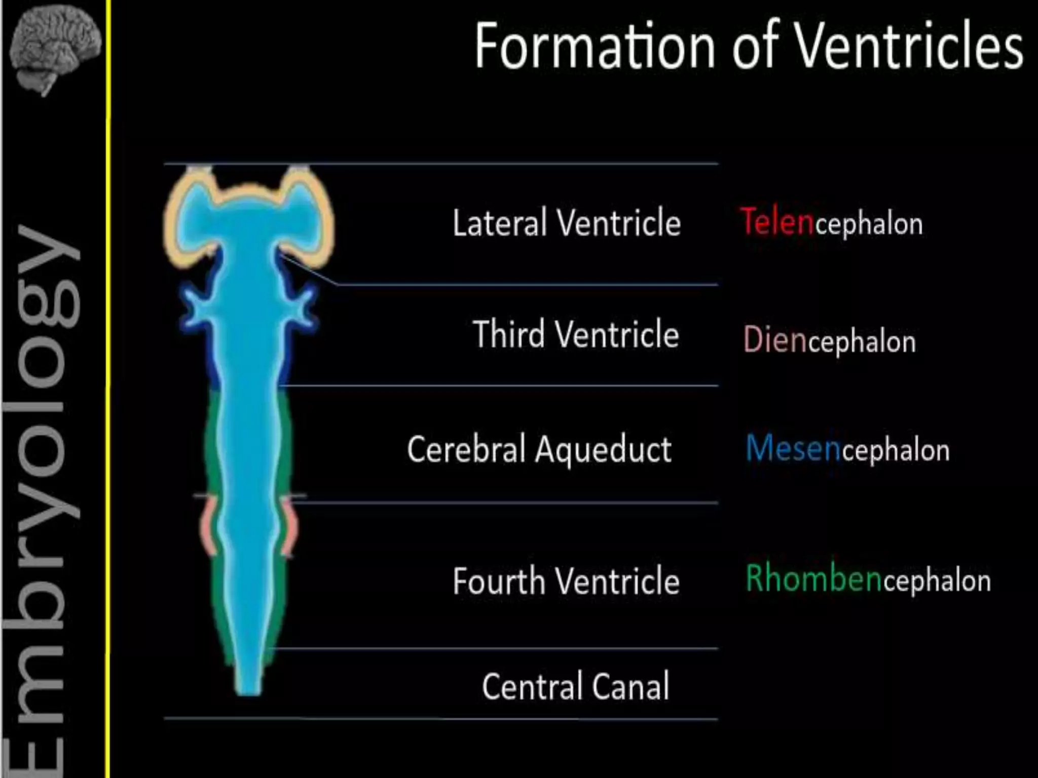
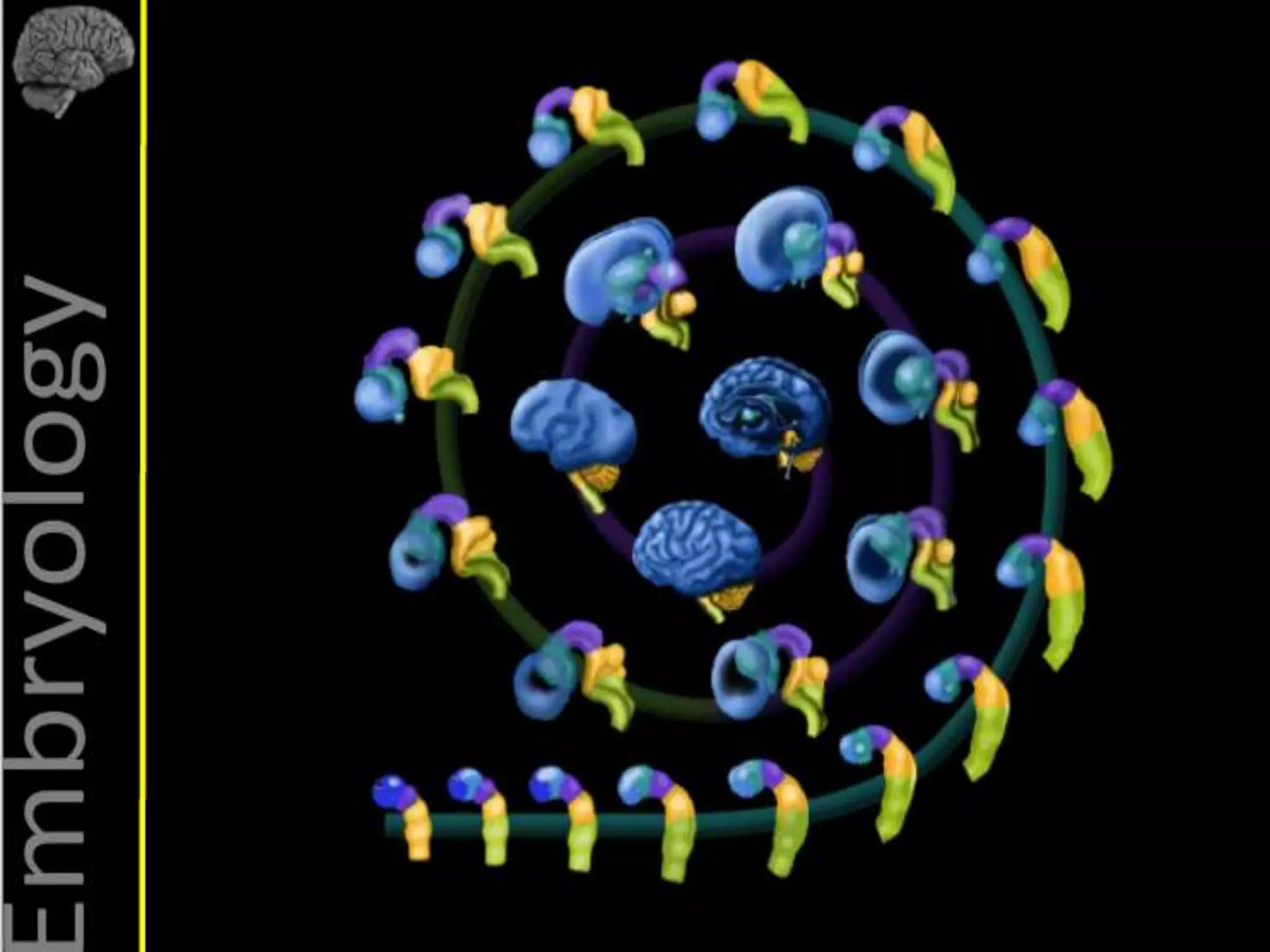
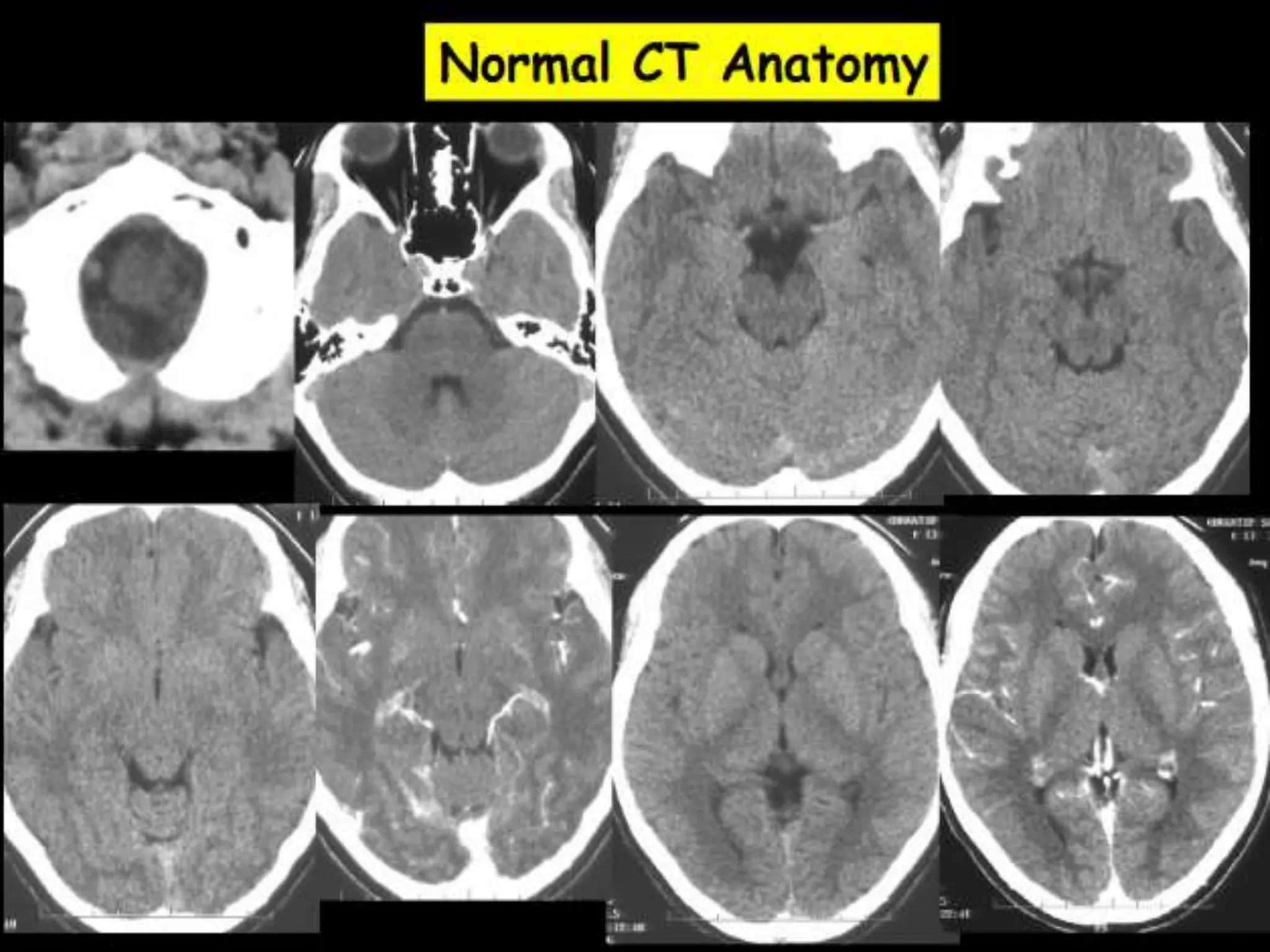
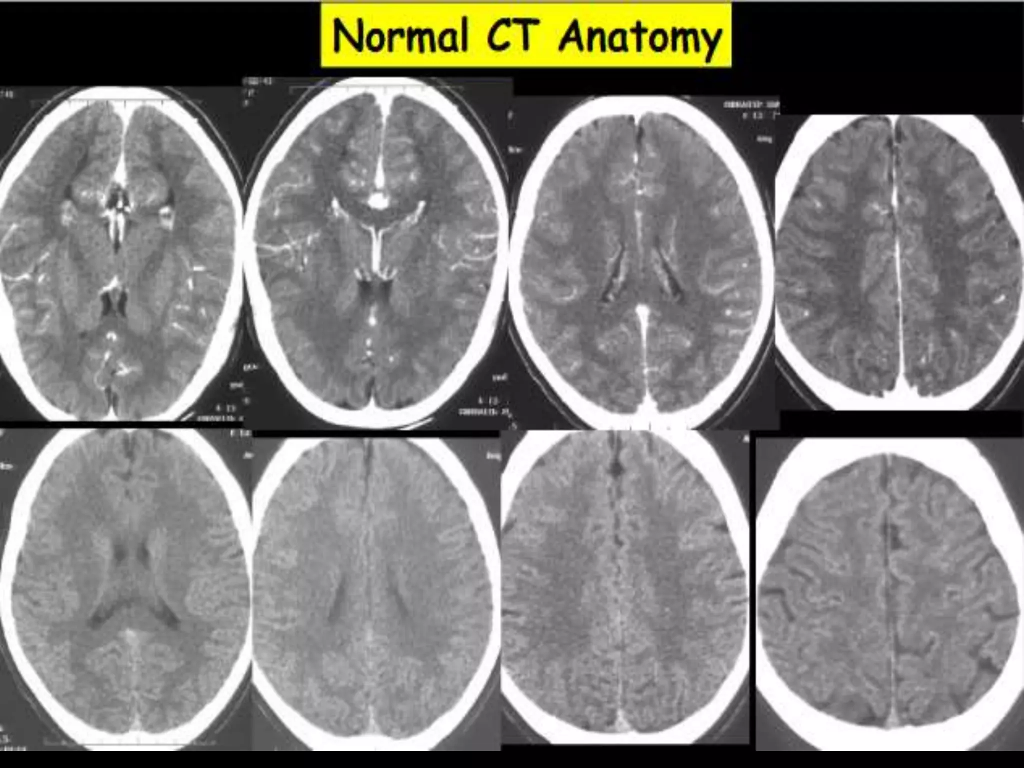
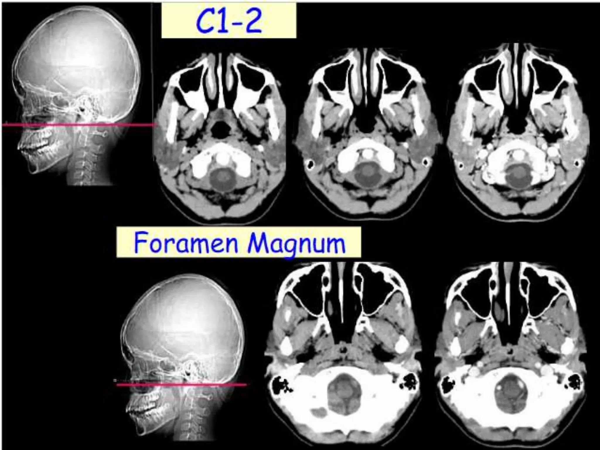
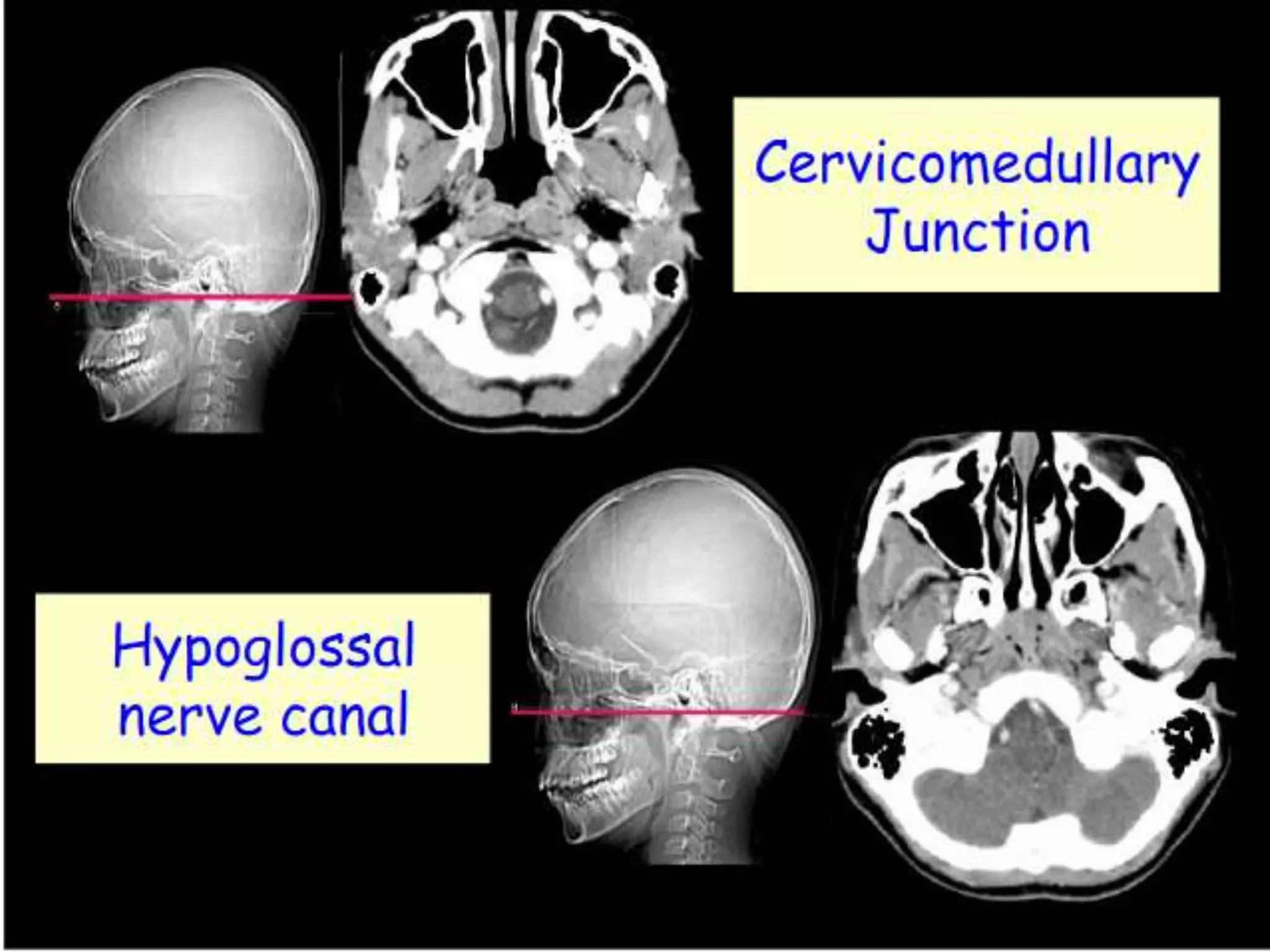
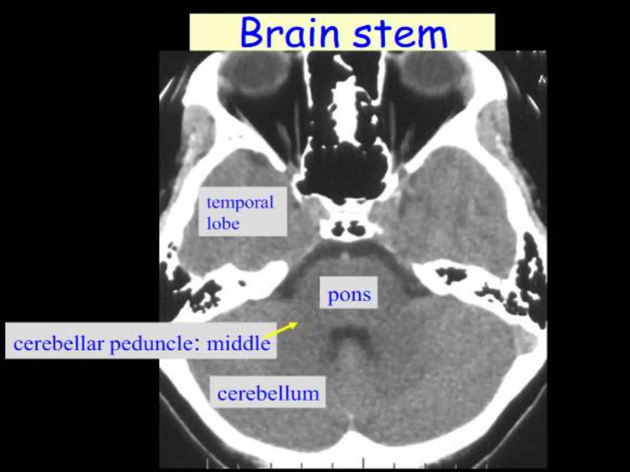
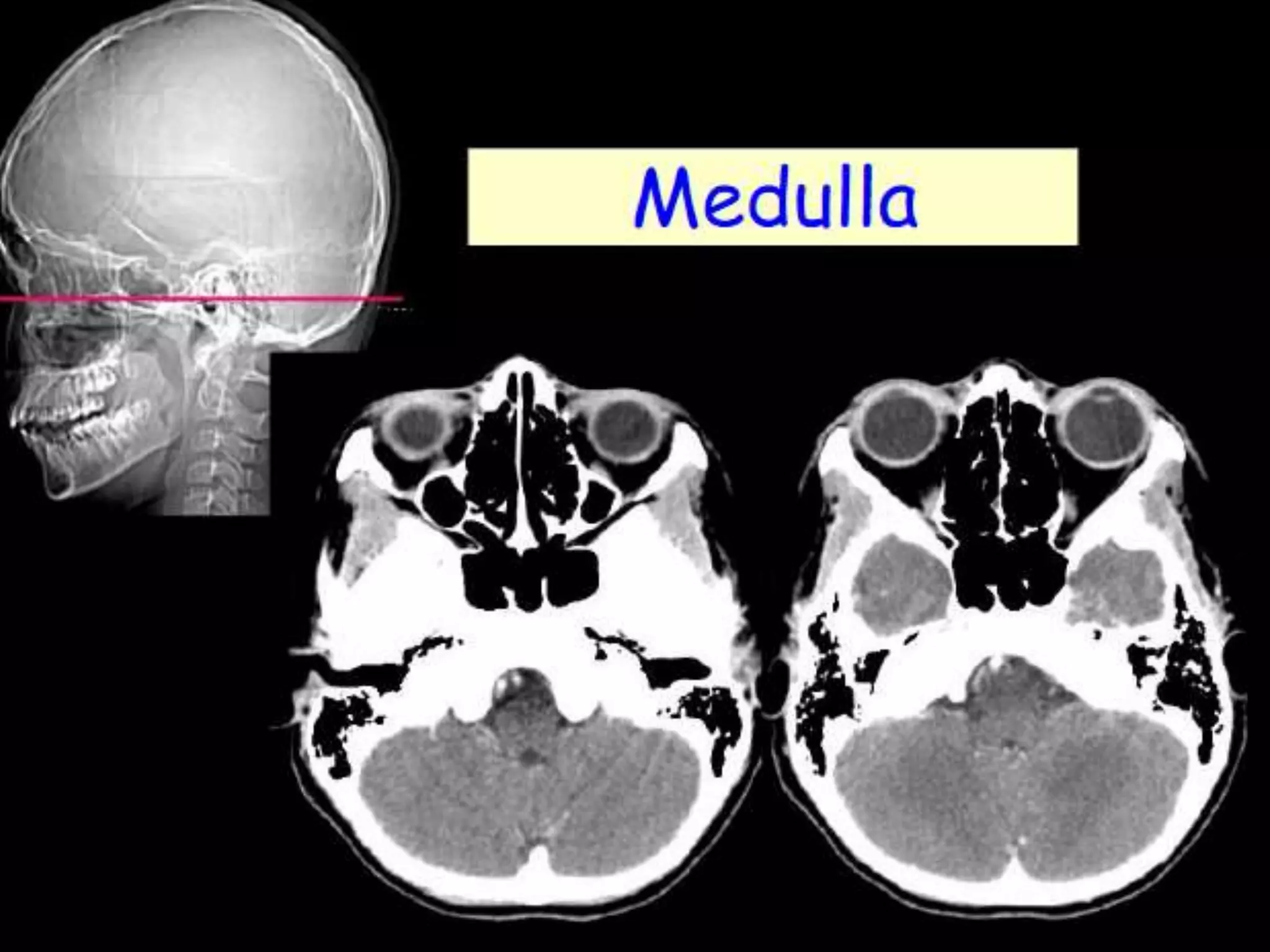
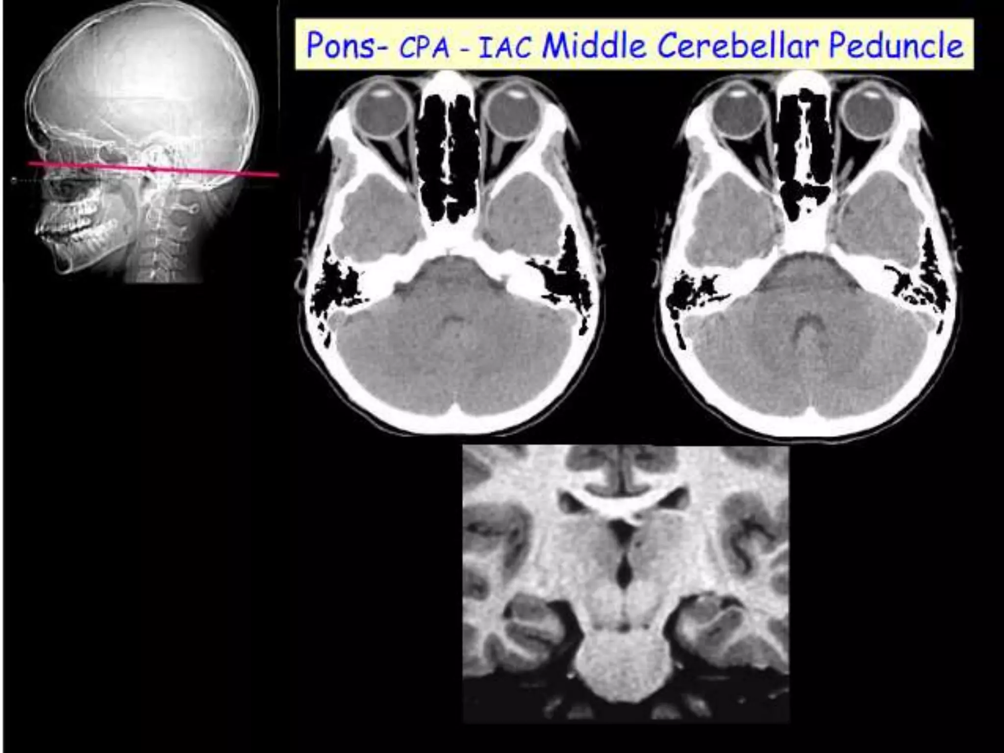
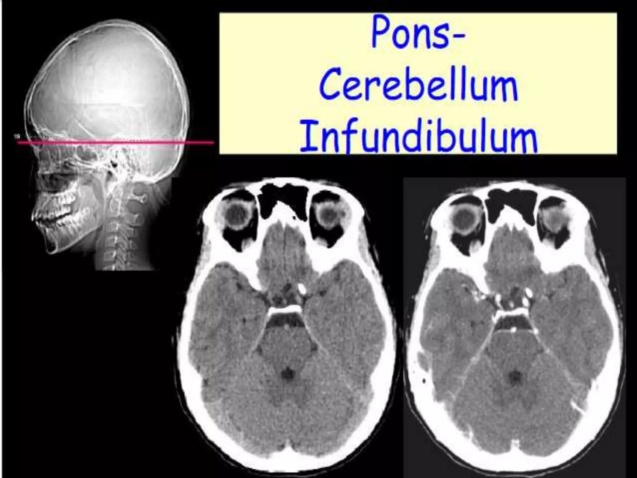
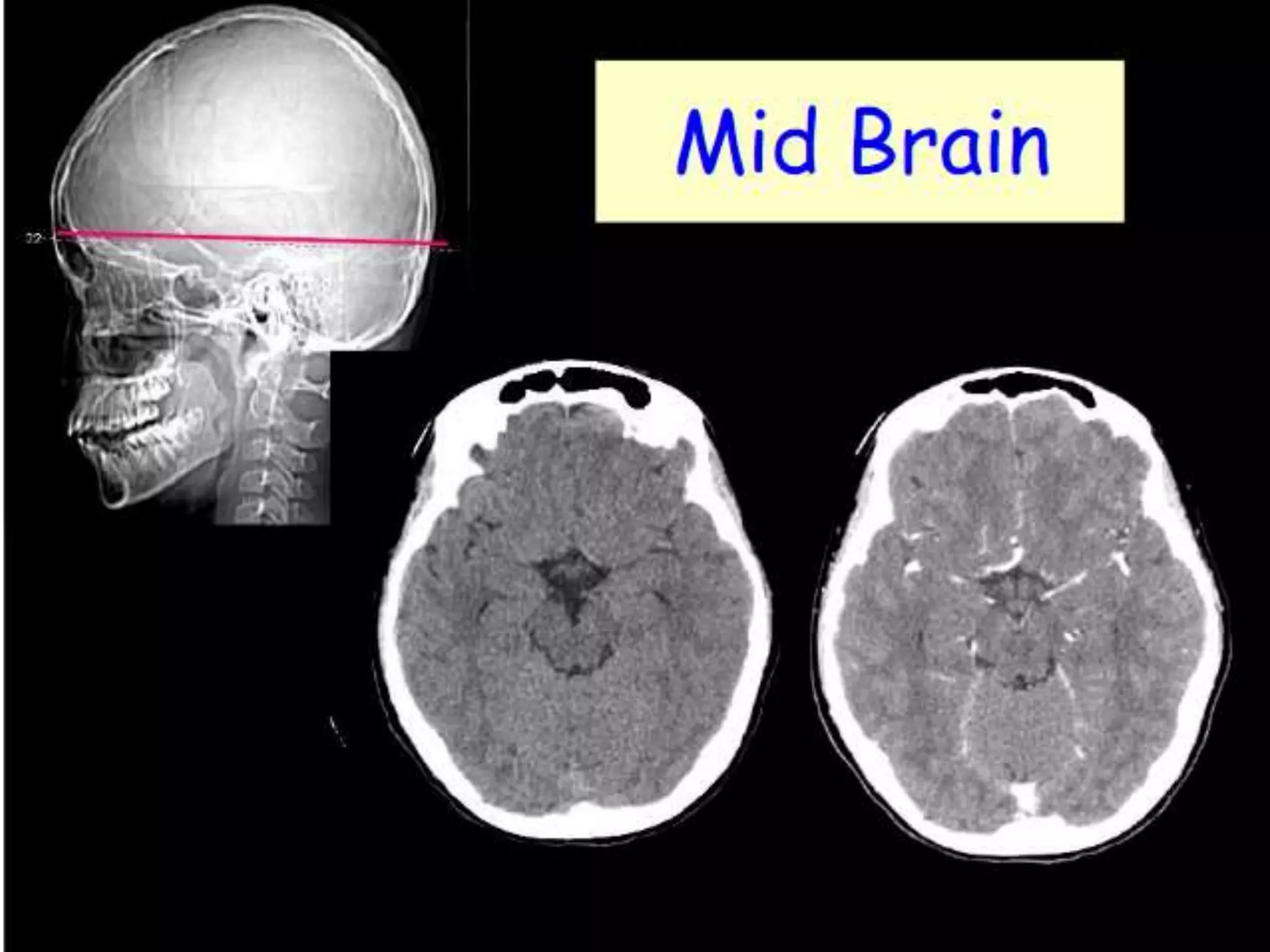
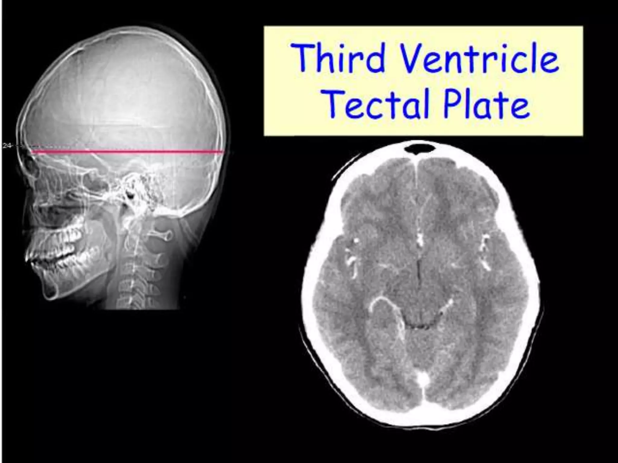
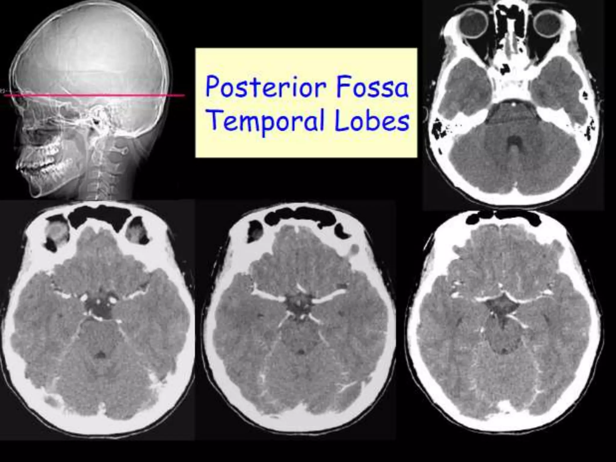
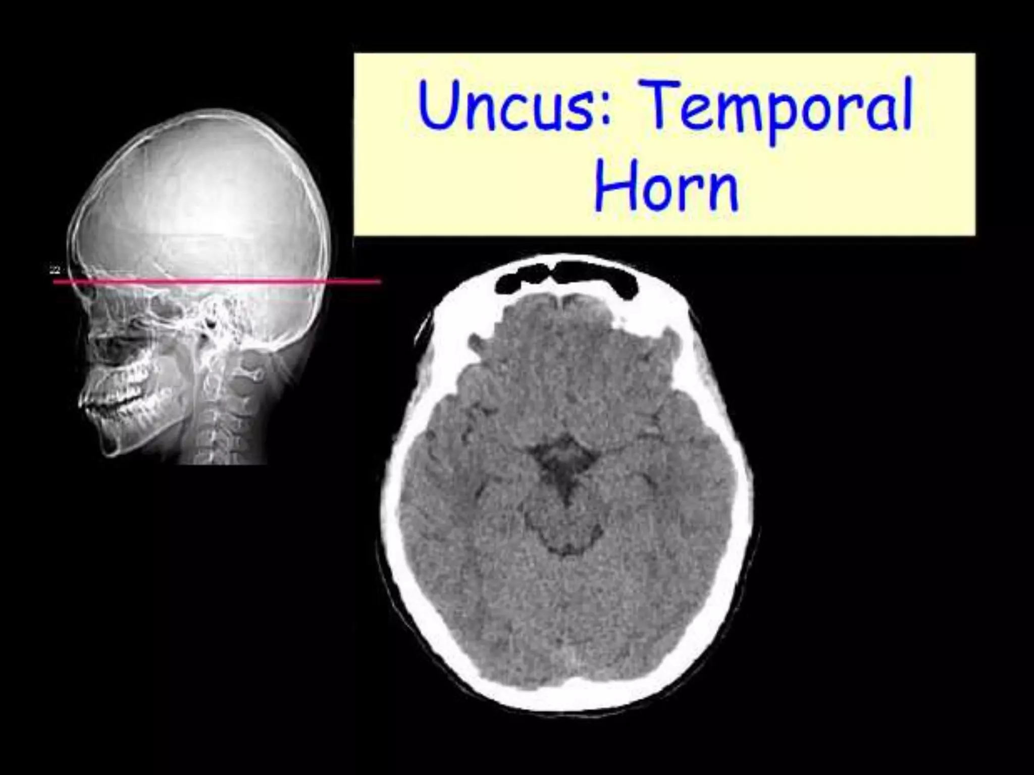
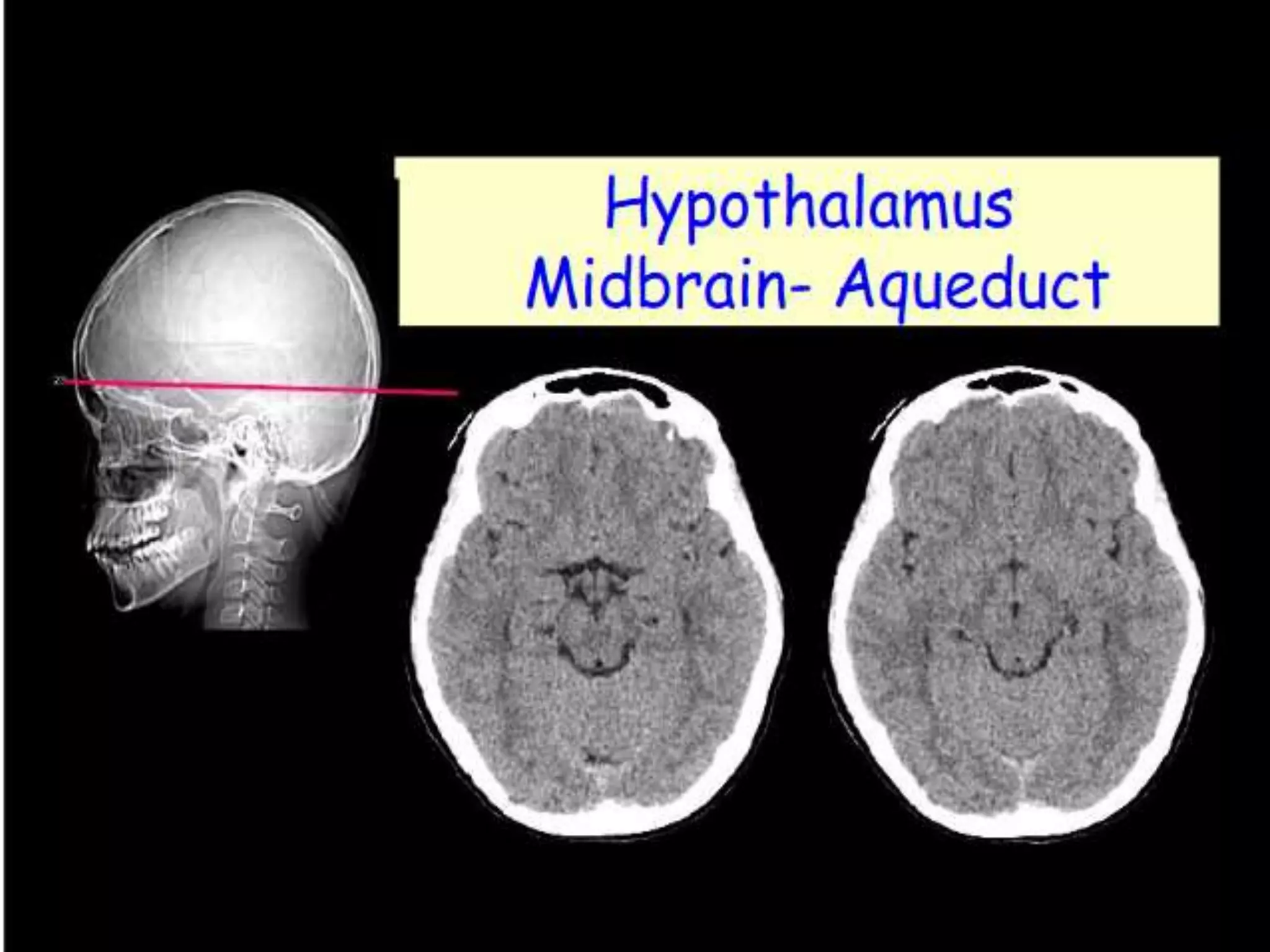
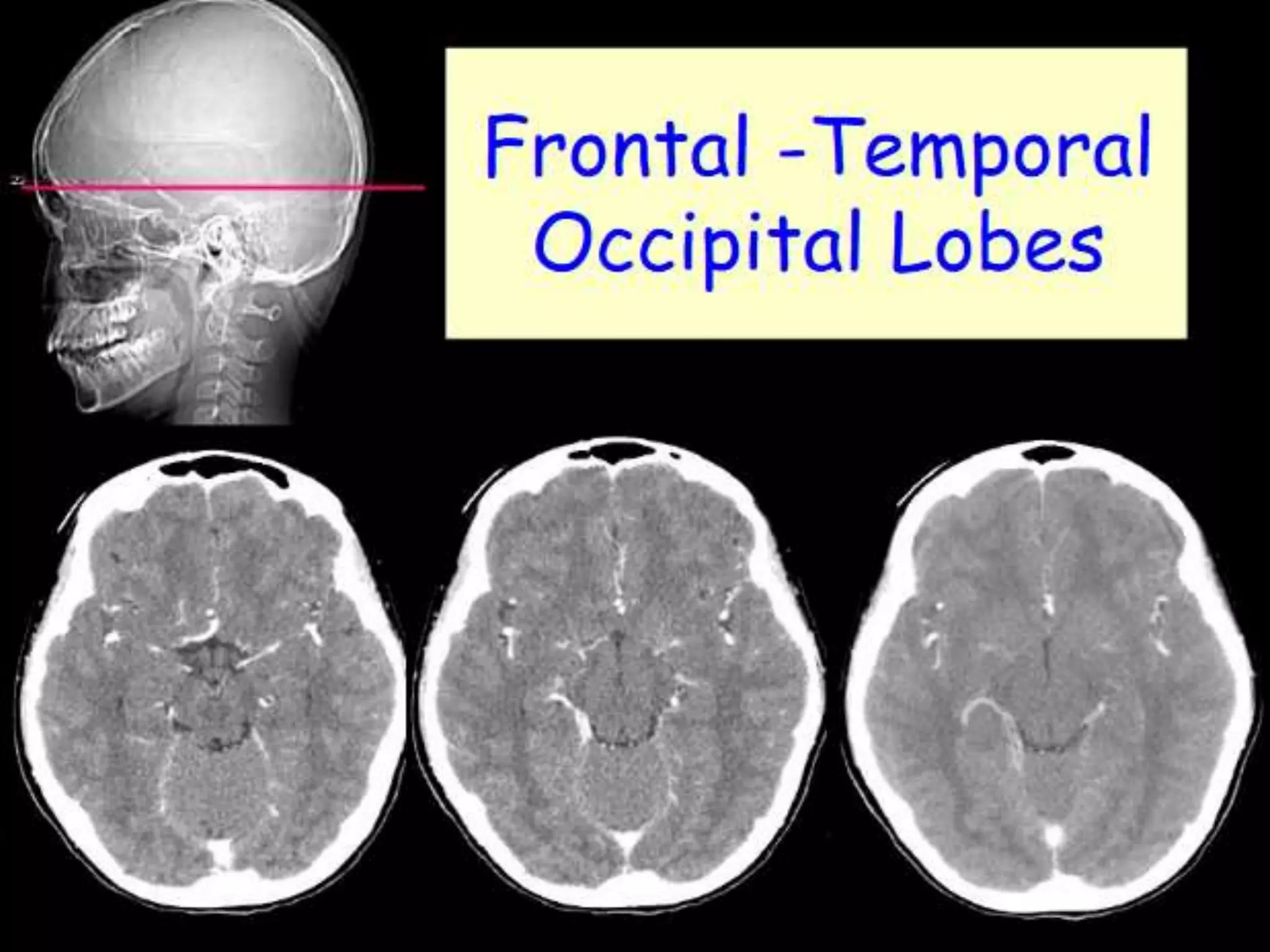
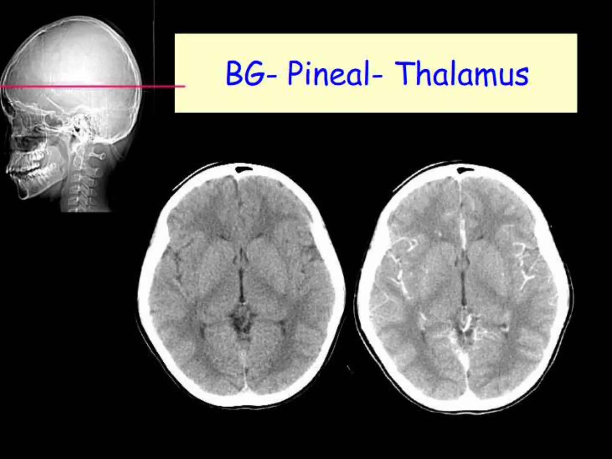
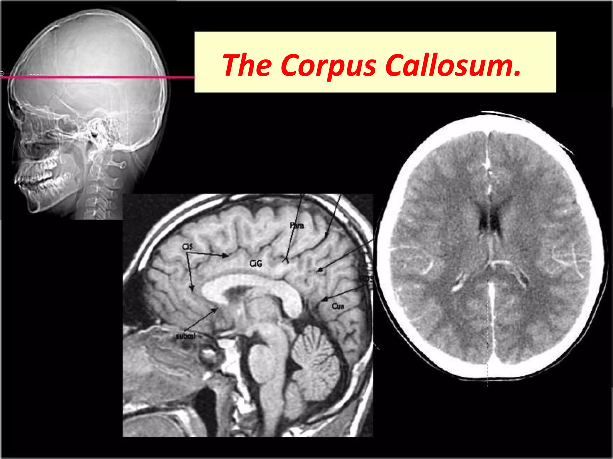
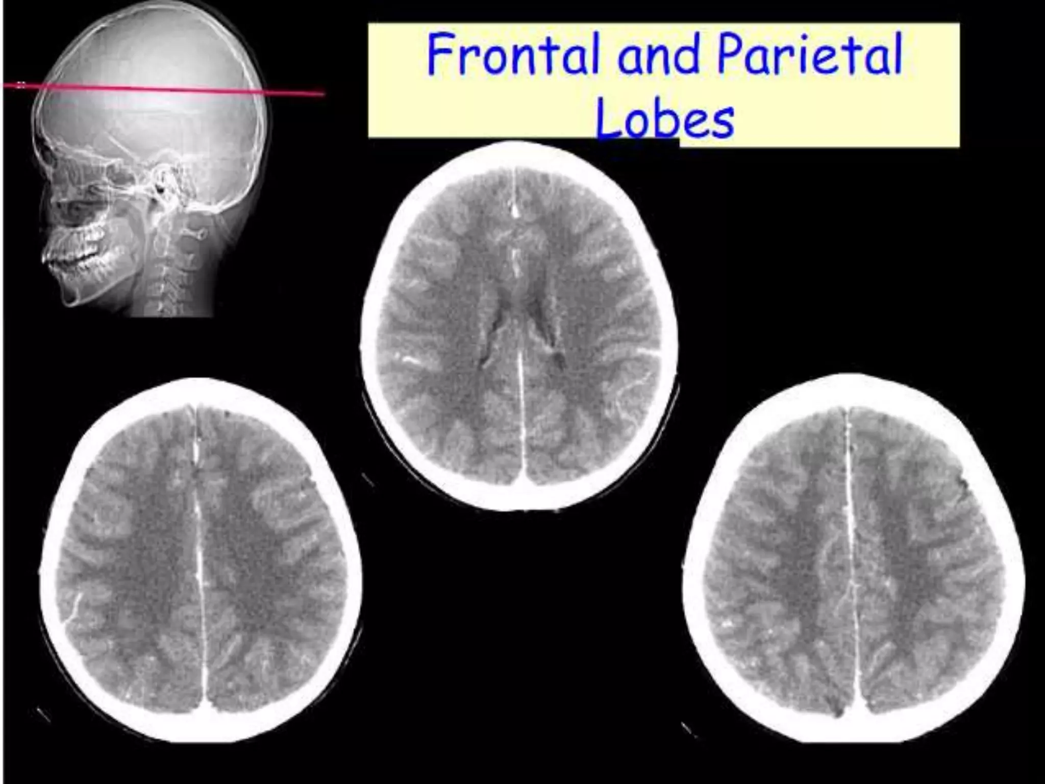
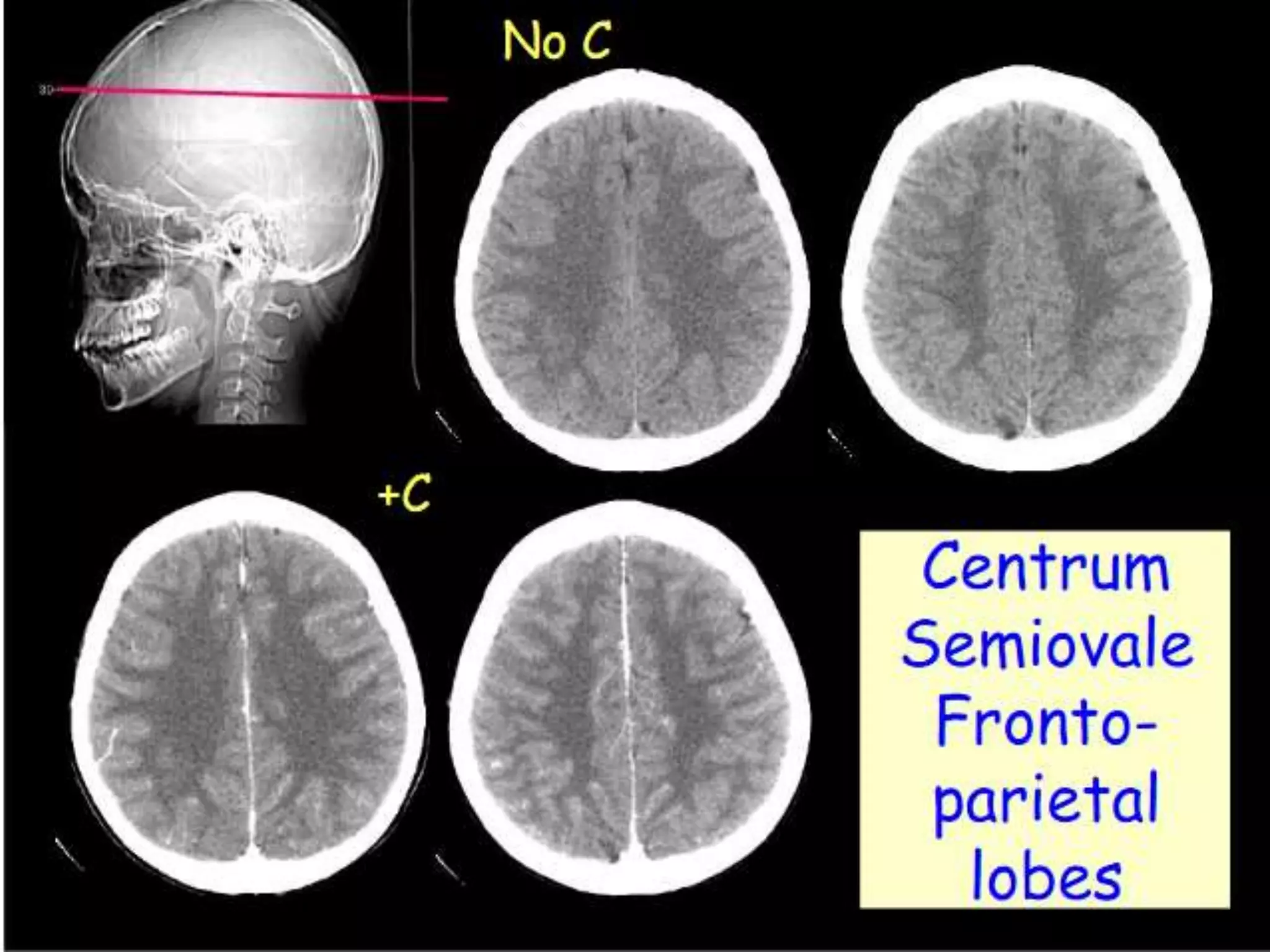
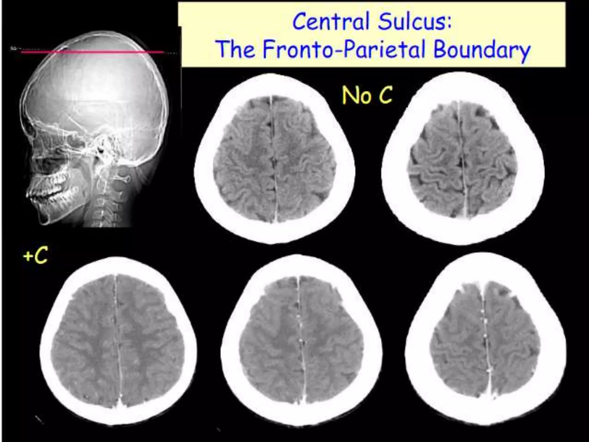
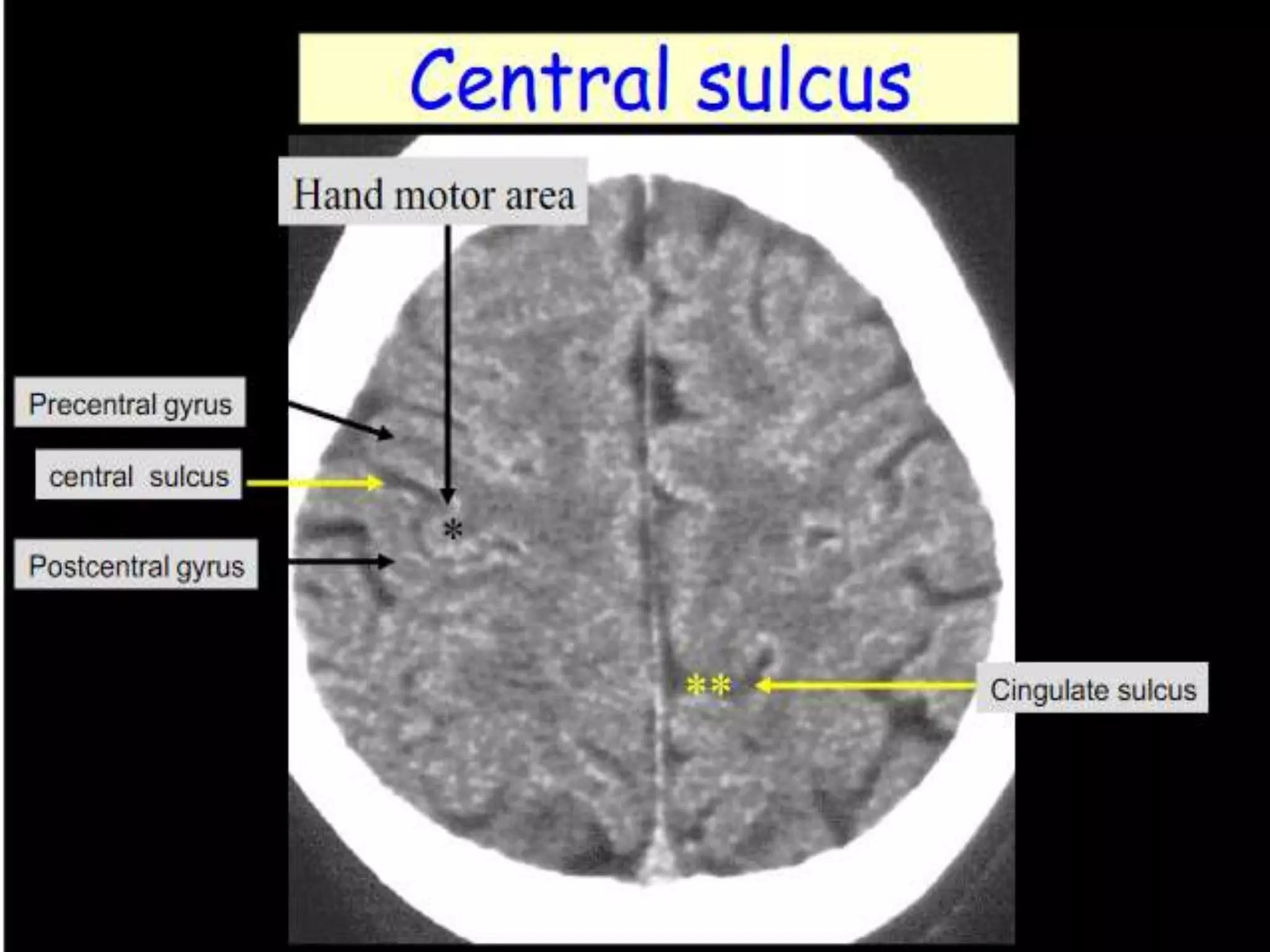
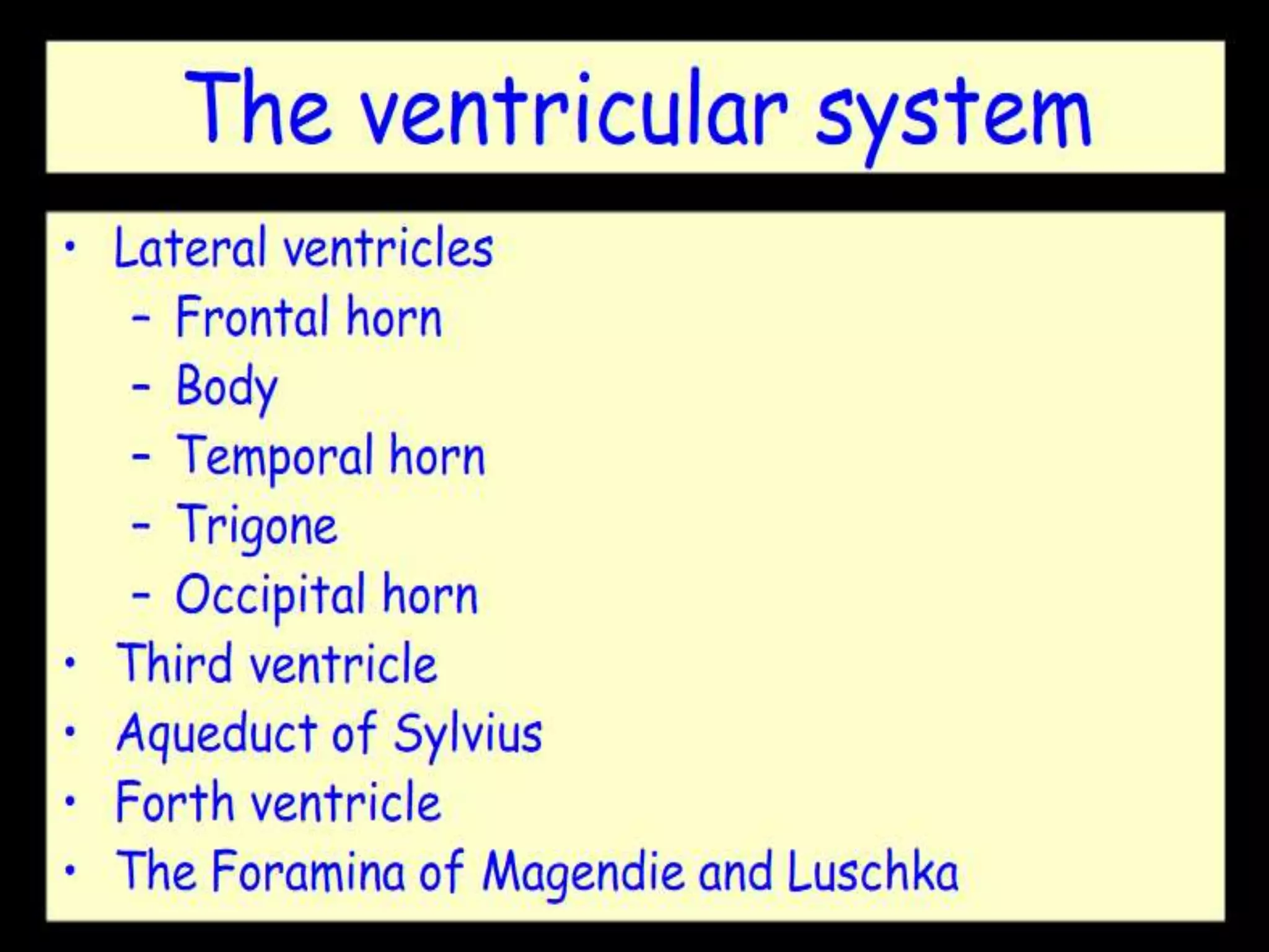
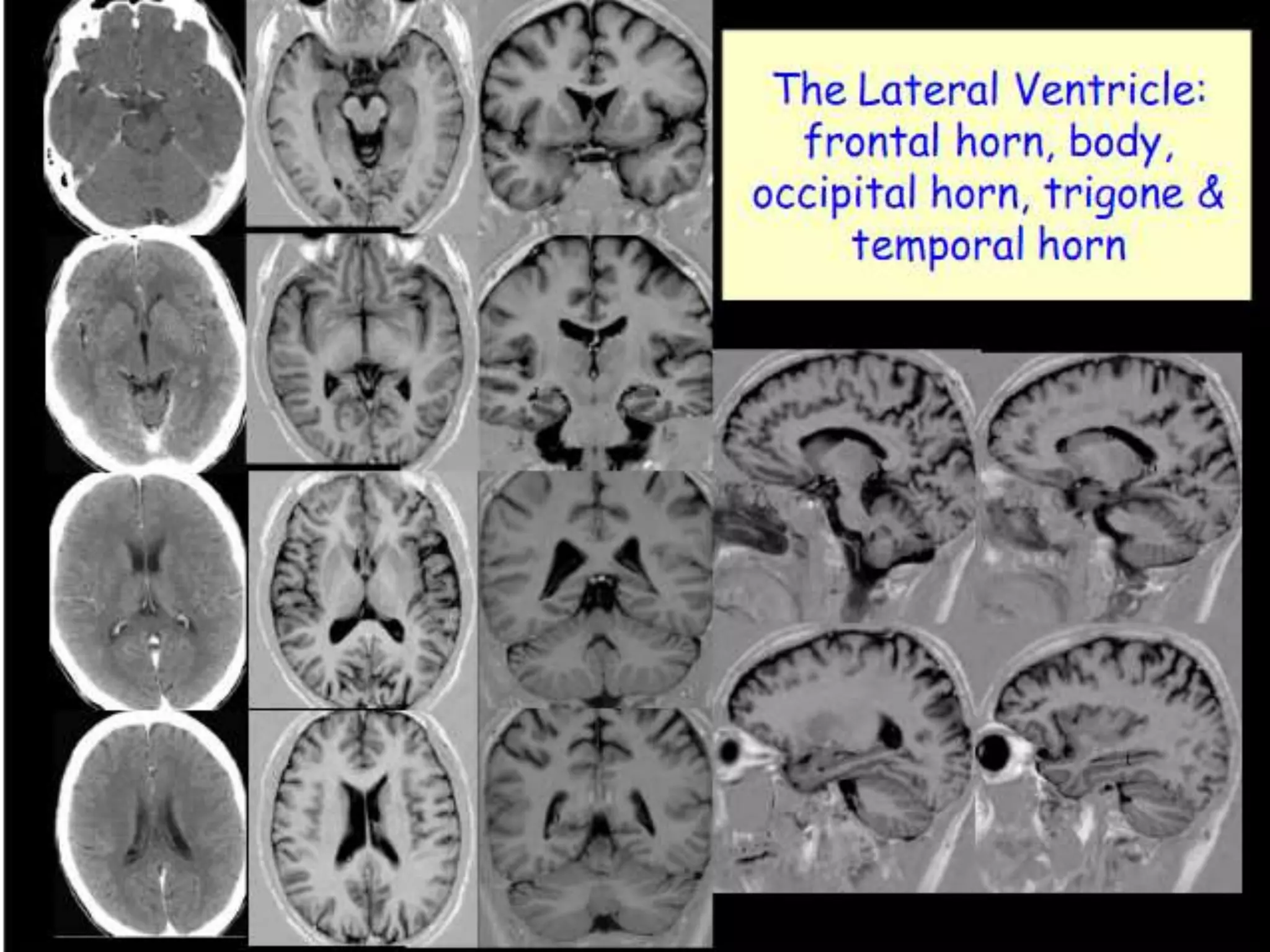
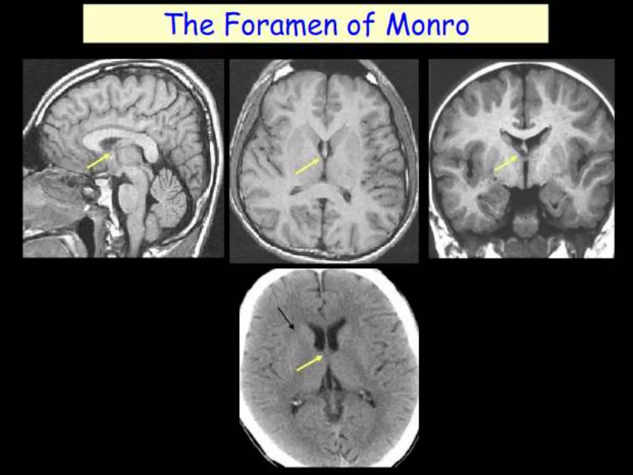
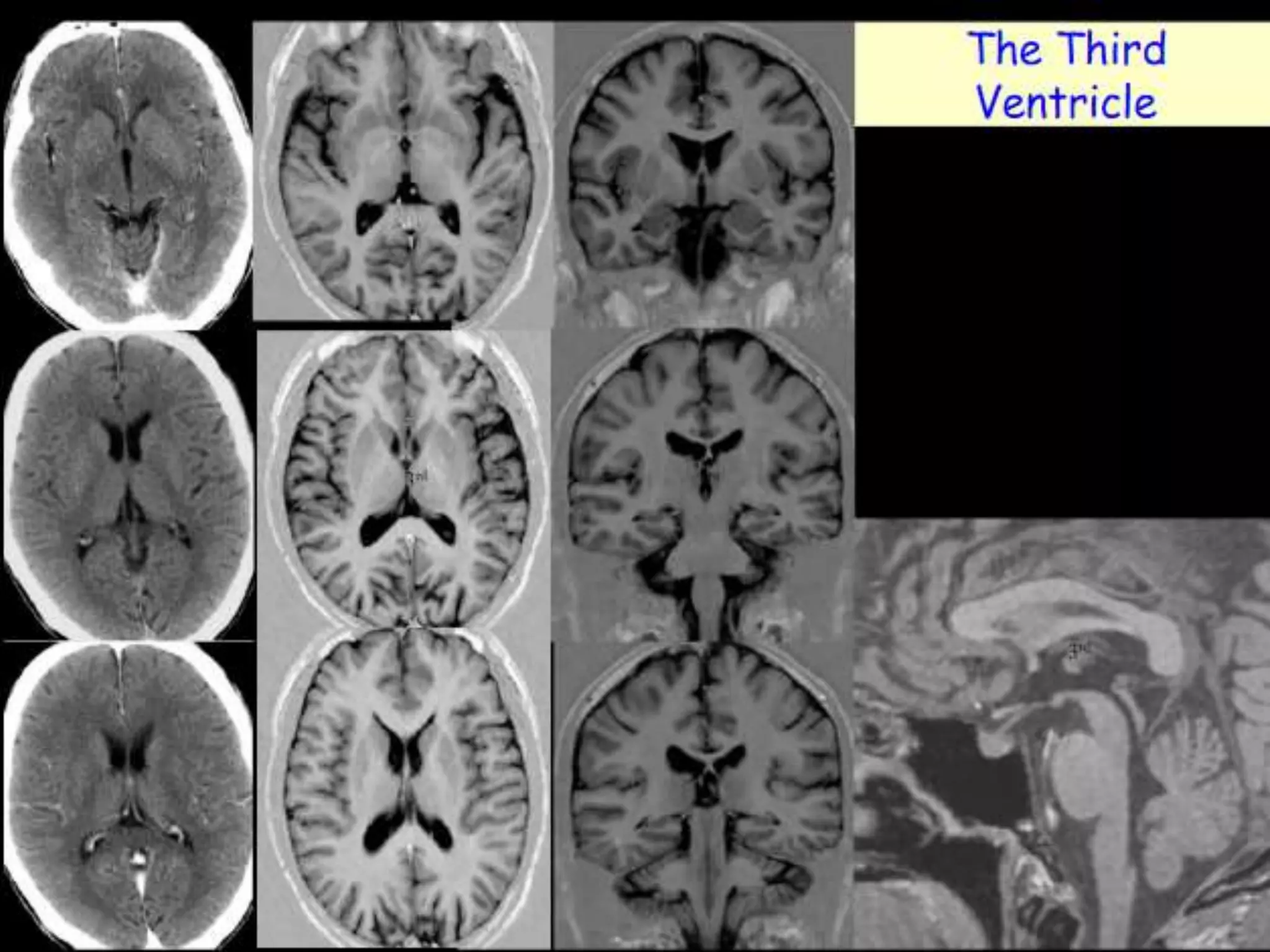
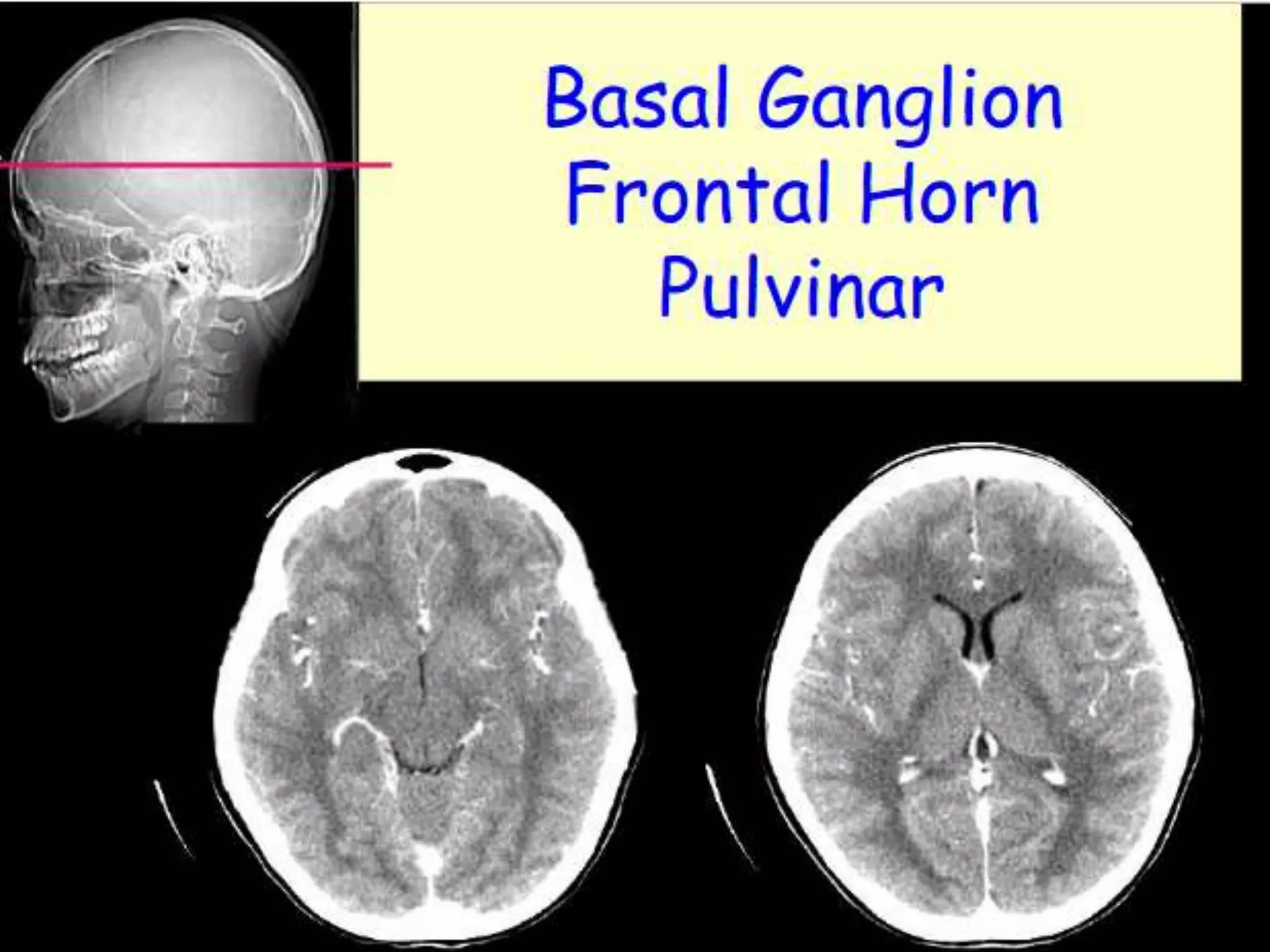
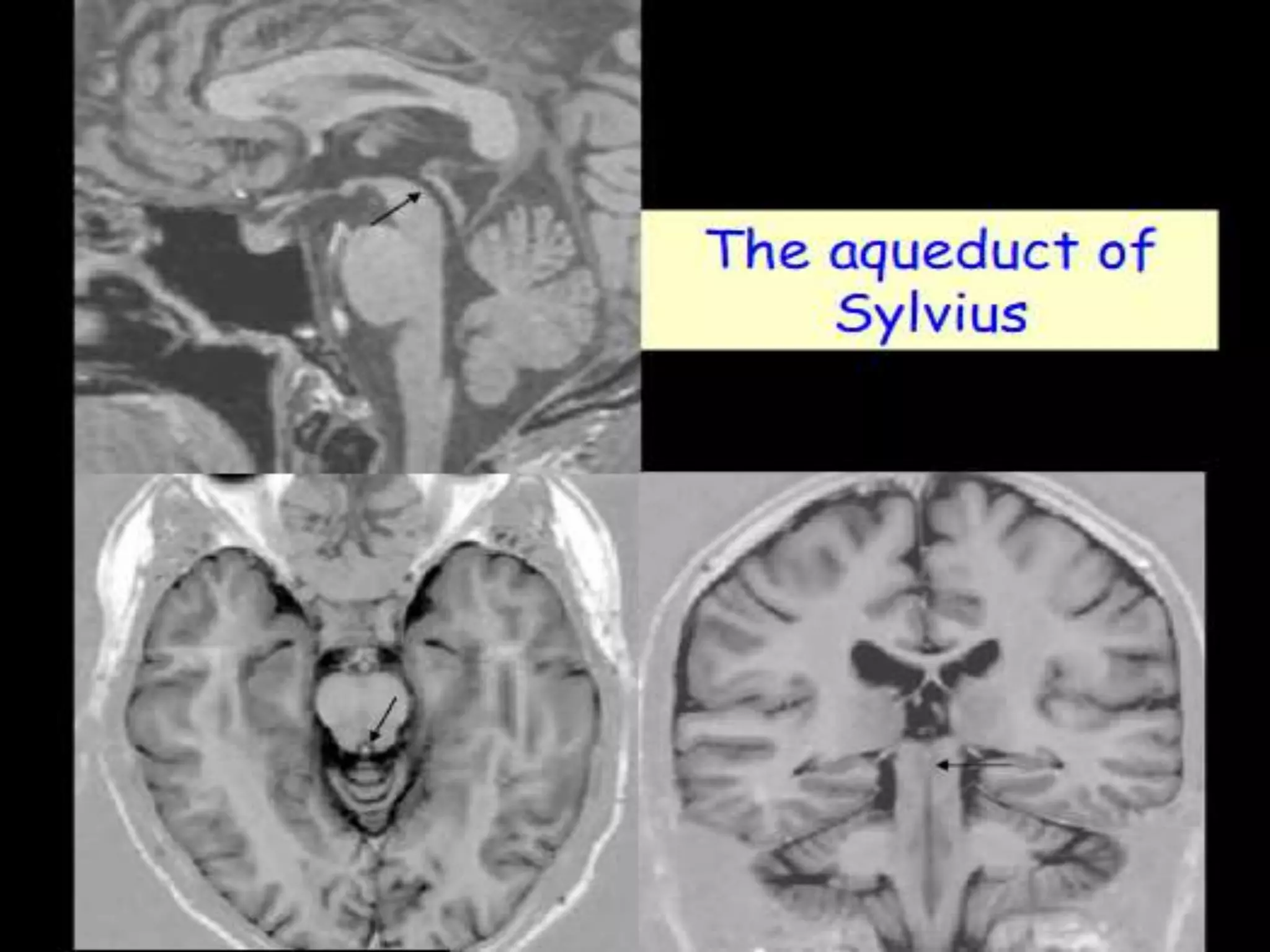
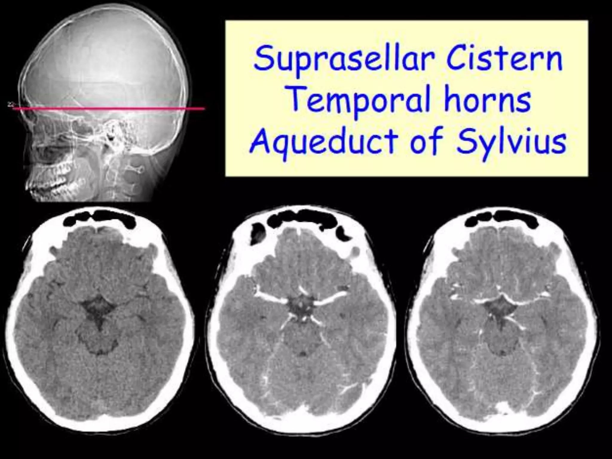
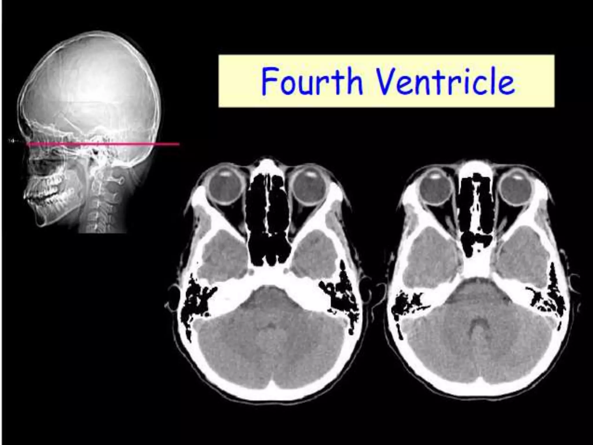
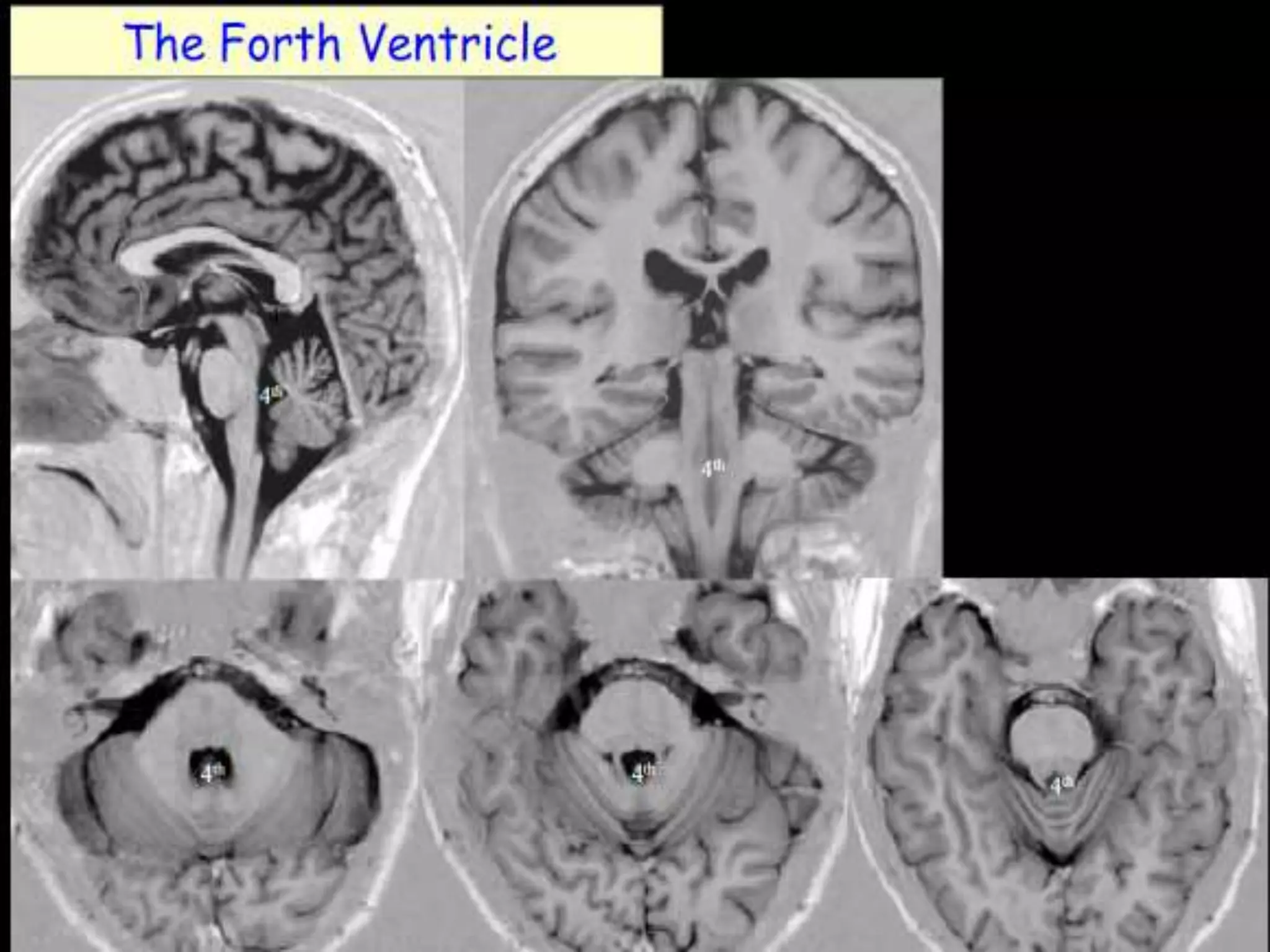
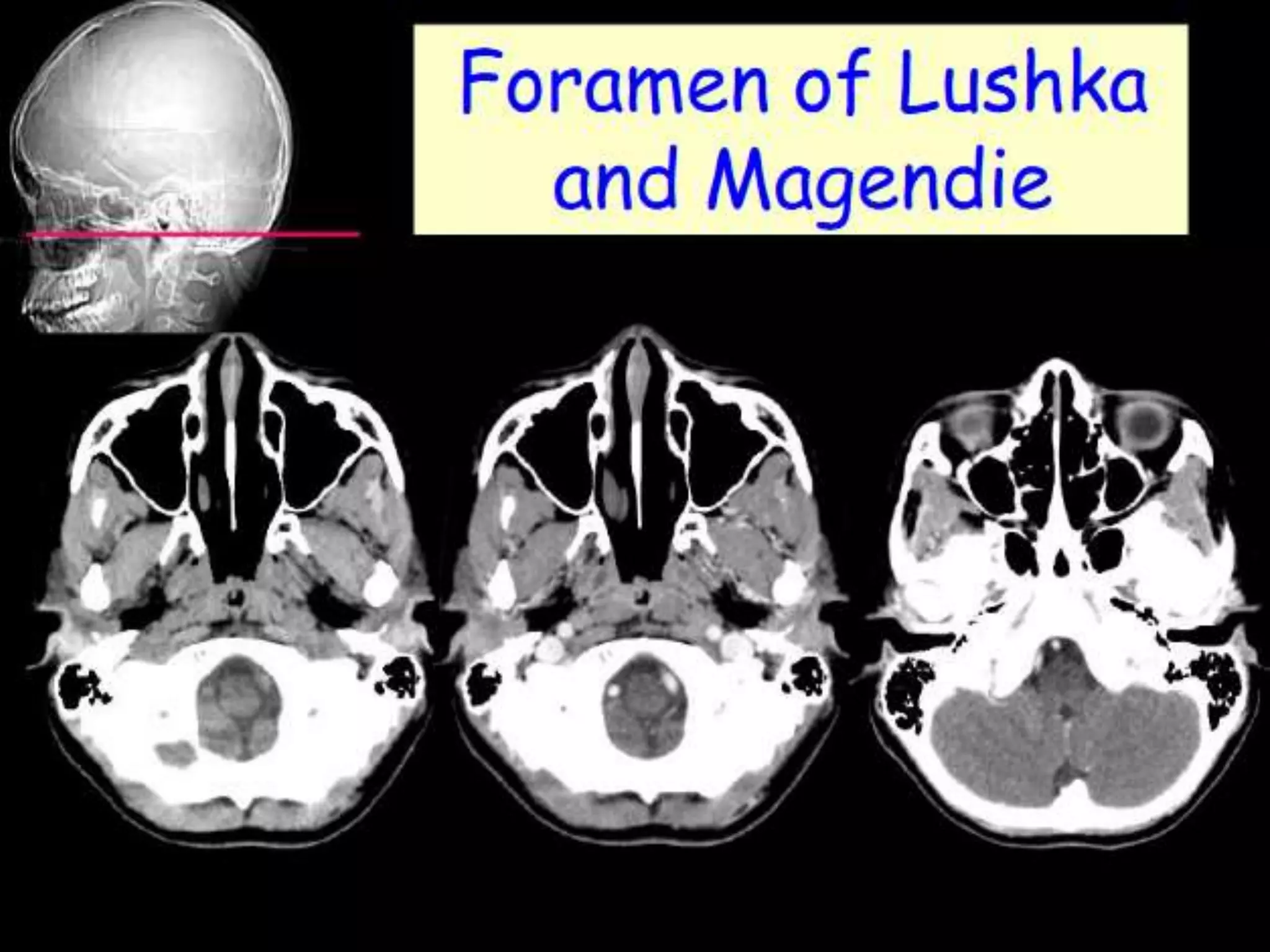
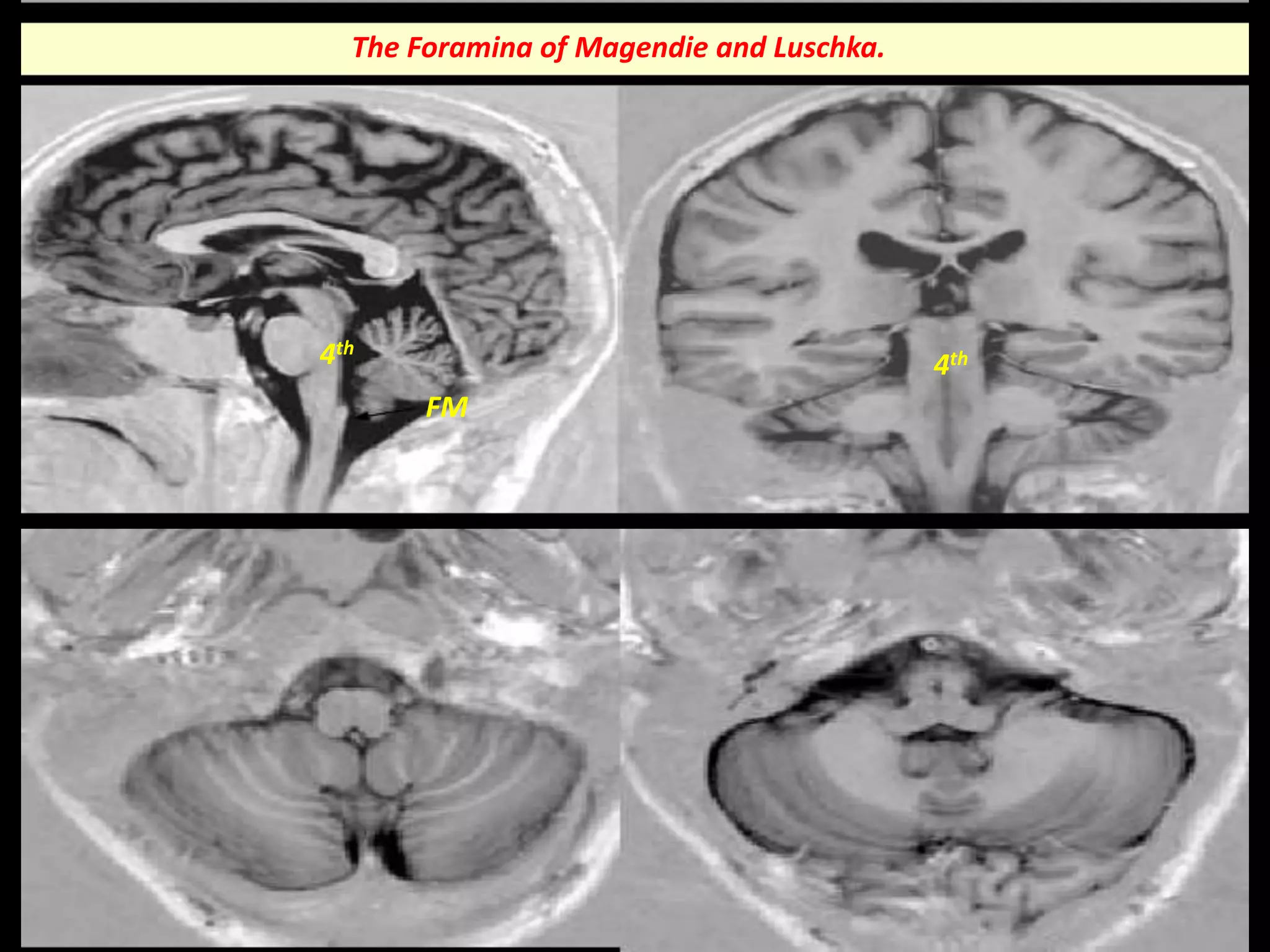
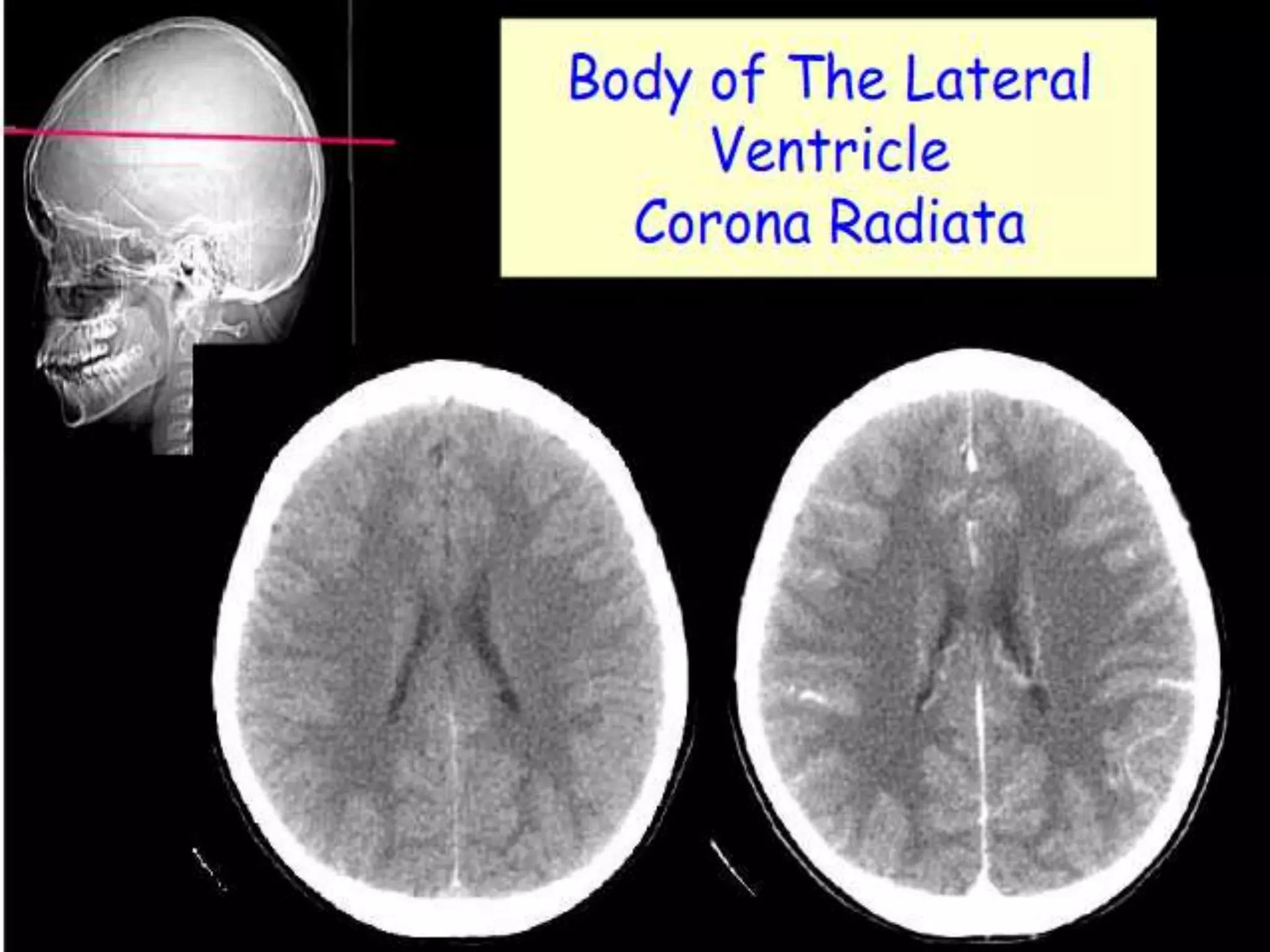
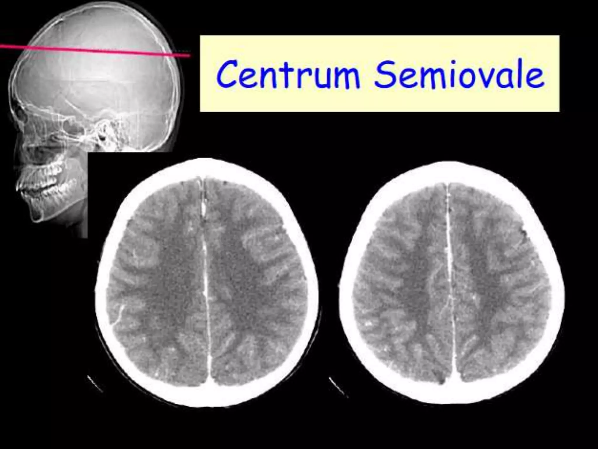
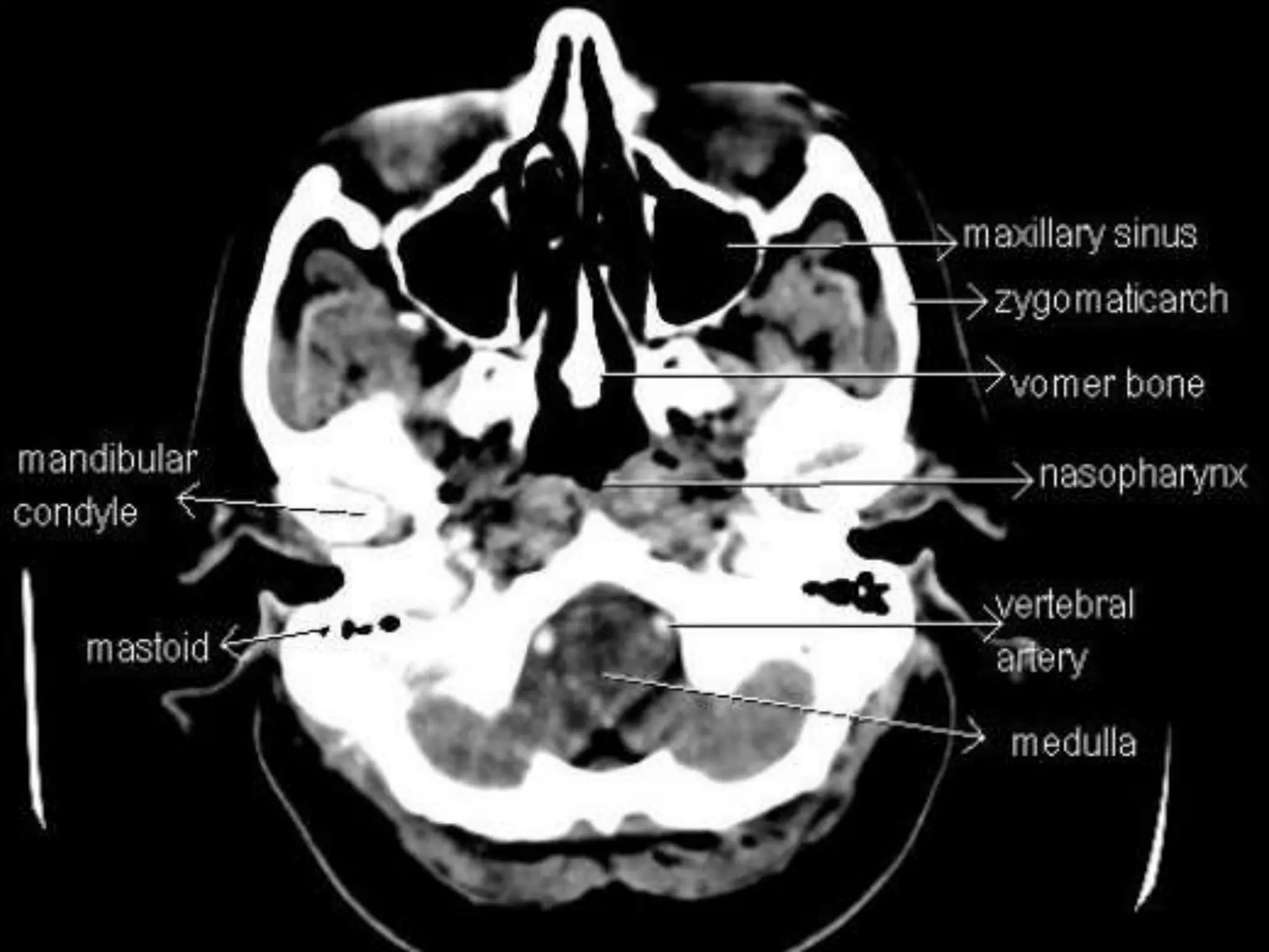
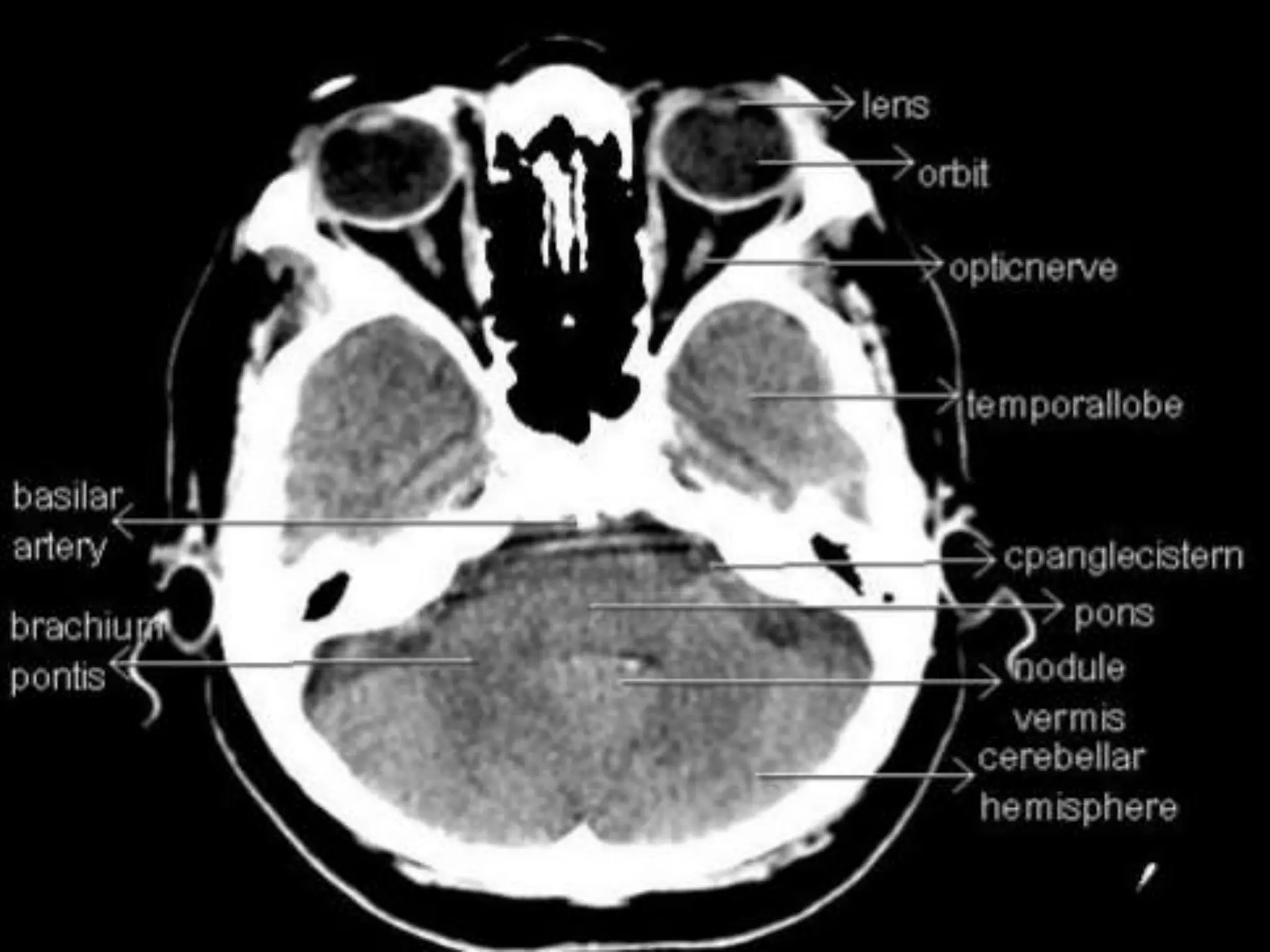
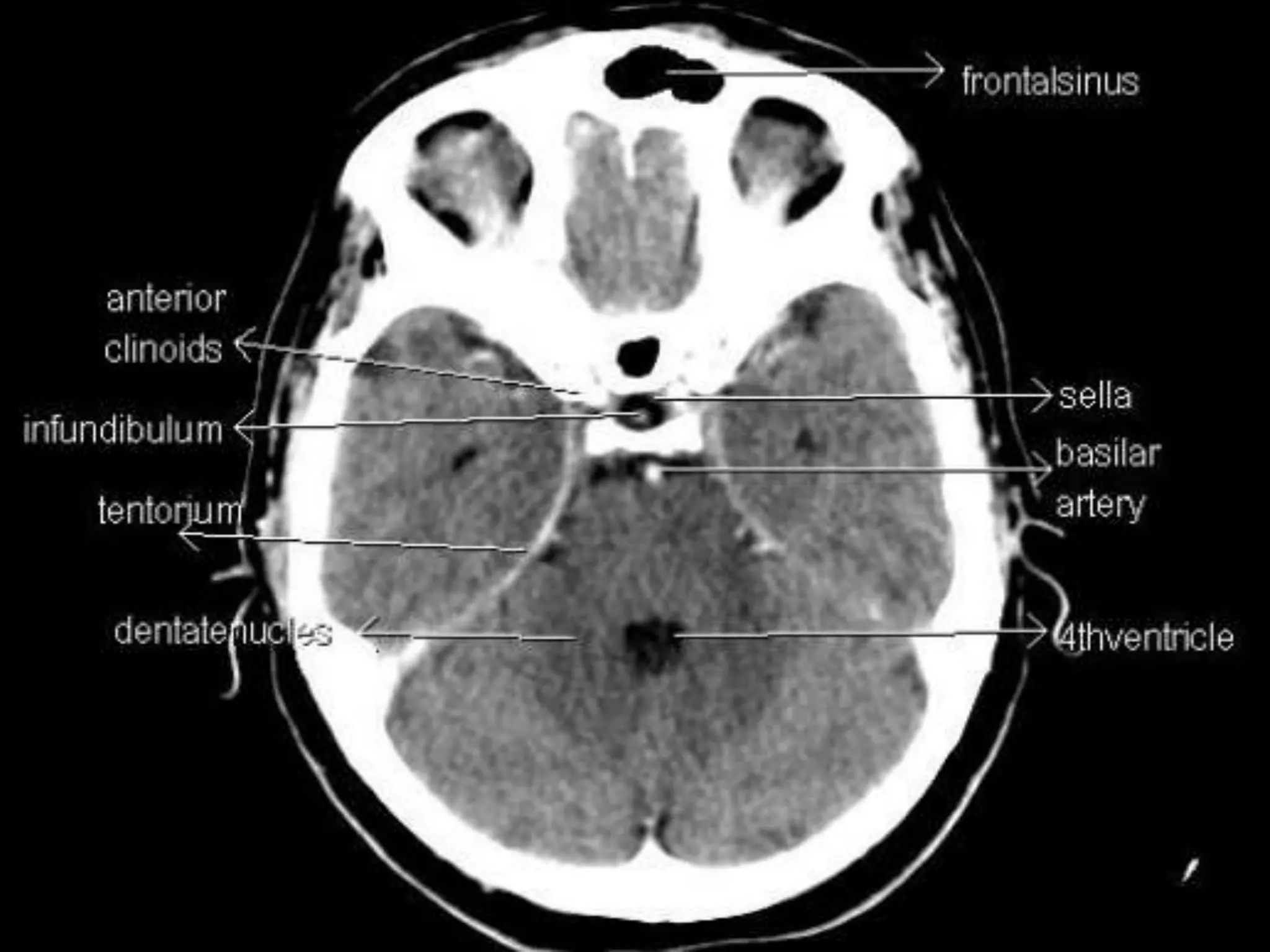
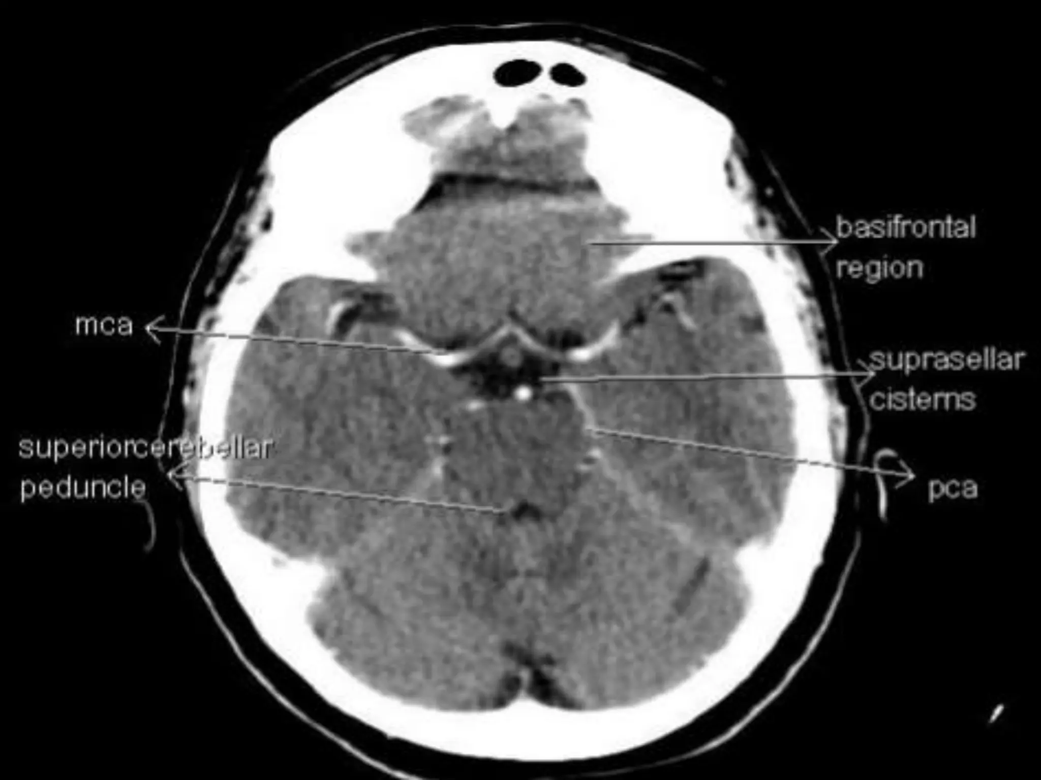
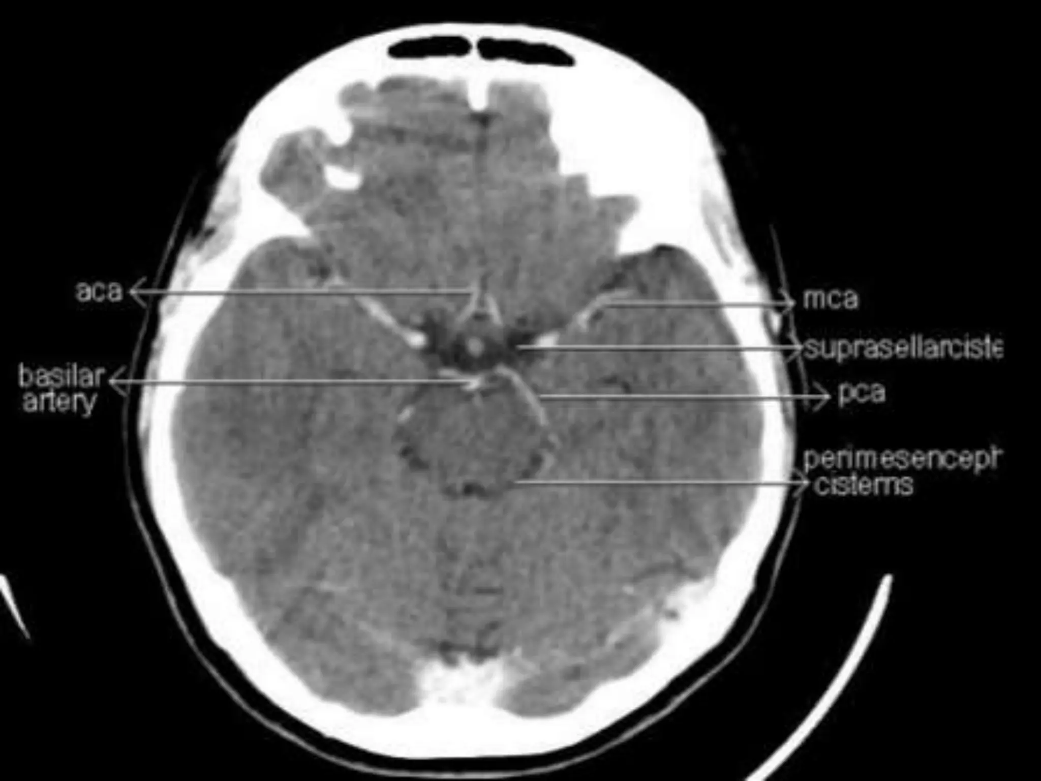
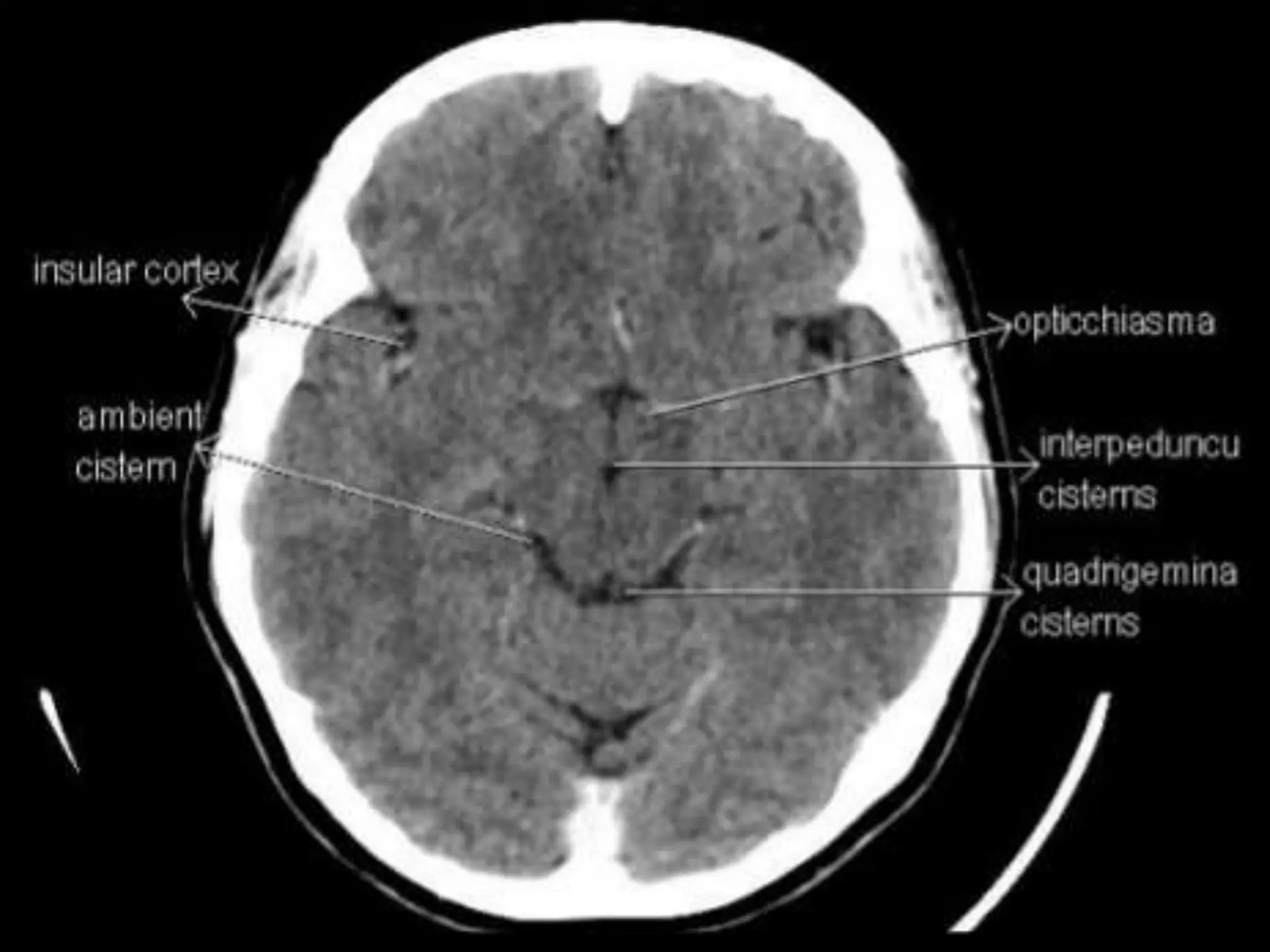
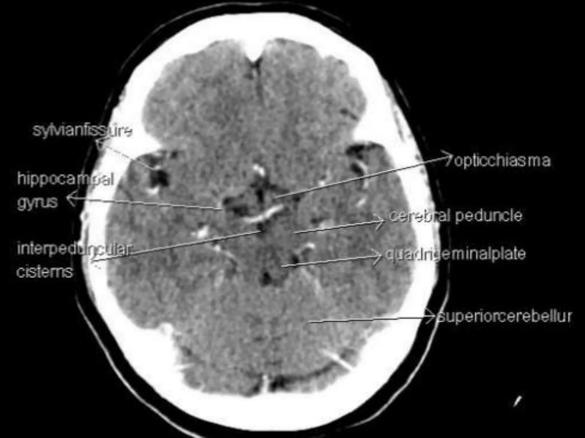
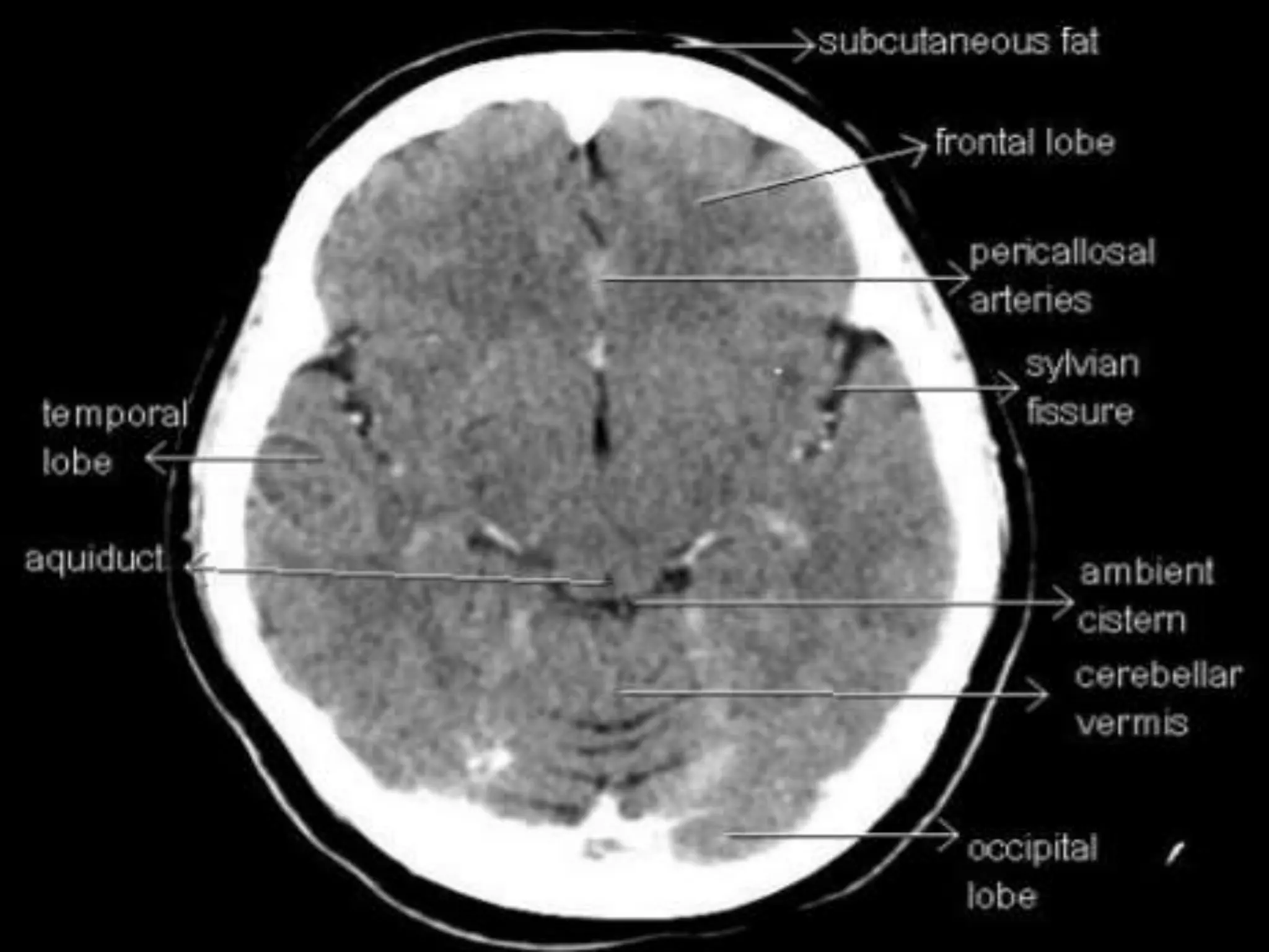
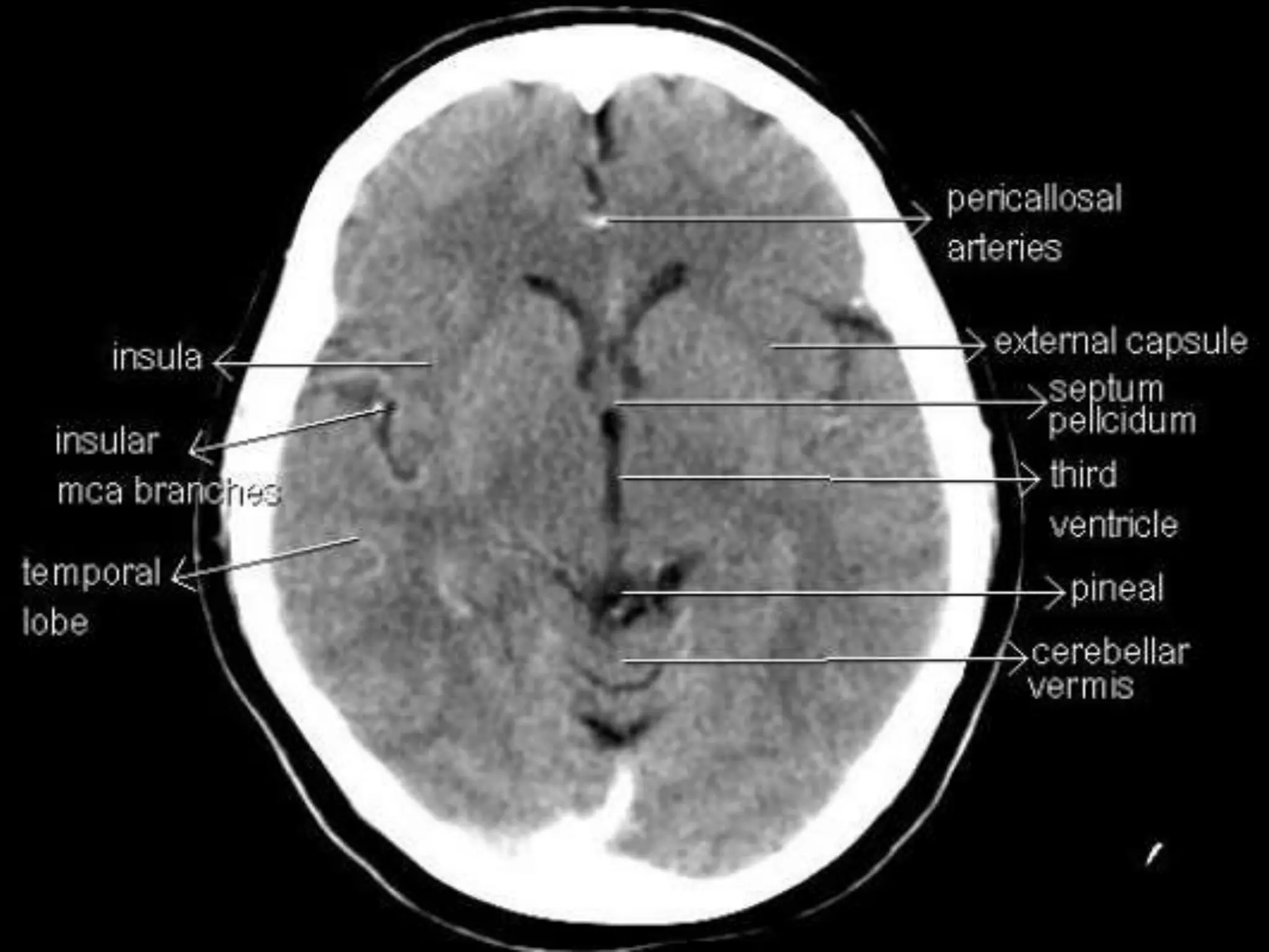
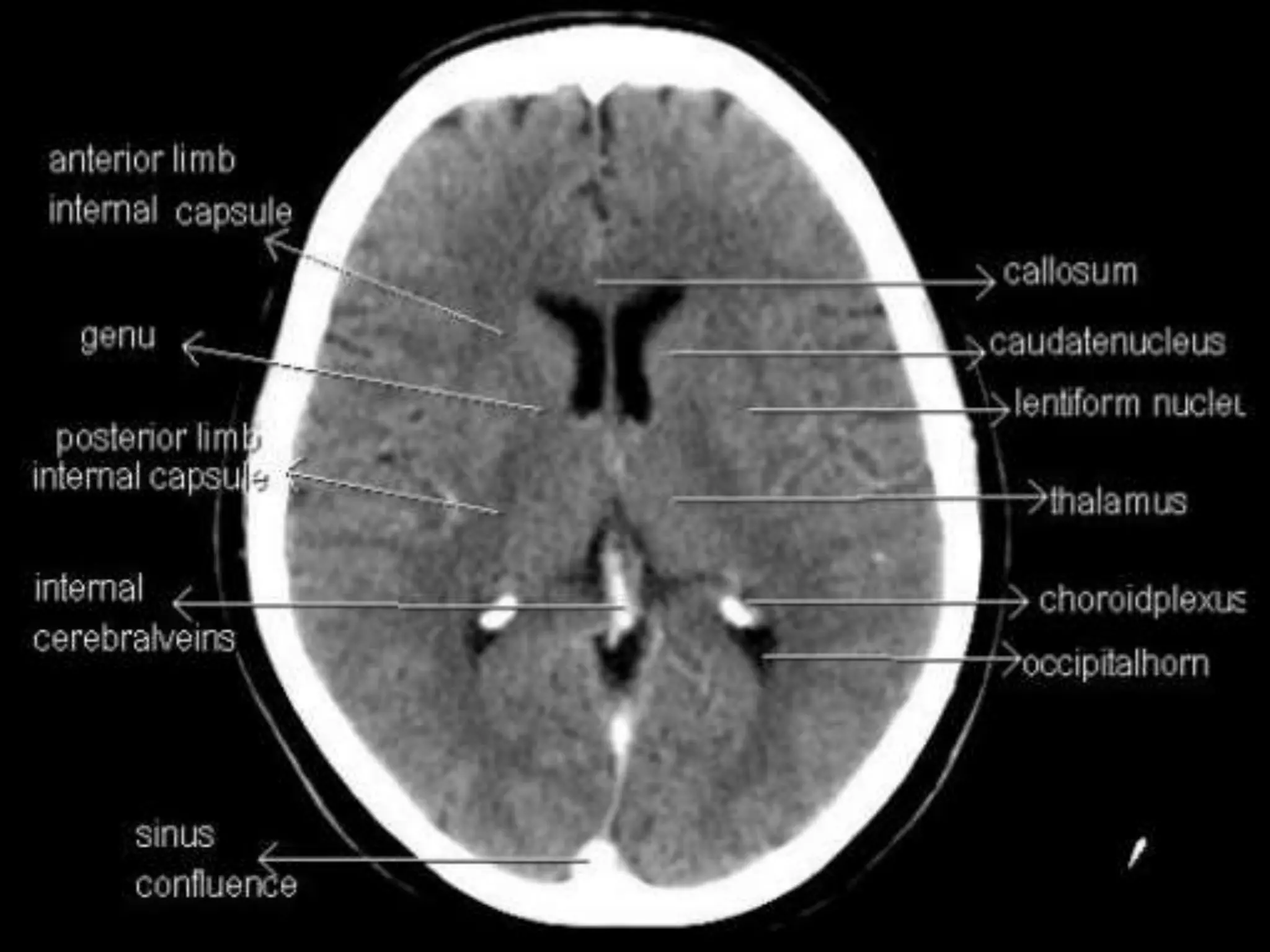
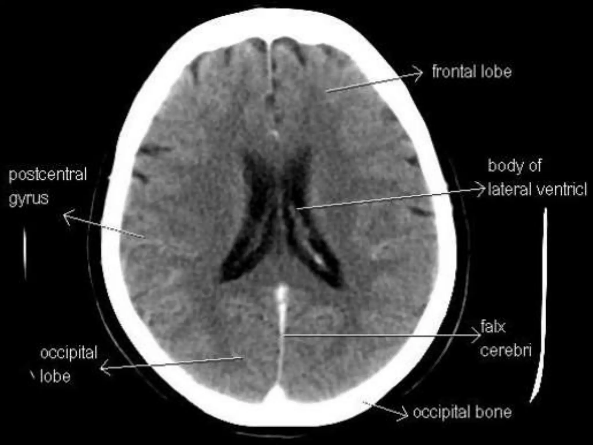
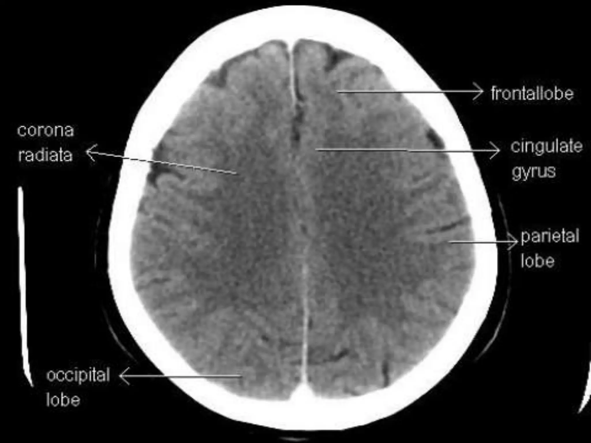
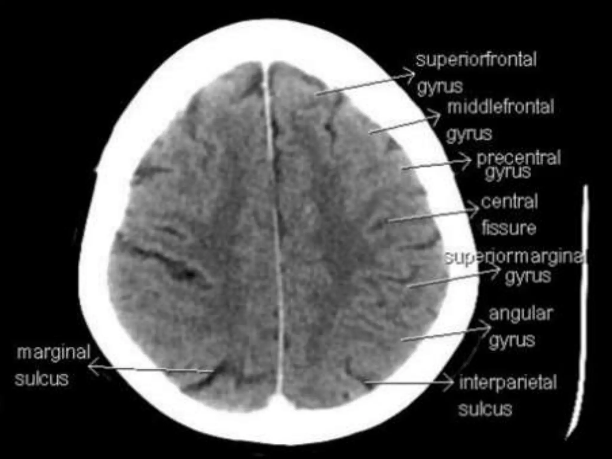
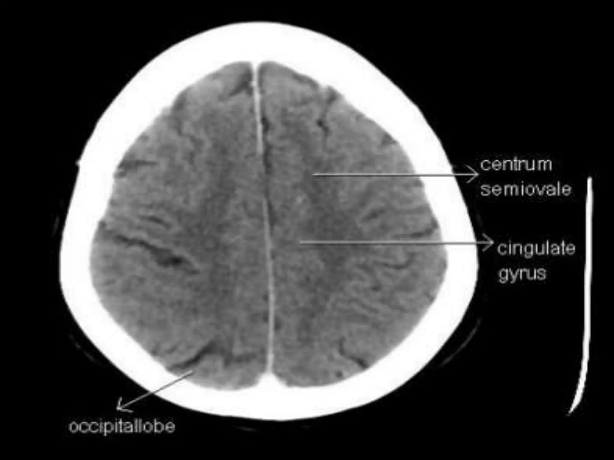
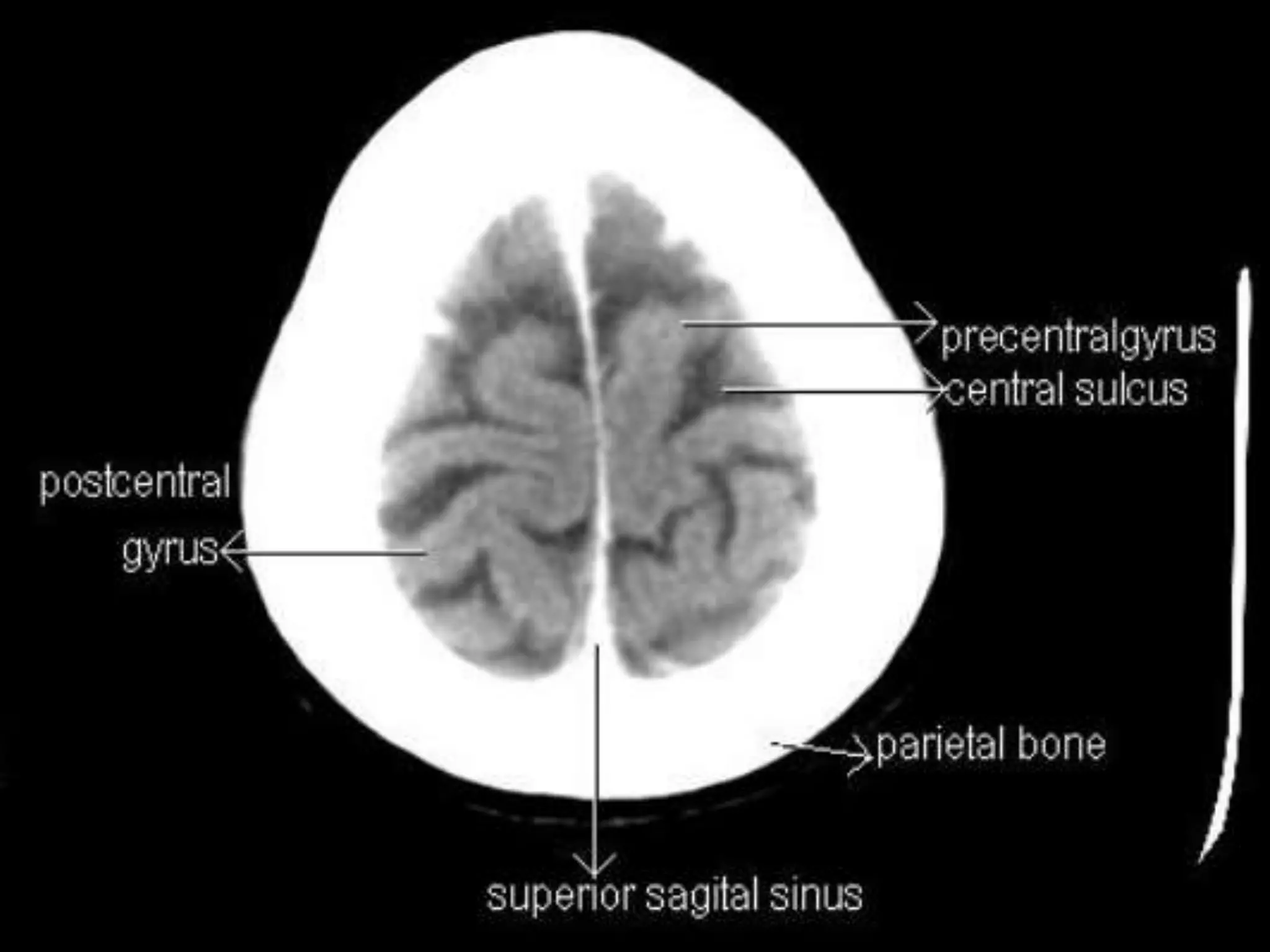
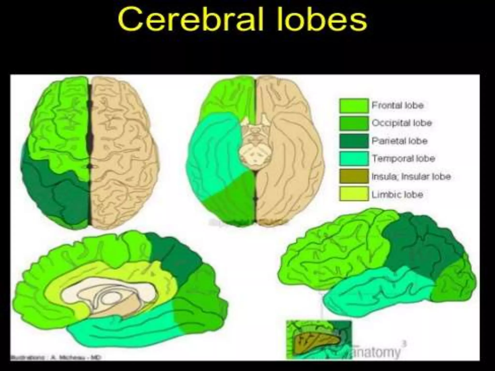
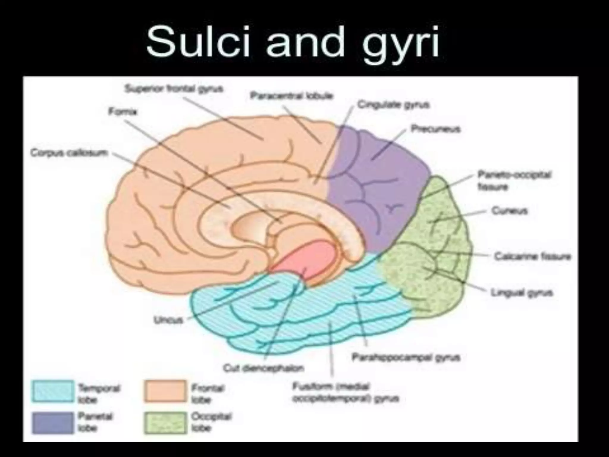
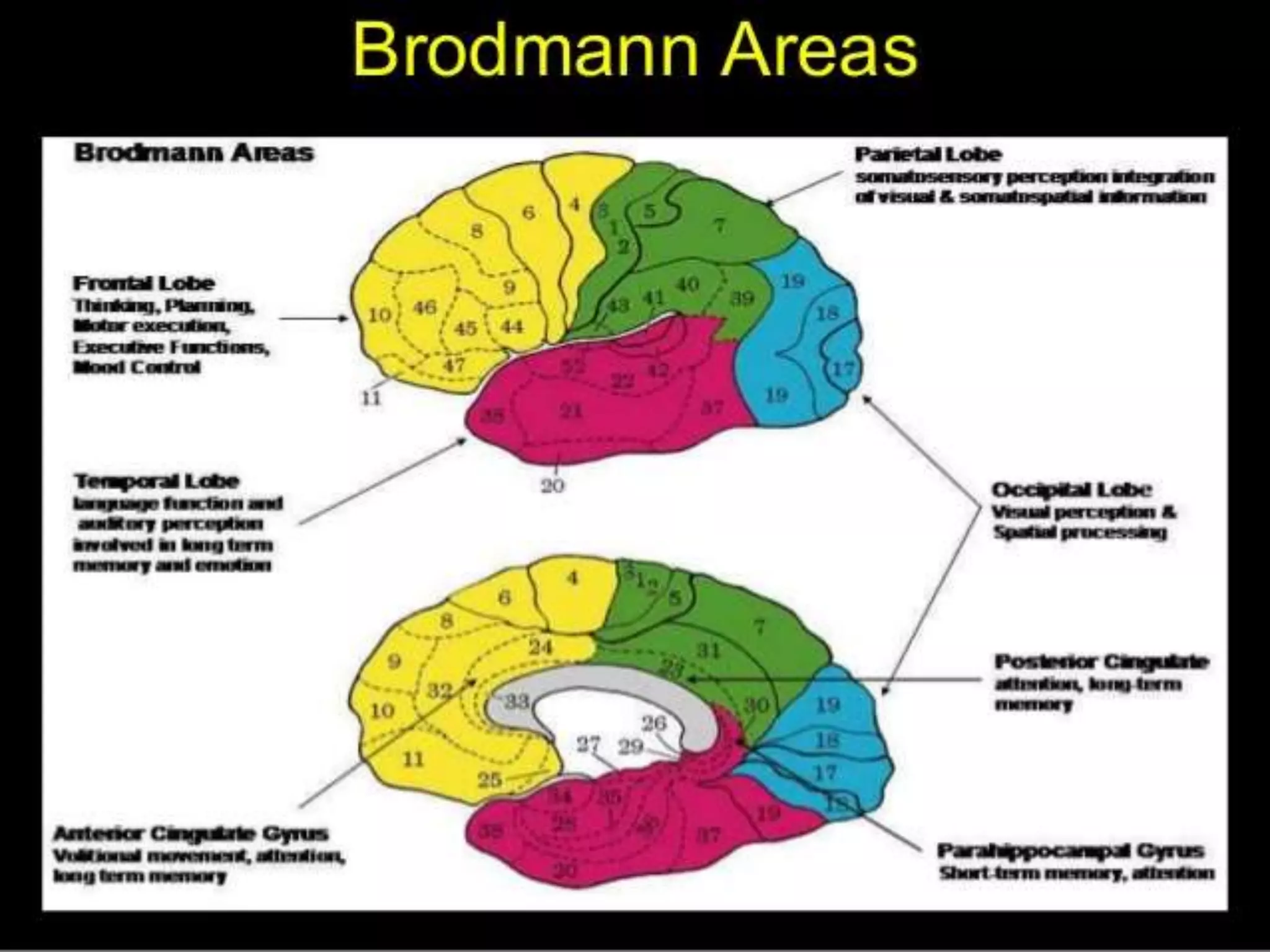
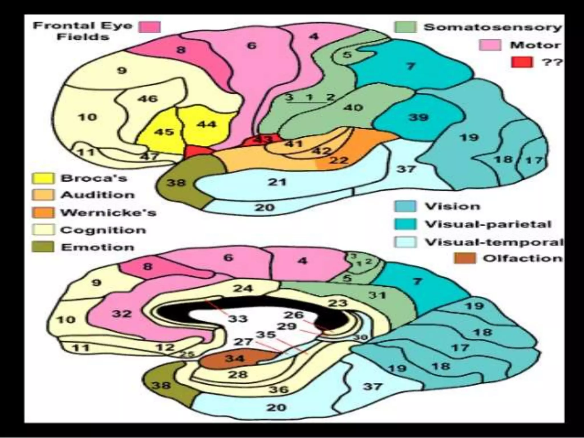
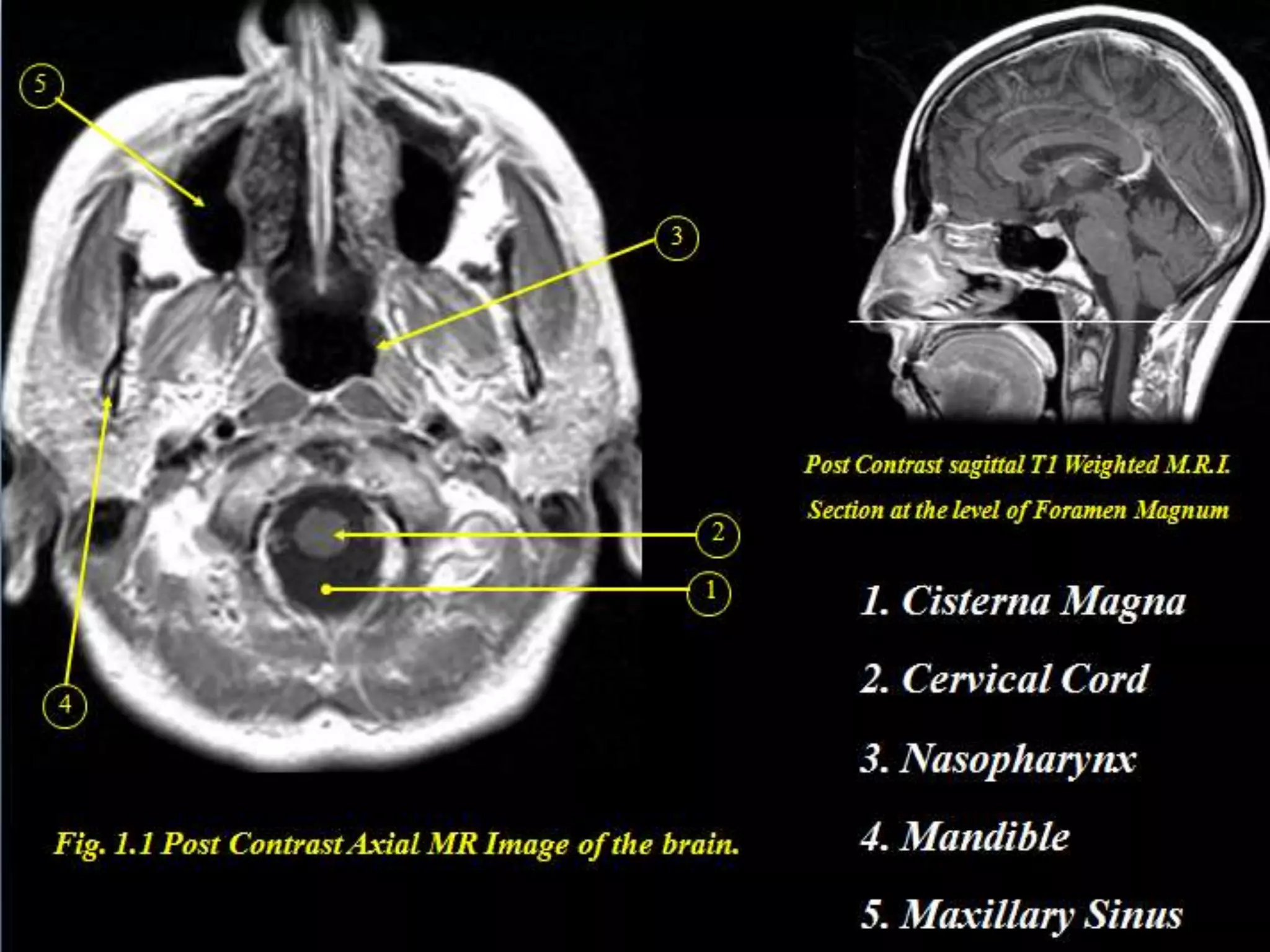
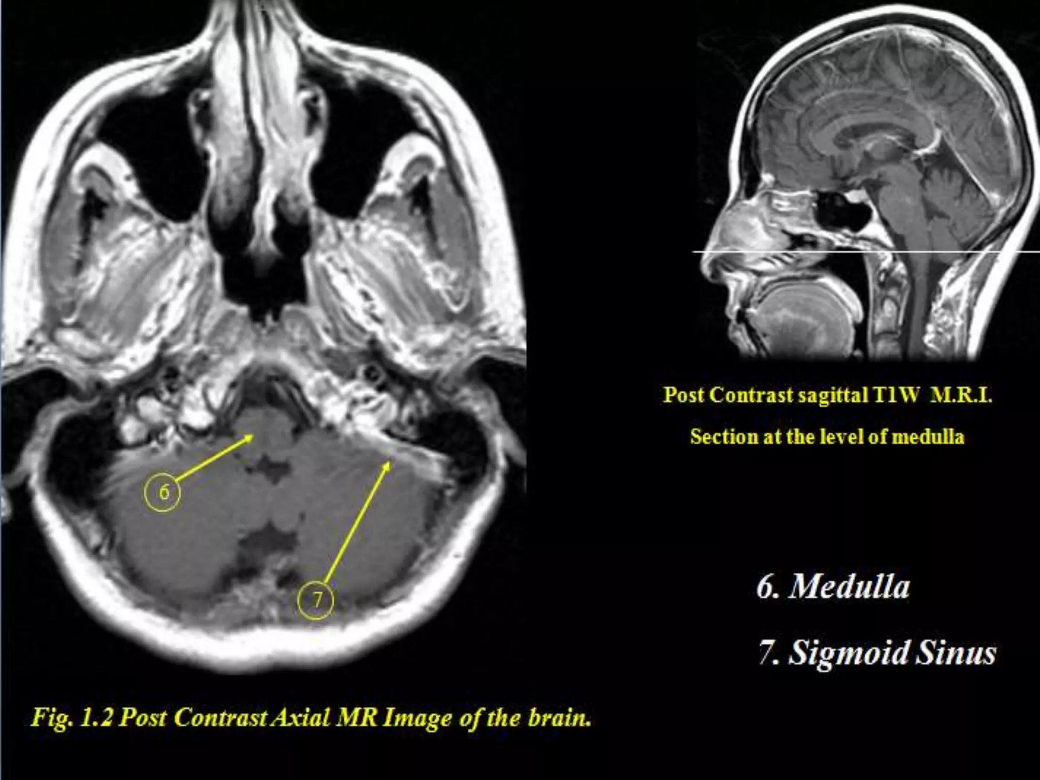
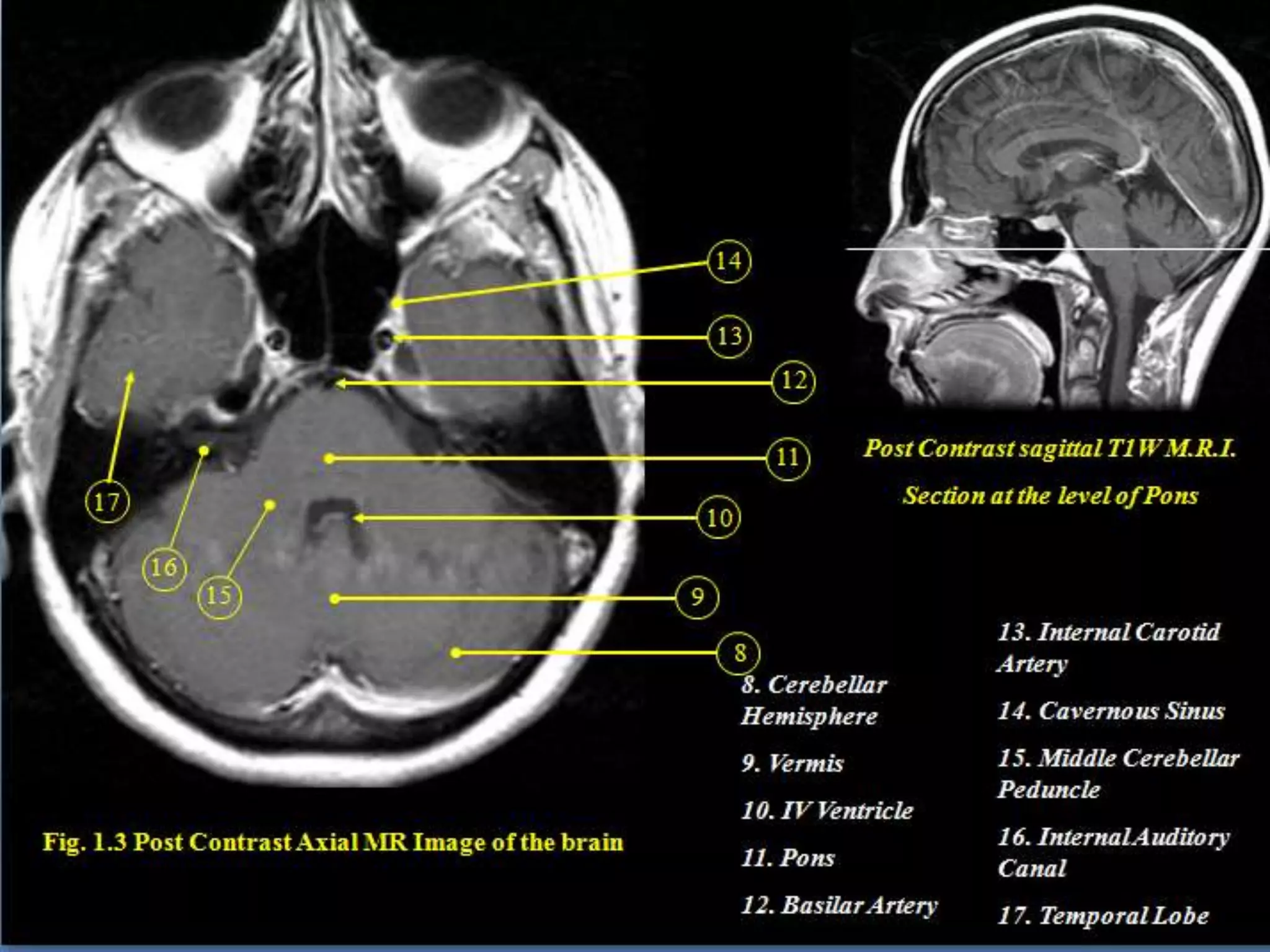
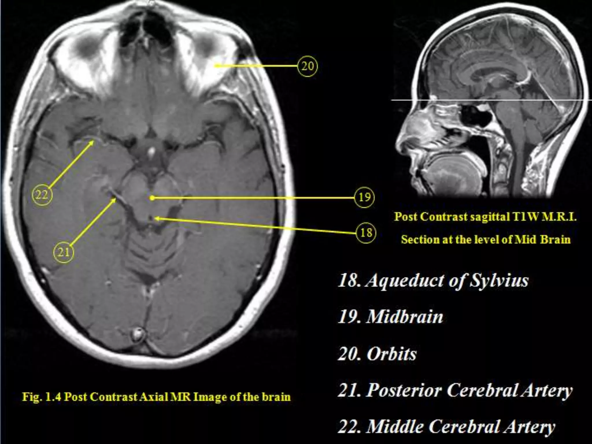
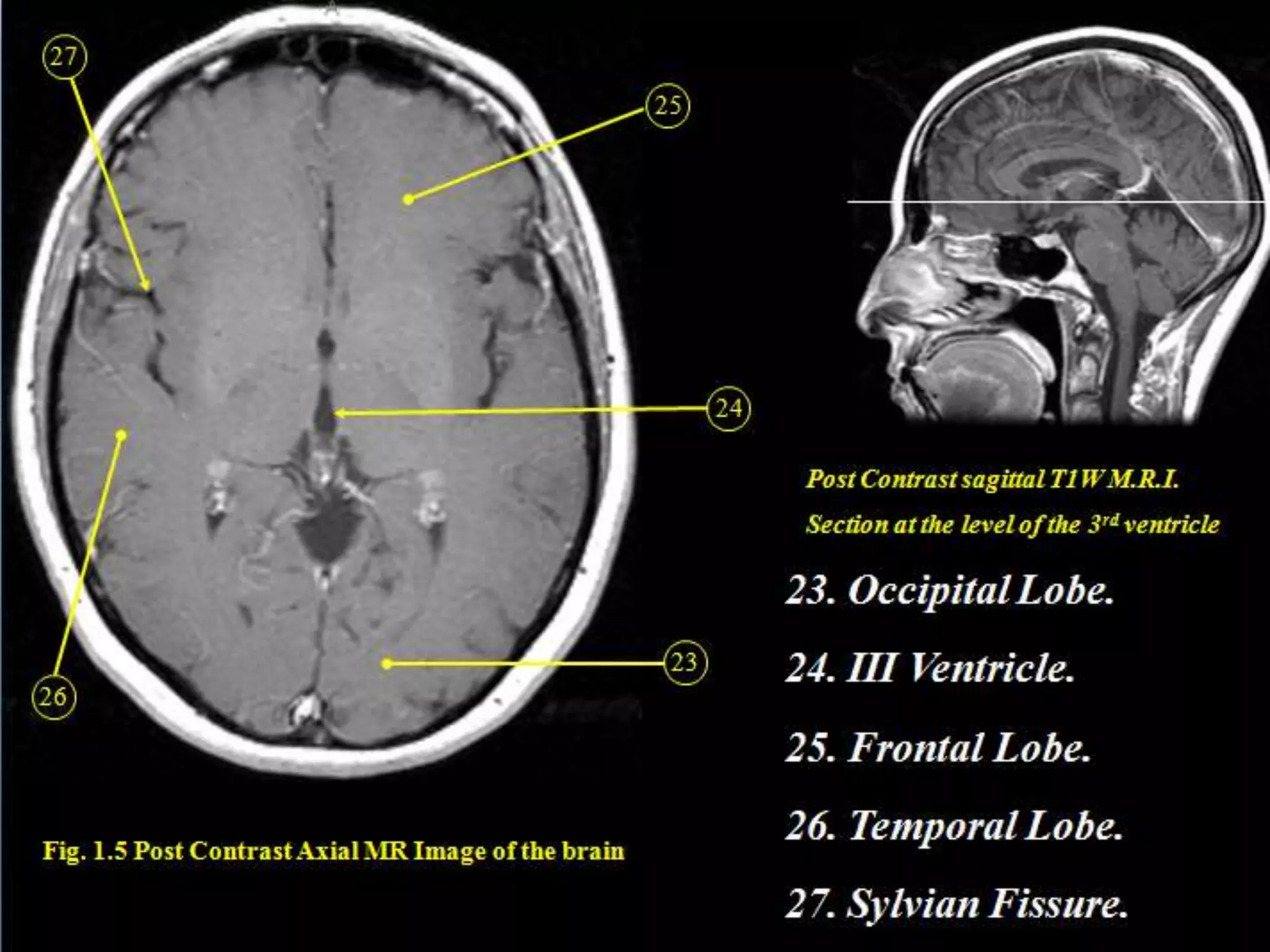
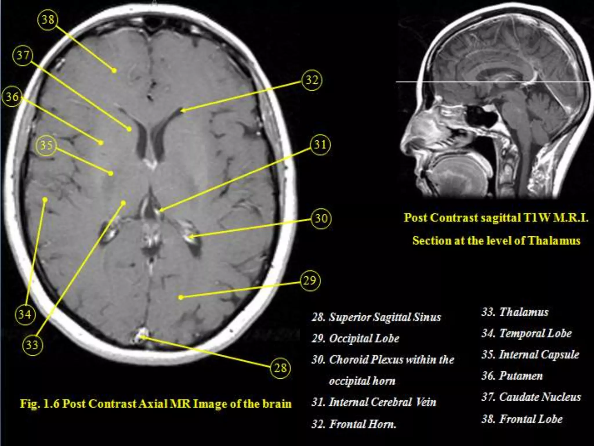
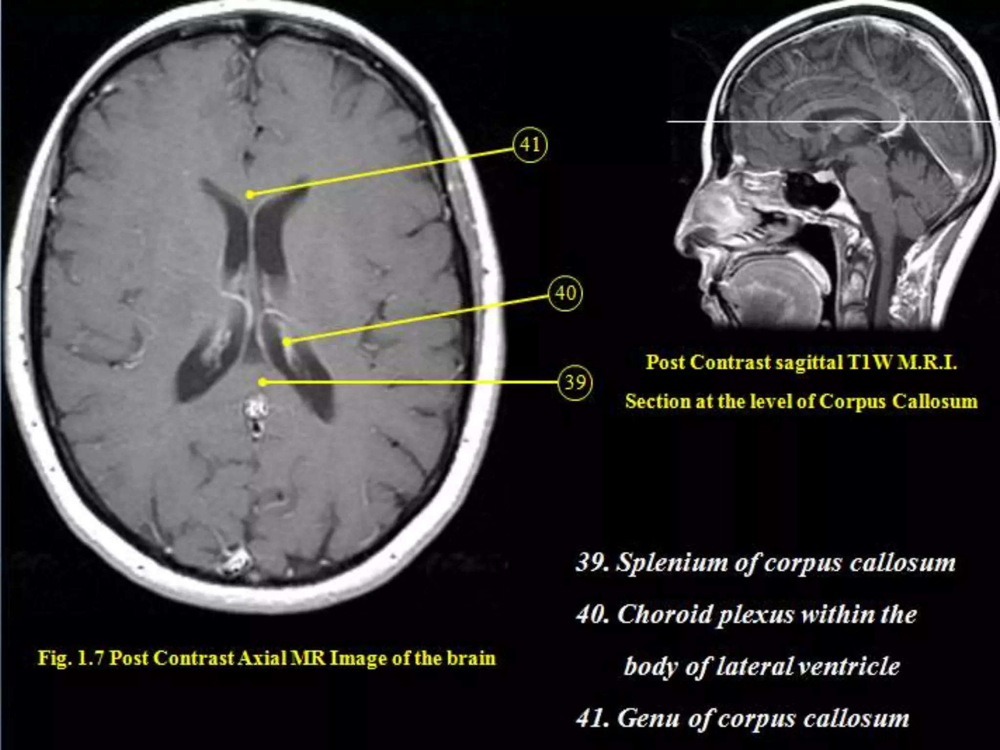

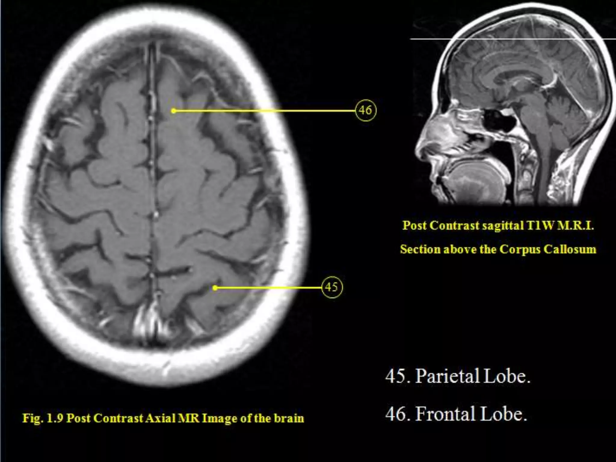
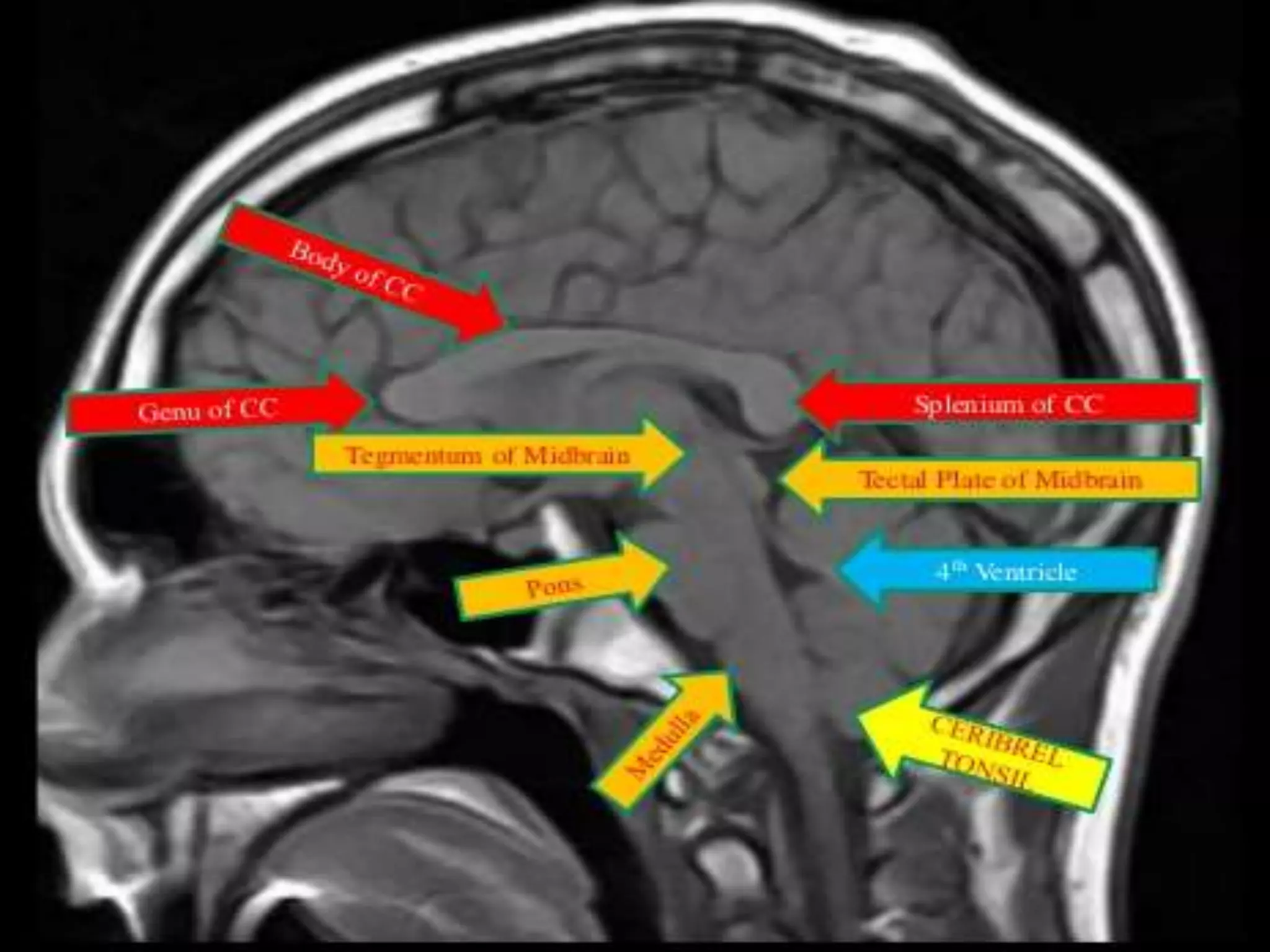
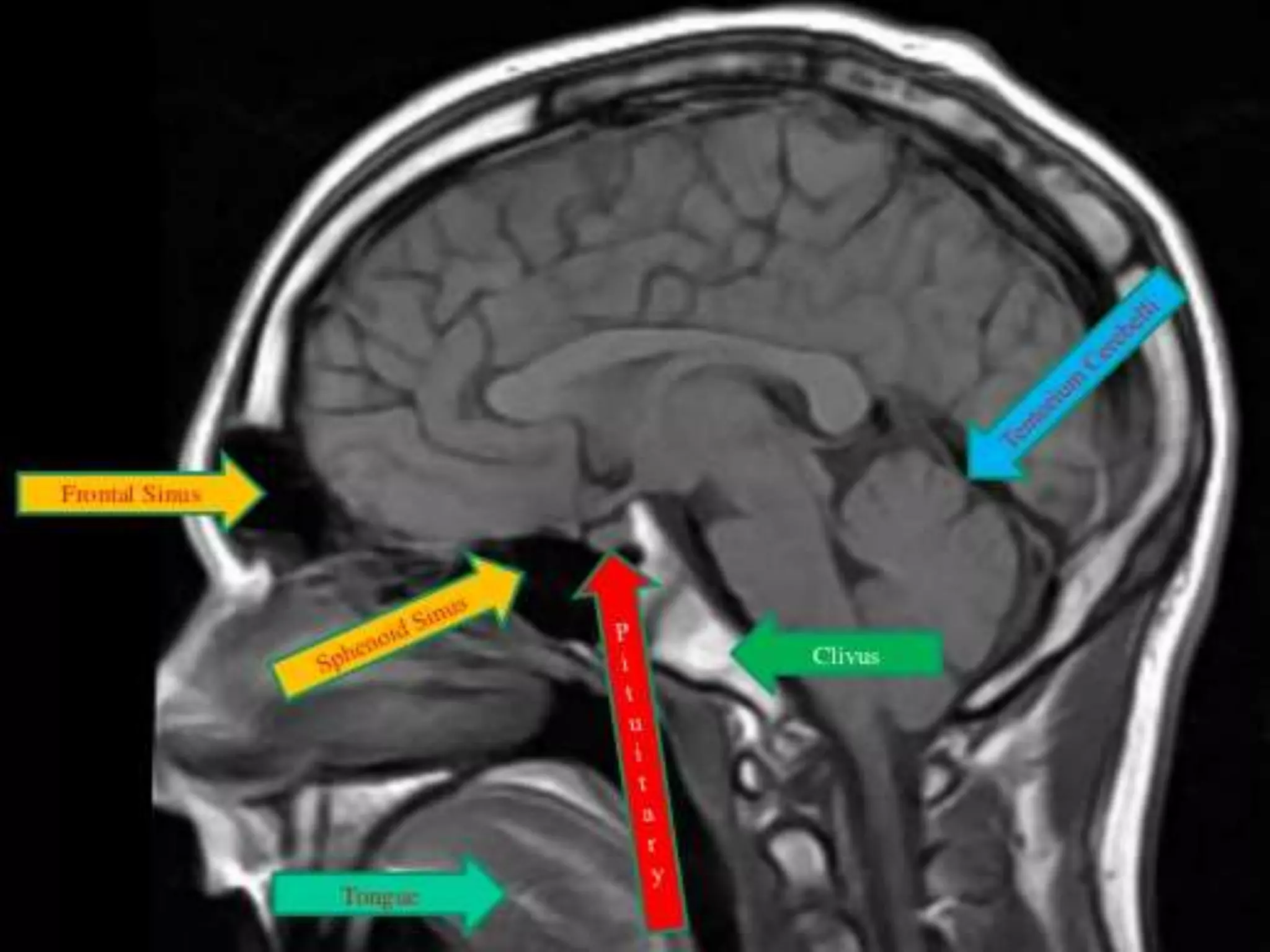
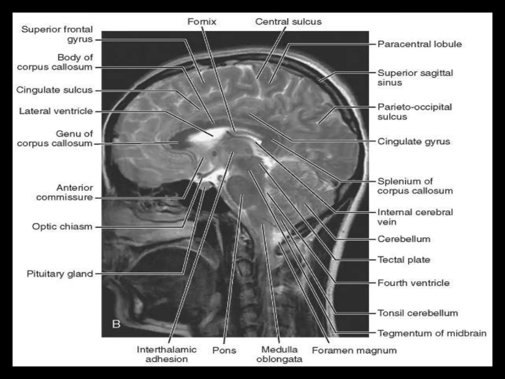

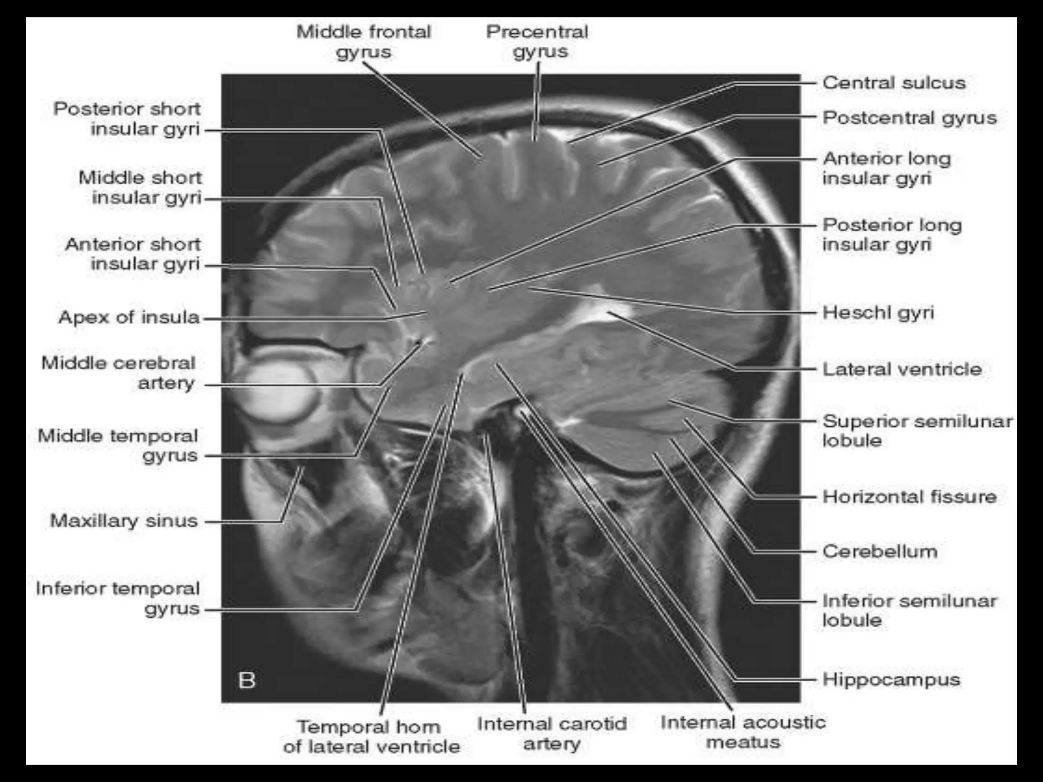
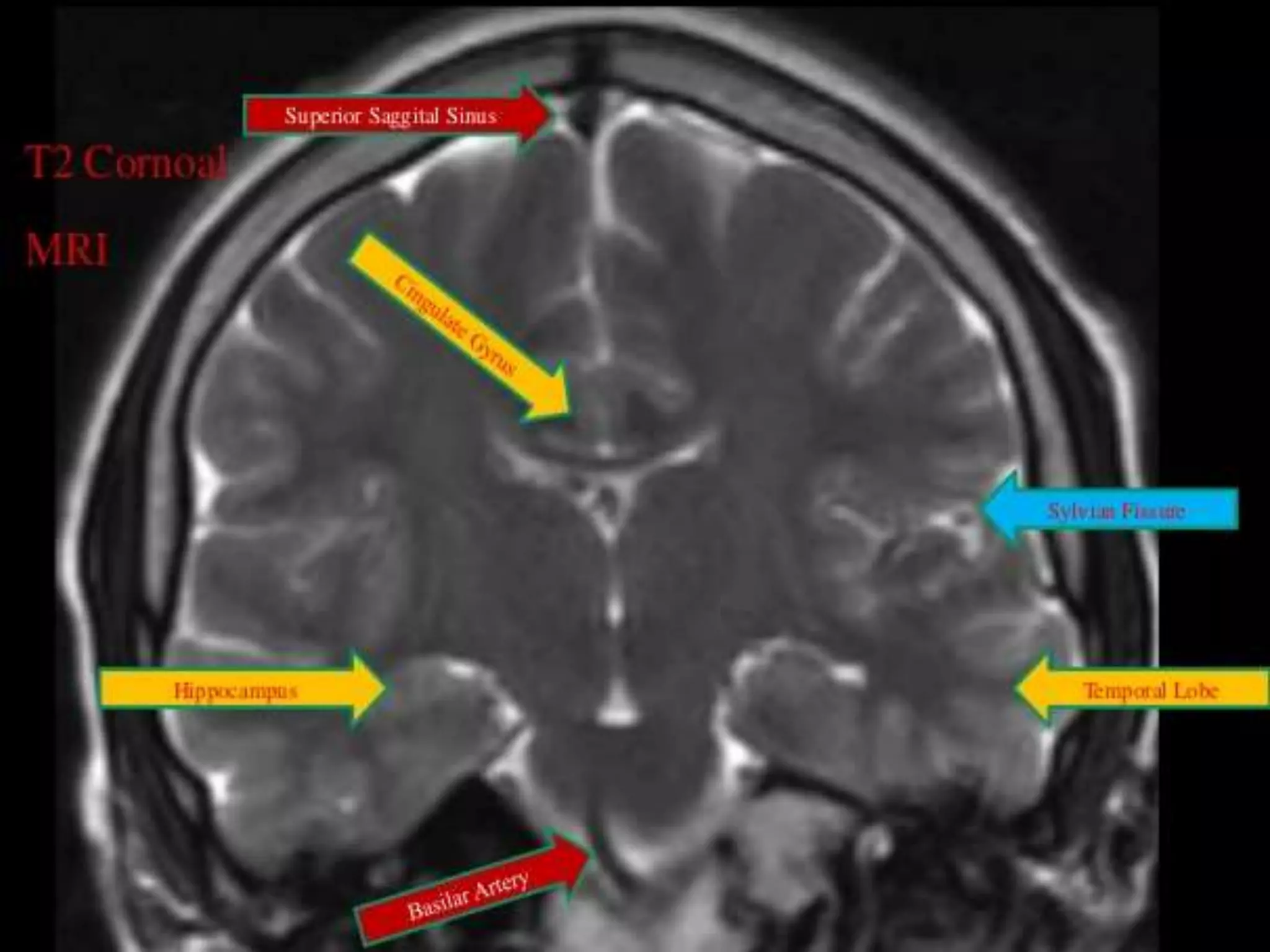
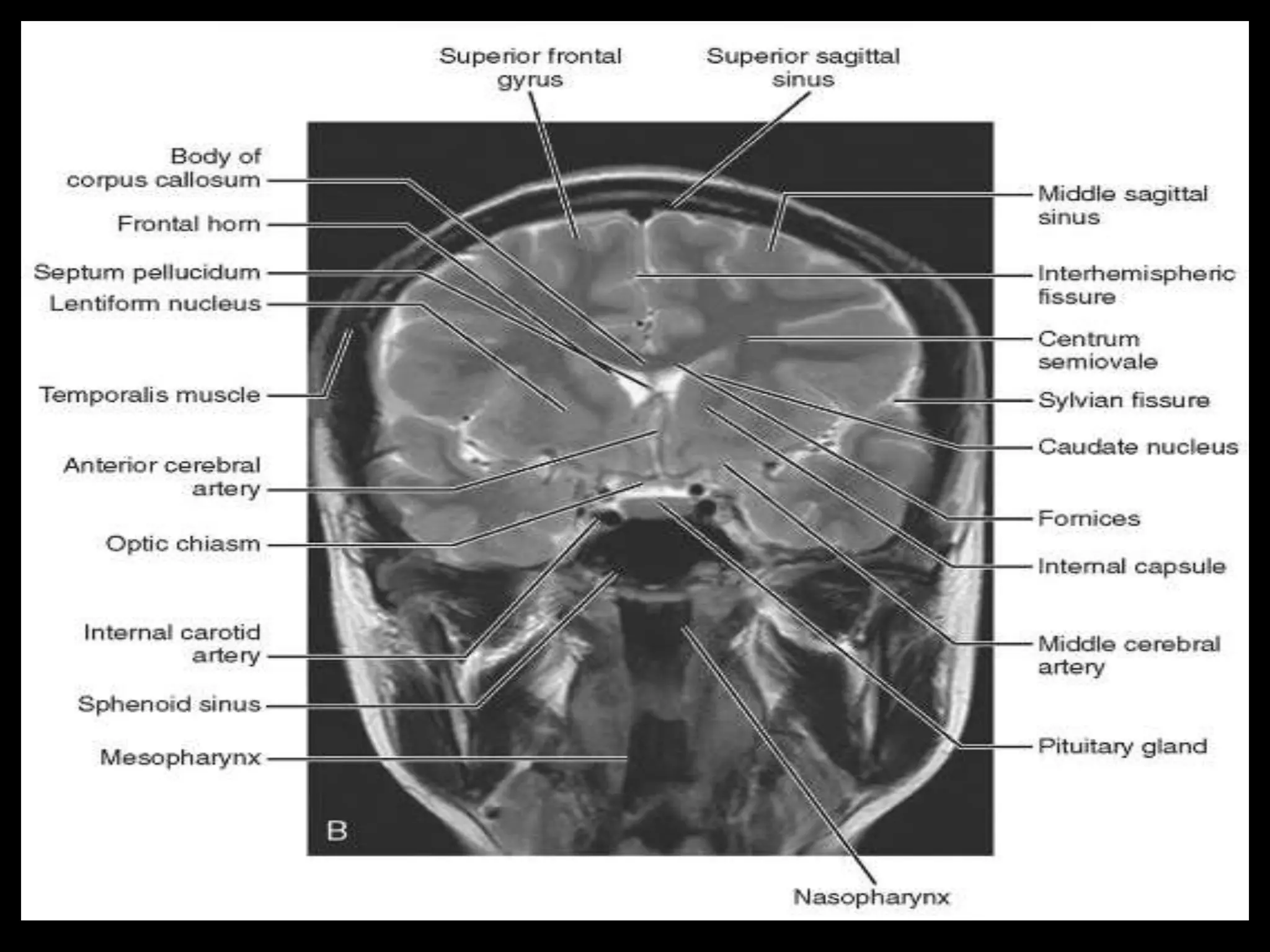

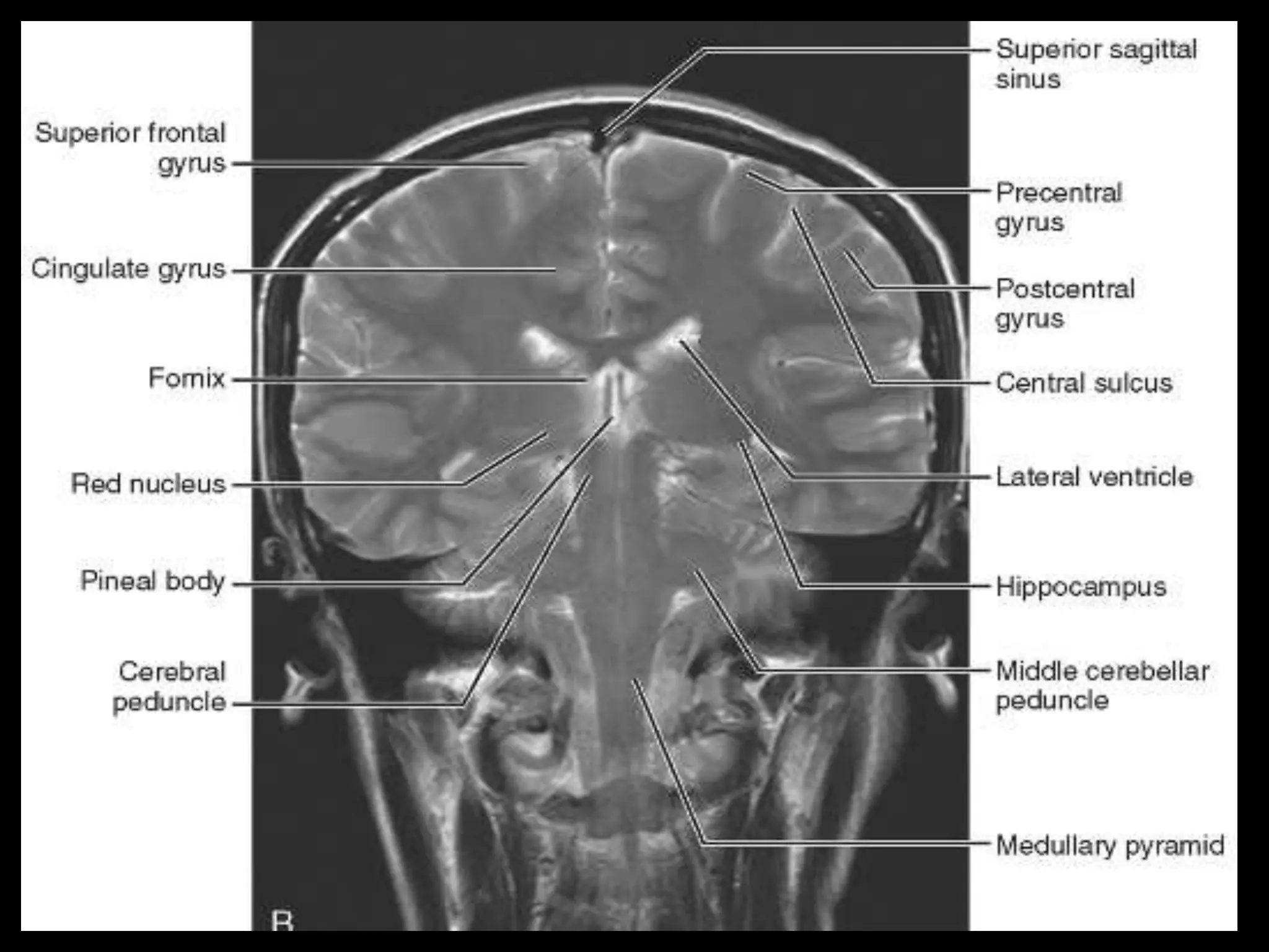
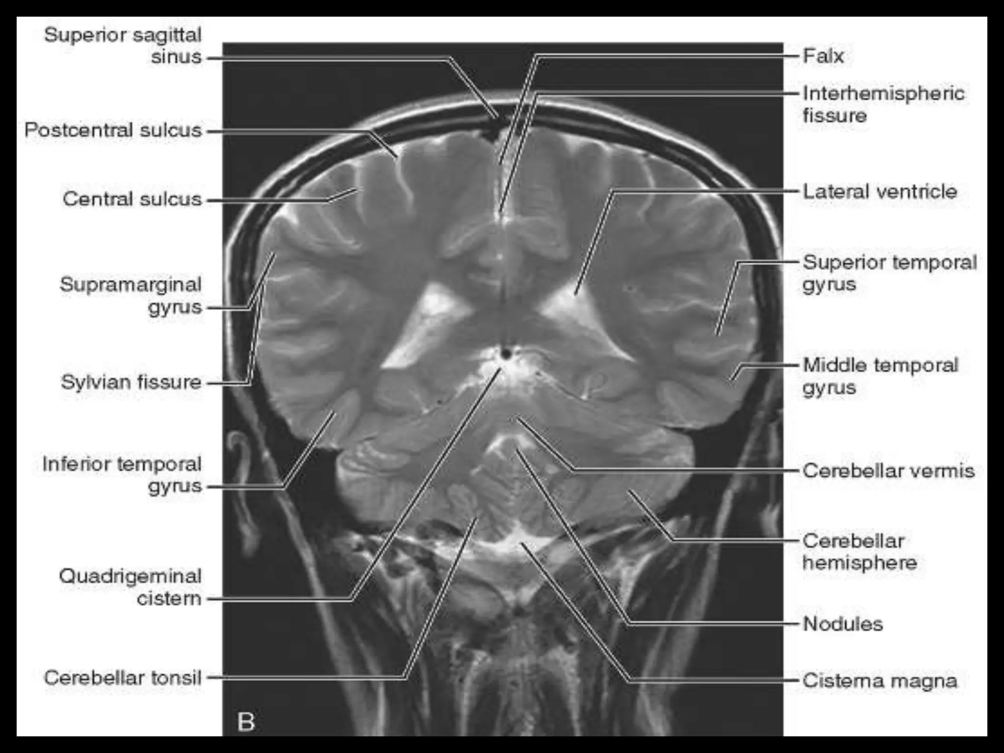
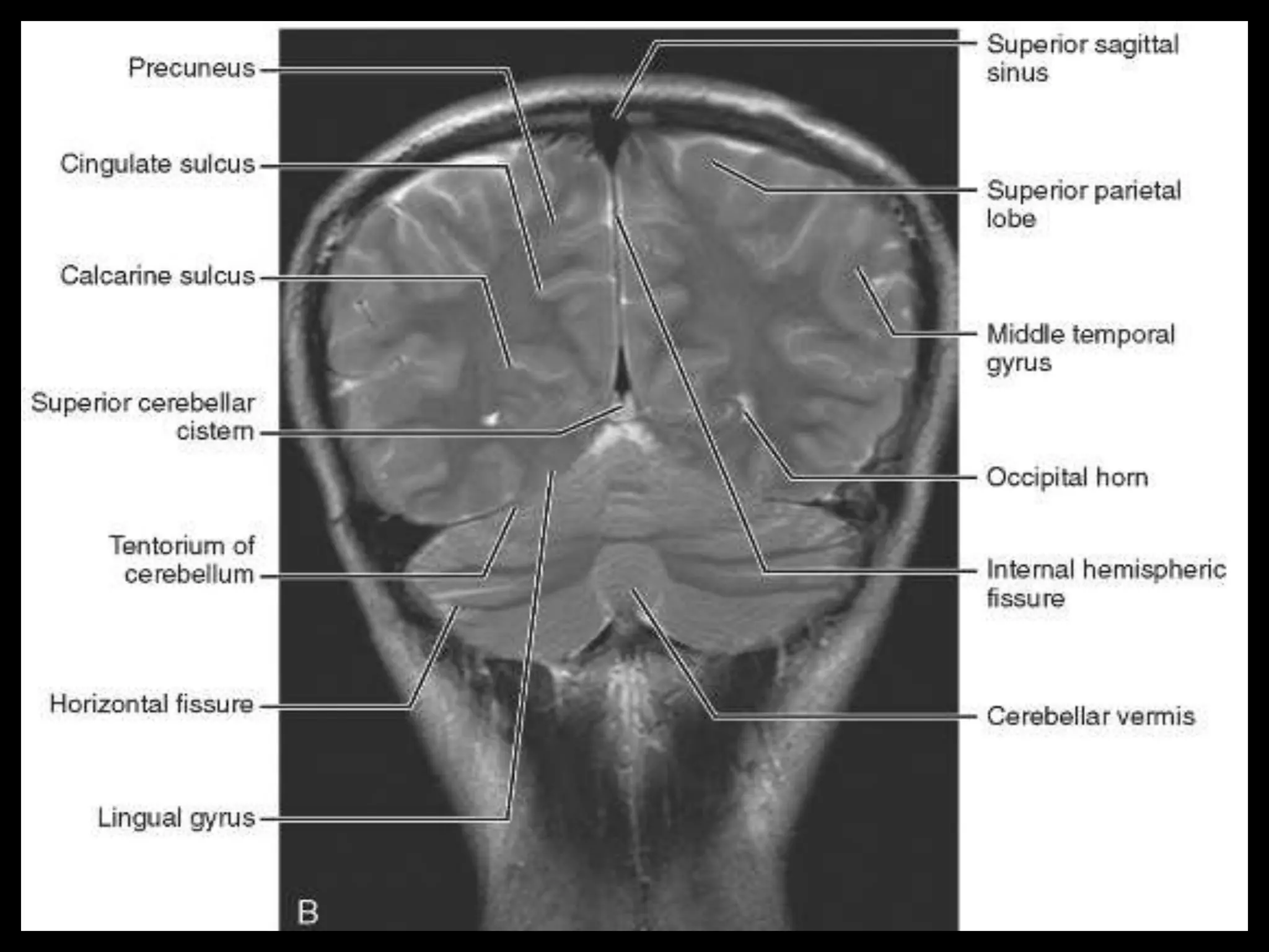
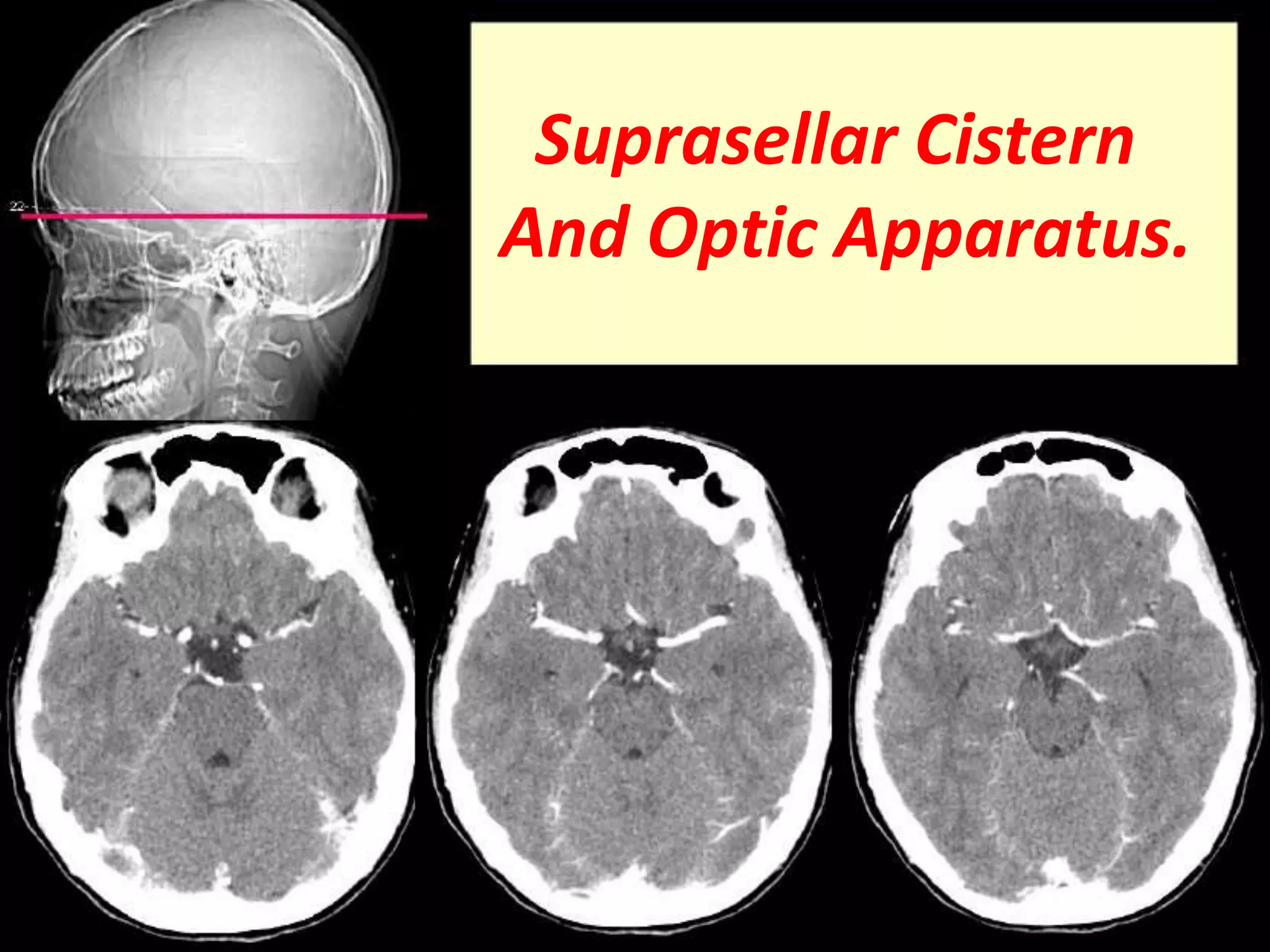
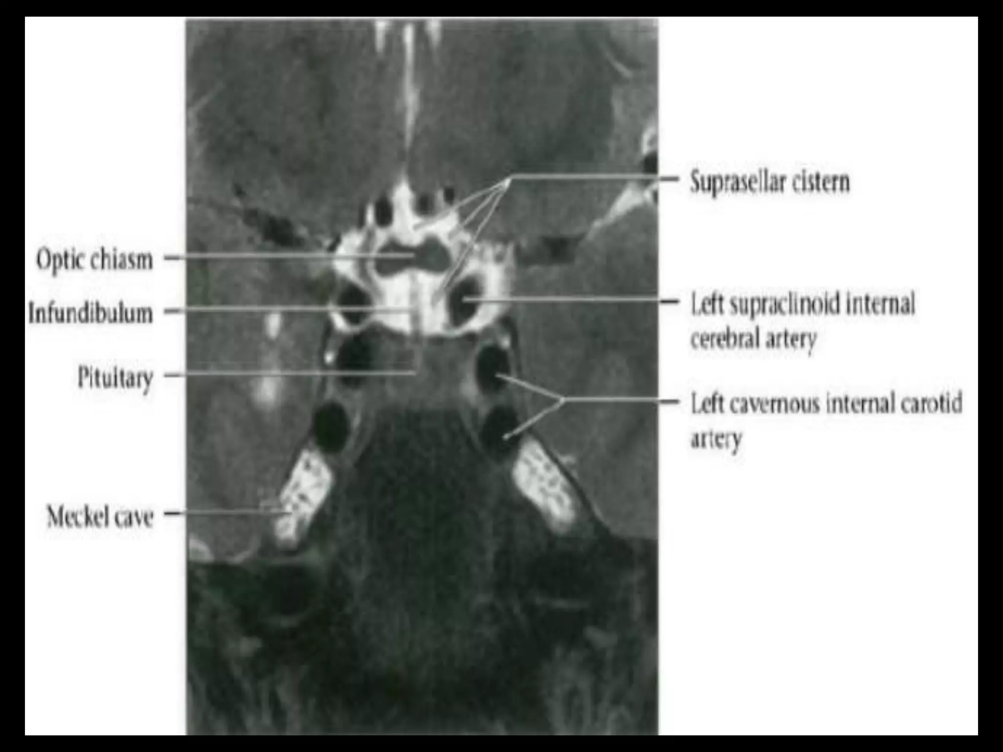
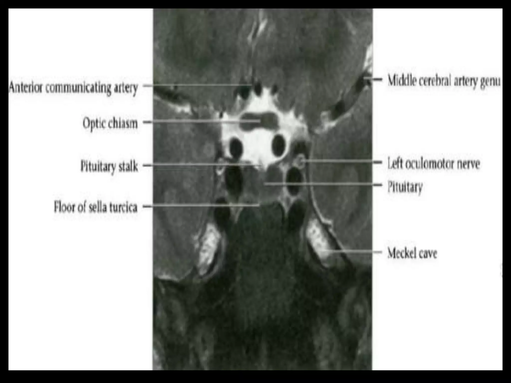
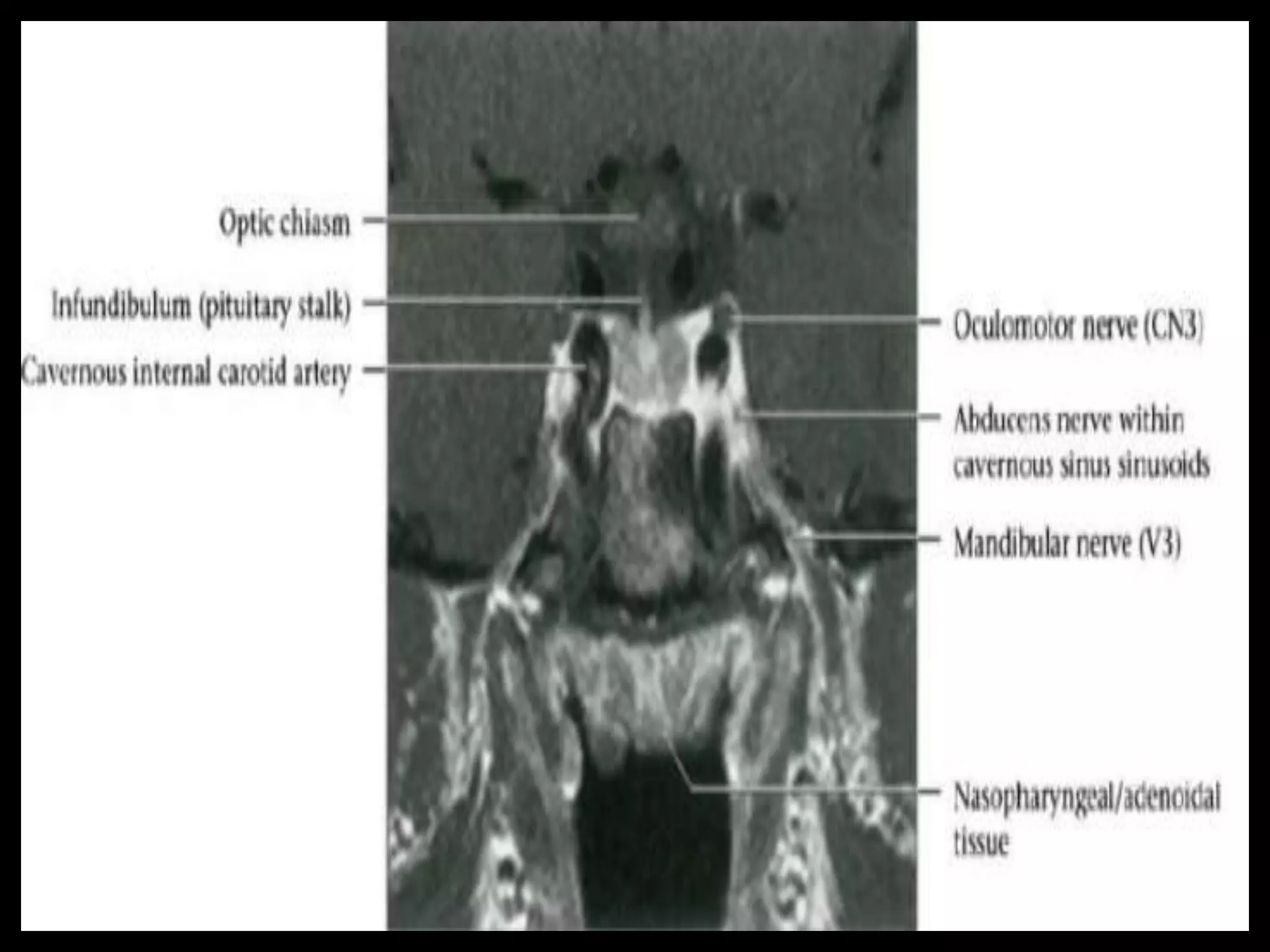
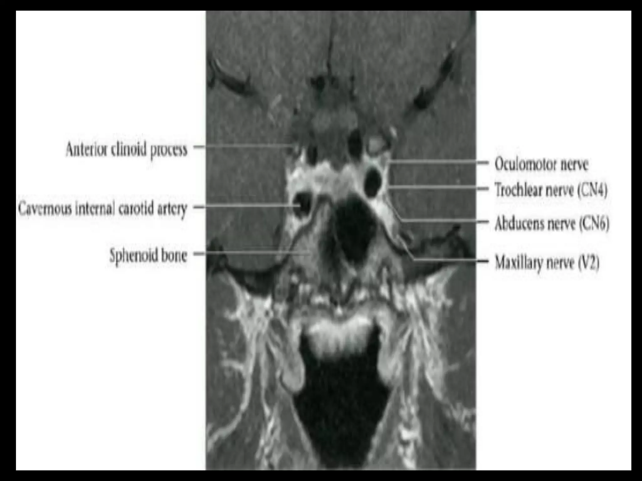
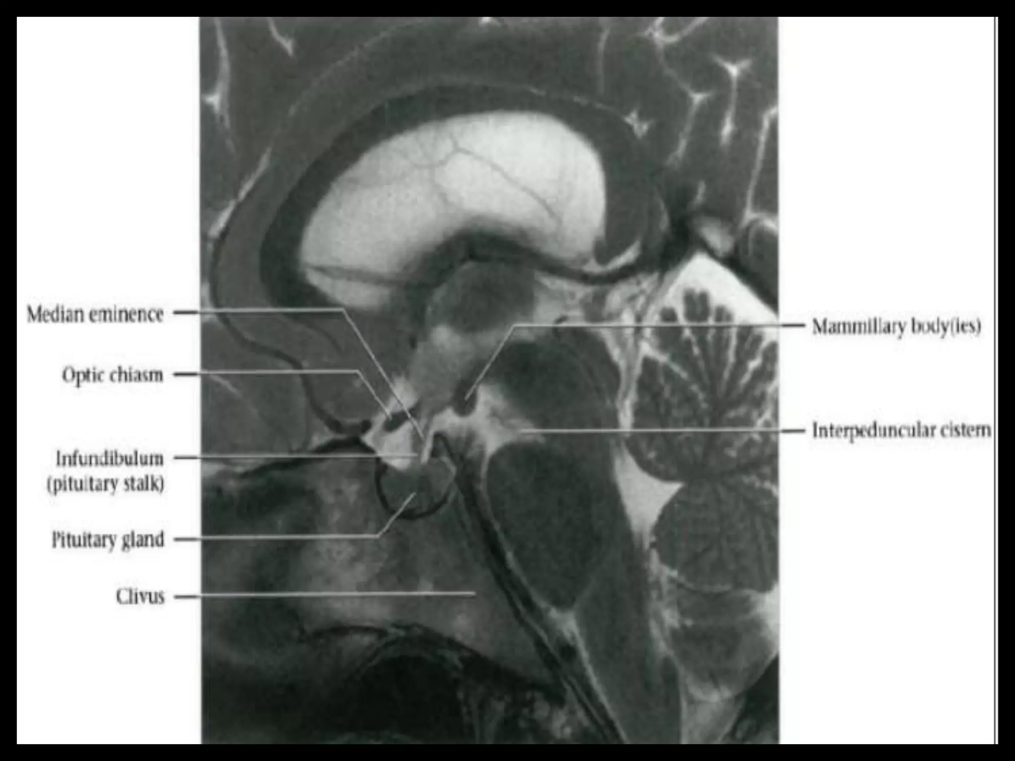
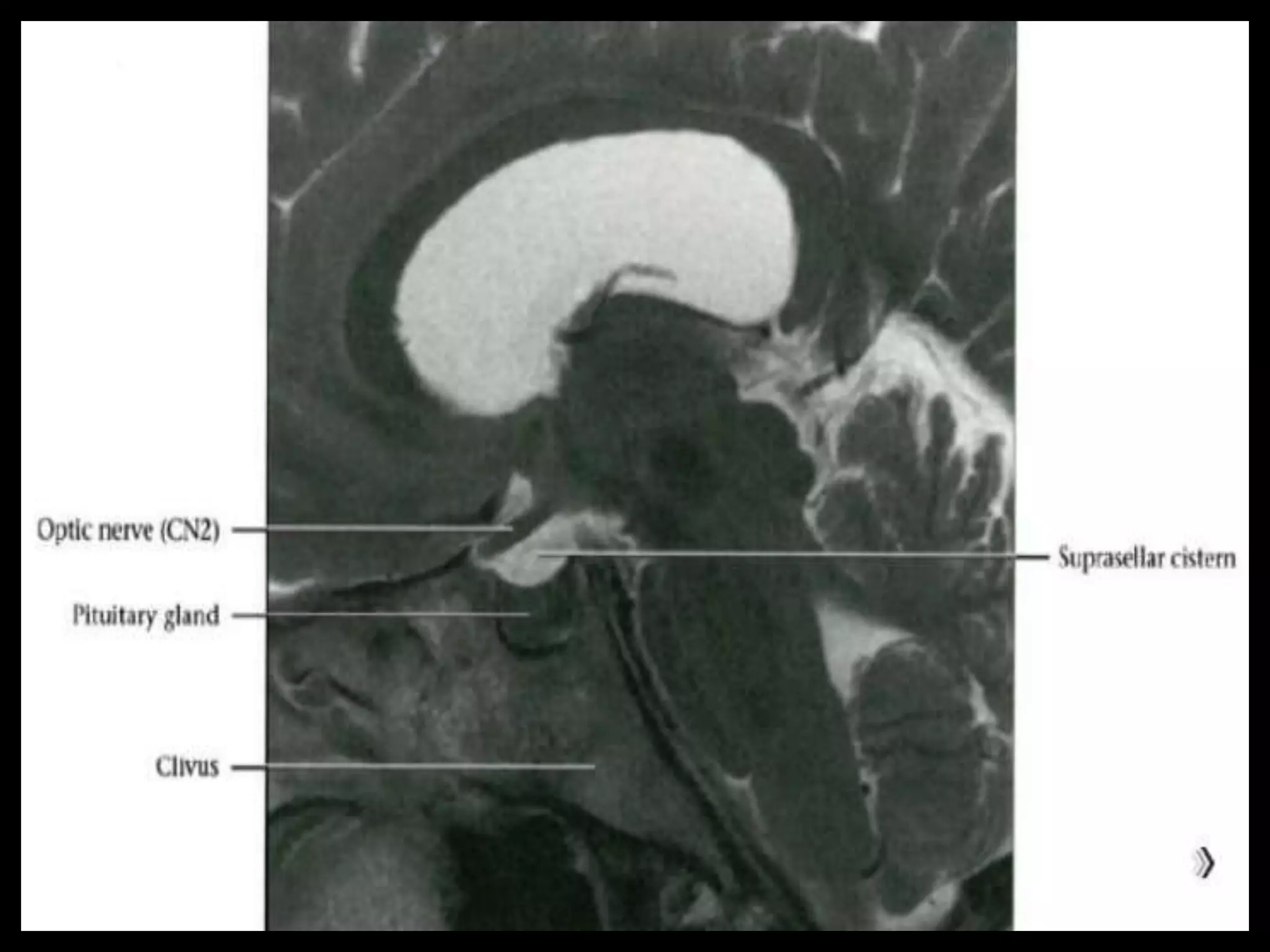
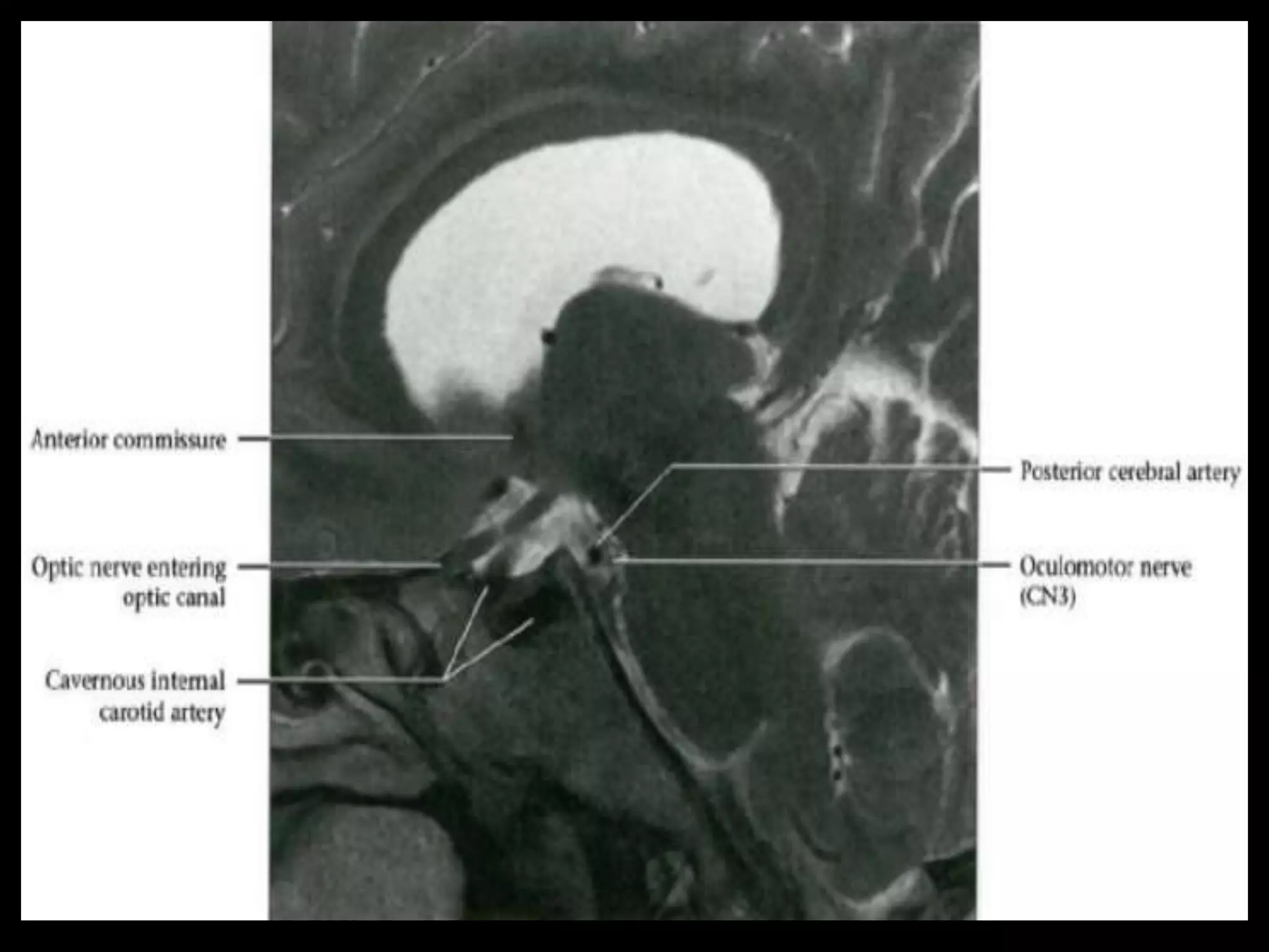
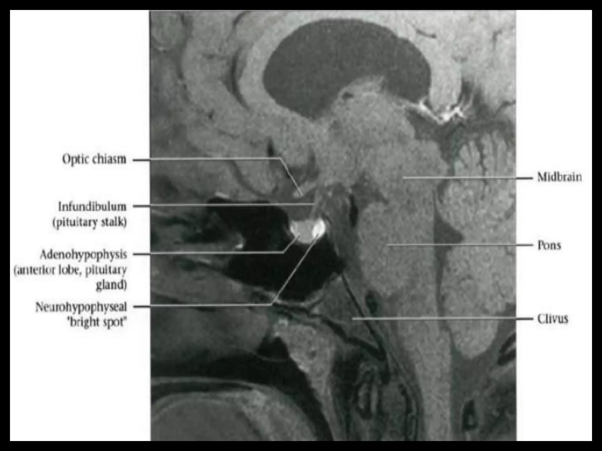


The document summarizes the normal radiological anatomy of the brain and pituitary gland as seen on computed tomography (CT) and magnetic resonance imaging (MRI). It describes the overall structure of the brain, including the cerebrum, cerebellum, brainstem, and four ventricles. It details the anatomy of the lateral, third, and fourth ventricles. It then outlines the major lobes and gyri of the cerebral hemispheres, including important motor and sensory areas. The document concludes by reviewing sectional anatomy as seen on axial CT and MRI scans.