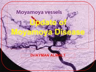
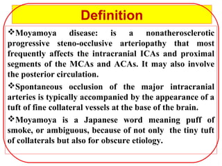
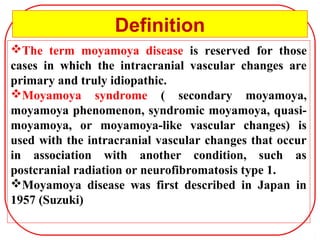
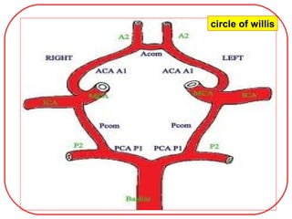
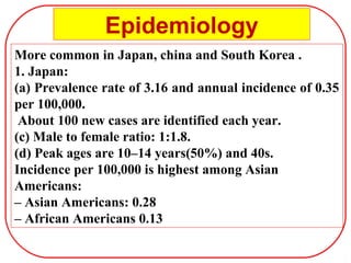
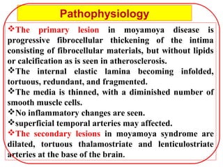
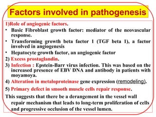
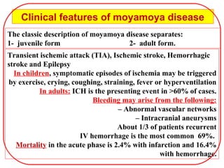
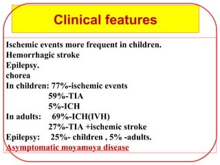
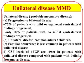
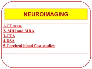
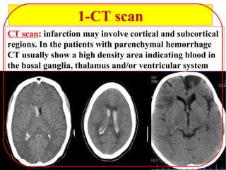
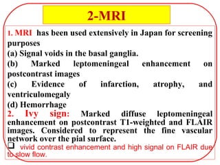
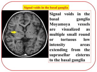
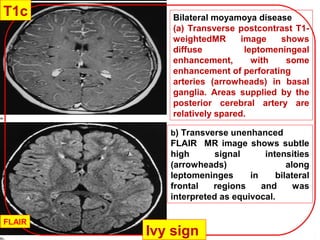
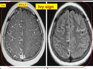
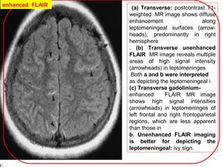
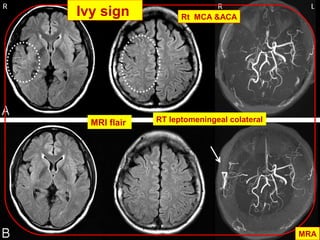
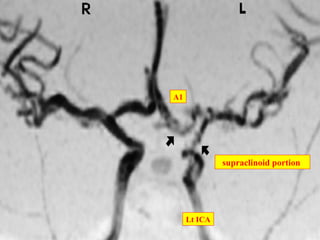
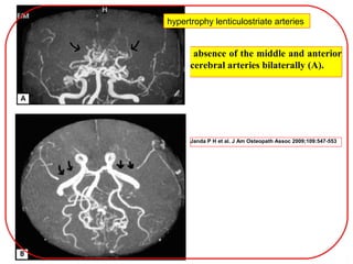
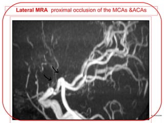
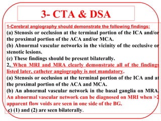
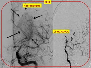
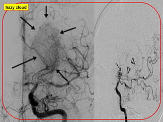
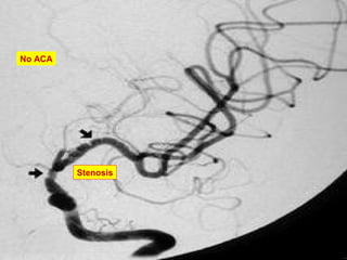
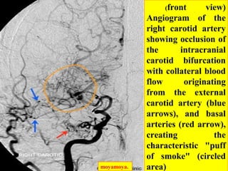
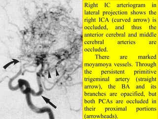
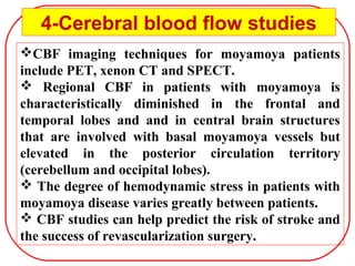
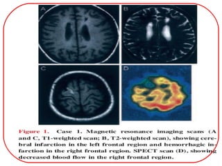
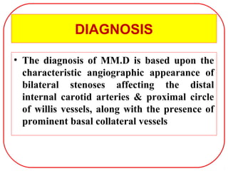
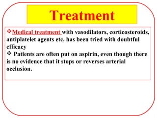
Update of
Moyamoya Disease
Dr/AYMAN ALMALT
Moyamoya Disease
1.Definition
2.Etiology
3.Epidemiology
4.Pathophysiology
5.Clinical presentation
6.Neuroimaging
7.Diagnosis
8.Treatment
9.Prognosis
Moyamoya disease: is a nonatherosclerotic
progressive steno-occlusive arteriopathy that most
frequently affects the intracranial ICAs and proximal
segments of the MCAs and ACAs. It may also involve
the posterior circulation.
Spontaneous occlusion of the major intracranial
arteries is typically accompanied by the appearance of a
tuft of fine collateral vessels at the base of the brain.
Moyamoya is a Japanese word meaning puff of
smoke, or ambiguous, because of not only the tiny tuft
of collaterals but also for obscure etiology.
Definition
The term moyamoya disease is reserved for those
cases in which the intracranial vascular changes are
primary and truly idiopathic.
Moyamoya syndrome ( secondary moyamoya,
moyamoya phenomenon, syndromic moyamoya, quasi-
moyamoya, or moyamoya-like vascular changes) is
used with the intracranial vascular changes that occur
in association with another condition, such as
postcranial radiation or neurofibromatosis type 1.
Moyamoya disease was first described in Japan in
1957 (Suzuki)
Definition
circle of willis
Etiology
• unknown.
• A genetic mode of inheritance is considered possible
because of the higher incidence of the disease in
Japan and Korea.
• wide spread use of MRI and &MRA have been used
in detecting the disease in asymptomatic familial
cases( 10%).
• Familial MMD has been linked to ch 3, 6, 8, 12and
17.
• Secondary: such as postcranial radiation,
neurofibromatosis type 1 or Epstein-Barr virus
infection .
1. when at least one first-degree relative is affected.
2. 10% are familial.
3. Earlier age of onset (10 & 40 IN SPORADIC)
4. Greater female>male (1:5 or 1:3 in sporadic).
5. AD with incomplete penetrance.
6. Familial MMD has been linked to ch 3, 6, 8, 12and 17.
7. Familial moyamoya disease is associated with
(a) SLE. (b) Basilar tip aneurysms.
8. Screening with MRA has been recommended for
family members of patients with MMD (30 fold )
Familial moyamoya disease
1. cranial radiotherapy
2. Atherosclerosis
3. phakomatoses
neurofibromatosis type 1(NF1)
tuberous sclerosis (TS)
4. infection
tuberculous meningitis
bacterial leptomeningitis, post-varicella vasculitis
5. connective tissue disorders
systemic lupus erythematosus (SLE)
6. haematological disorders
sickle cell disease, anti phospholipid syndrome
Moyamoya syndrome
More common in Japan, china and South Korea .
1. Japan:
(a) Prevalence rate of 3.16 and annual incidence of 0.35
per 100,000.
About 100 new cases are identified each year.
(c) Male to female ratio: 1:1.8.
(d) Peak ages are 10–14 years(50%) and 40s.
Incidence per 100,000 is highest among Asian
Americans:
– Asian Americans: 0.28
– African Americans 0.13
Epidemiology
The primary lesion in moyamoya disease is
progressive fibrocellular thickening of the intima
consisting of fibrocellular materials, but without lipids
or calcification as is seen in atherosclerosis.
The internal elastic lamina becoming infolded,
tortuous, redundant, and fragmented.
The media is thinned, with a diminished number of
smooth muscle cells.
No inflammatory changes are seen.
superficial temporal arteries may affected.
The secondary lesions in moyamoya syndrome are
dilated, tortuous thalamostriate and lenticulostriate
arteries at the base of the brain.
Pathophysiology
Factors involved in pathogenesis
1)Role of angiogenic factors.
• Basic Fibroblast growth factor: mediator of the neovascular
response.
• Transforming growth beta factor 1 (TGF beta 1), a factor
involved in angiogenesis
• Hepatocyte growth factor, an angiogenic factor
2) Excess prostaglandin.
3) Infection : Epstein-Barr virus infection. This was based on the
increased presence of EBV DNA and antibody in patients with
moyamoya.
4) Alteration in metaloproteinase gene expression (remodeling).
5) Primary defect in smooth muscle cells repair response.
This suggests that there be a derangement in the vessel wall
repair mechanism that leads to long-term proliferation of cells
and progressive occlusion of the vessel lumen.
The classic description of moyamoya disease separates:
1- juvenile form 2- adult form.
Clinical features of moyamoya disease
Transient ischemic attack (TIA), Ischemic stroke, Hemorrhagic
stroke and Epilepsy
In children, symptomatic episodes of ischemia may be triggered
by exercise, crying, coughing, straining, fever or hyperventilation
In adults: ICH is the presenting event in >60% of cases.
Bleeding may arise from the following:
– Abnormal vascular networks
– Intracranial aneurysms
About 1/3 of patients recurrent
IV hemorrhage is the most common 69%.
Mortality in the acute phase is 2.4% with infarction and 16.4%
with hemorrhage.
Clinical features
Ischemic events more frequent in children.
Hemorrhagic stroke
Epilepsy.
chorea
In children: 77%-ischemic events
59%-TIA
5%-ICH
In adults: 69%-ICH(IVH)
27%-TIA +ischemic stroke
Epilepsy: 25%- children , 5% -adults.
Asymptomatic moyamoya disease
Unilateral disease ( probable moyamoya disease):
(a) Progression to bilateral disease:
–75% of patients with mild or equivocal contralateral
findings progressed,
only 10% of patients with no initial contralateral
findings progressed.
(b) Unilateral disease common adults >children.
(c) Familial occurrence is less common in patients with
unilateral disease,
d) CSF levels of bFGF are lower in patients with
unilateral disease compared with patients with definite
moyamoya disease.
Unilateral disease MMD
NEUROIMAGING
1-CT scan.
2- MRI and MRA
3-CTA
4-DSA
5-Cerebral blood flow studies
1-CT scan
CT scan: infarction may involve cortical and subcortical
regions. In the patients with parenchymal hemorrhage
CT usually show a high density area indicating blood in
the basal ganglia, thalamus and/or ventricular system
1. MRI has been used extensively in Japan for screening
purposes
(a) Signal voids in the basal ganglia.
(b) Marked leptomeningeal enhancement on
postcontrast images
(c) Evidence of infarction, atrophy, and
ventriculomegaly
(d) Hemorrhage
2. Ivy sign: Marked diffuse leptomeningeal
enhancement on postcontrast T1-weighted and FLAIR
images. Considered to represent the fine vascular
network over the pial surface.
 vivid contrast enhancement and high signal on FLAIR due
to slow flow.
2-MRI
Signal voids in the basal ganglia
Signal voids in the
basal ganglia
Moyamoya vessels
are visualized as
multiple small round
or tortuous low
intensity areas
extending from the
suprasellar cisterns
to the basal ganglia
Bilateral moyamoya disease
(a) Transverse postcontrast T1-
weightedMR image shows
diffuse leptomeningeal
enhancement, with some
enhancement of perforating
arteries (arrowheads) in basal
ganglia. Areas supplied by the
posterior cerebral artery are
relatively spared.
b) Transverse unenhanced
FLAIR MR image shows subtle
high signal intensities
(arrowheads) along
leptomeninges in bilateral
frontal regions and was
interpreted as equivocal.
Ivy sign
T1c
FLAIR
RT>LT
Ivy signT1c
(a) Transverse: postcontrast T1-
weighted MR image shows diffuse
enhancement along
leptomeningeal surfaces (arrow-
heads), predominantly in right
hemisphere
. (b) Transverse unenhanced
FLAIR MR image reveals multiple
areas of high signal intensity
(arrowheads) in leptomeninges
Both a and b were interpreted
as depicting the leptomeningeal i
(c) Transverse gadolinium-
enhanced FLAIR MR image
shows high signal intensities
(arrowheads) in leptomeninges of
left frontal and right frontoparietal
regions, which are less apparent
than those in
b. Unenhanced FLAIR imaging
is better for depicting the
leptomeningeal: ivy sign.
enhanced FLAIR
MRI flair
MRA
Rt MCA &ACA
RT leptomeningeal colateral
Ivy sign
supraclinoid portion
A1
Lt ICA
absence of the middle and anterior
cerebral arteries bilaterally (A).
Janda P H et al. J Am Osteopath Assoc 2009;109:547-553
hypertrophy lenticulostriate arteries
Lateral MRA proximal occlusion of the MCAs &ACAs
3- CTA & DSA
1-Cerebral angiography should demonstrate the following findings:
(a) Stenosis or occlusion at the terminal portion of the ICA and/or
the proximal portion of the ACA and/or MCA.
(b) Abnormal vascular networks in the vicinity of the occlusive or
stenotic lesions.
(c) These findings should be present bilaterally.
2. When MRI and MRA clearly demonstrate all of the findings
listed later, catheter angiography is not mandatory.
(a) Stenosis or occlusion at the terminal portion of the ICA and at
the proximal portion of the ACA and MCA.
(b) An abnormal vascular network in the basal ganglia on MRA.
An abnormal vascular network can be diagnosed on MRI when >2
apparent flow voids are seen in one side of the BG.
c) (1) and (2) are seen bilaterally.
Puff of smoke
DSA
LT MCA&ACA
hazy cloud
Stenosis
No ACA
moyamoya.
(front view)
Angiogram of the
right carotid artery
showing occlusion of
the intracranial
carotid bifurcation
with collateral blood
flow originating
from the external
carotid artery (blue
arrows), and basal
arteries (red arrow),
creating the
characteristic "puff
of smoke" (circled
area)
Right IC arteriogram in
lateral projection shows the
right ICA (curved arrow) is
occluded, and thus the
anterior cerebral and middle
cerebral arteries are
occluded.
There are marked
moyamoya vessels. Through
the persistent primitive
trigeminal artery (straight
arrow), the BA and its
branches are opacified, but
both PCAs are occluded in
their proximal portions
(arrowheads).
CBF imaging techniques for moyamoya patients
include PET, xenon CT and SPECT.
 Regional CBF in patients with moyamoya is
characteristically diminished in the frontal and
temporal lobes and and in central brain structures
that are involved with basal moyamoya vessels but
elevated in the posterior circulation territory
(cerebellum and occipital lobes).
 The degree of hemodynamic stress in patients with
moyamoya disease varies greatly between patients.
 CBF studies can help predict the risk of stroke and
the success of revascularization surgery.
4-Cerebral blood flow studies
DIAGNOSIS
• The diagnosis of MM.D is based upon the
characteristic angiographic appearance of
bilateral stenoses affecting the distal
internal carotid arteries & proximal circle
of willis vessels, along with the presence of
prominent basal collateral vessels
Medical treatment with vasodilators, corticosteroids,
antiplatelet agents etc. has been tried with doubtful
efficacy
 Patients are often put on aspirin, even though there
is no evidence that it stops or reverses arterial
occlusion.
Treatment
Surgery for moyamoya
• create collateralization on the brain surface.
• Indirect revascularization procedures such as EDAS
(encephaloduroarterio synangiosis), pial synangiosis,
indirect revasularization using muscle flaps etc.
• Direct revascularization procedures such as
superficial temporal-middle cerebral artery bypass .
• indirect revascularization is preferred in treatment
of children.
• The decision is based on angiography and cerebral
blood flow studies.
• TlAs reduce in frequency and patients do not
develop new strokes in successful cases.
Prognosis
The natural history tends to be progressive with
extensive intracranial large artery occlusion and
collateral circulation.
The natural history may be more benign in US
compared to Asian population.
Moyamoya D. is one of the D.D. of stroke in children
and young adults.
Cerebral blood flow and
metabolism in moyamoya
• The morbidity of moyamoya is directly related to cerebral blood flow.
• This was demonstrated in earlier studies using Xenon-133 inhalation. The
cerebral blood flow was decreased most in the frontal region with relatively
normal flow in the temporal and occipital region. After hyperventilation the
blood flow was reduced in all regions.
• Positron emission tomographic studies have shown an increase in total blood
volume, especially in the striatum and increased transit time. The
cerebrovascular response to hypercapnia was shown to be impaired. These
changes were reversed after reperfusion surgery.
• PET studies have also demonstrated the vasodilatation in normal areas after
the termination of hyperventilation. This may cause a steal response increasing
hypoperfusion.
• These studies may help to understand the effects of chronic cerebral occlusive
disease. Xenon computed tomography has been used for pre and post surgical
evaluation.
• These studies were found to correlate with angiographic studies and have been
claimed to be superior in the study of basal ganglia and posterior circulation.
• Diffusion weighted imaging and perfusion magnetic resonance imagine using
contrast have been used in the study of ischemic episodes.
• Serial studies have also shown the decrease in cerebral blood flow with
advancing age
Angiographic features of moyamoya
• The development of the moyamoya network may be seen
at different sites
• The formation of network of vessels at the frontal base with
blood supply from the branches of the ophthalmic artery is
known as ethmoidal moyamoya.
• Dilatation of the basilar artery and formation of moyamoya
network by perforating branches of the posterior cerebral
artery is known as posterior basal moyamoya.
• Vault moyamoya is due to development of extra and
intracranial transdural leptomeningeal collaterals between pial
vessel and branches of the external carotid artery.
• A well-developed posterior callosal artery is seen. The large
and proliferating irregular vessels and transdiploic collaterals
of the external carotid artery that supplies the ischemic regions
of the brain essentially cause the moyamoya network.

Moyamoya disease

  • 1.
  • 2.
  • 3.
    Moyamoya disease: isa nonatherosclerotic progressive steno-occlusive arteriopathy that most frequently affects the intracranial ICAs and proximal segments of the MCAs and ACAs. It may also involve the posterior circulation. Spontaneous occlusion of the major intracranial arteries is typically accompanied by the appearance of a tuft of fine collateral vessels at the base of the brain. Moyamoya is a Japanese word meaning puff of smoke, or ambiguous, because of not only the tiny tuft of collaterals but also for obscure etiology. Definition
  • 4.
    The term moyamoyadisease is reserved for those cases in which the intracranial vascular changes are primary and truly idiopathic. Moyamoya syndrome ( secondary moyamoya, moyamoya phenomenon, syndromic moyamoya, quasi- moyamoya, or moyamoya-like vascular changes) is used with the intracranial vascular changes that occur in association with another condition, such as postcranial radiation or neurofibromatosis type 1. Moyamoya disease was first described in Japan in 1957 (Suzuki) Definition
  • 5.
  • 6.
    Etiology • unknown. • Agenetic mode of inheritance is considered possible because of the higher incidence of the disease in Japan and Korea. • wide spread use of MRI and &MRA have been used in detecting the disease in asymptomatic familial cases( 10%). • Familial MMD has been linked to ch 3, 6, 8, 12and 17. • Secondary: such as postcranial radiation, neurofibromatosis type 1 or Epstein-Barr virus infection .
  • 7.
    1. when atleast one first-degree relative is affected. 2. 10% are familial. 3. Earlier age of onset (10 & 40 IN SPORADIC) 4. Greater female>male (1:5 or 1:3 in sporadic). 5. AD with incomplete penetrance. 6. Familial MMD has been linked to ch 3, 6, 8, 12and 17. 7. Familial moyamoya disease is associated with (a) SLE. (b) Basilar tip aneurysms. 8. Screening with MRA has been recommended for family members of patients with MMD (30 fold ) Familial moyamoya disease
  • 8.
    1. cranial radiotherapy 2.Atherosclerosis 3. phakomatoses neurofibromatosis type 1(NF1) tuberous sclerosis (TS) 4. infection tuberculous meningitis bacterial leptomeningitis, post-varicella vasculitis 5. connective tissue disorders systemic lupus erythematosus (SLE) 6. haematological disorders sickle cell disease, anti phospholipid syndrome Moyamoya syndrome
  • 9.
    More common inJapan, china and South Korea . 1. Japan: (a) Prevalence rate of 3.16 and annual incidence of 0.35 per 100,000. About 100 new cases are identified each year. (c) Male to female ratio: 1:1.8. (d) Peak ages are 10–14 years(50%) and 40s. Incidence per 100,000 is highest among Asian Americans: – Asian Americans: 0.28 – African Americans 0.13 Epidemiology
  • 10.
    The primary lesionin moyamoya disease is progressive fibrocellular thickening of the intima consisting of fibrocellular materials, but without lipids or calcification as is seen in atherosclerosis. The internal elastic lamina becoming infolded, tortuous, redundant, and fragmented. The media is thinned, with a diminished number of smooth muscle cells. No inflammatory changes are seen. superficial temporal arteries may affected. The secondary lesions in moyamoya syndrome are dilated, tortuous thalamostriate and lenticulostriate arteries at the base of the brain. Pathophysiology
  • 11.
    Factors involved inpathogenesis 1)Role of angiogenic factors. • Basic Fibroblast growth factor: mediator of the neovascular response. • Transforming growth beta factor 1 (TGF beta 1), a factor involved in angiogenesis • Hepatocyte growth factor, an angiogenic factor 2) Excess prostaglandin. 3) Infection : Epstein-Barr virus infection. This was based on the increased presence of EBV DNA and antibody in patients with moyamoya. 4) Alteration in metaloproteinase gene expression (remodeling). 5) Primary defect in smooth muscle cells repair response. This suggests that there be a derangement in the vessel wall repair mechanism that leads to long-term proliferation of cells and progressive occlusion of the vessel lumen.
  • 12.
    The classic descriptionof moyamoya disease separates: 1- juvenile form 2- adult form. Clinical features of moyamoya disease Transient ischemic attack (TIA), Ischemic stroke, Hemorrhagic stroke and Epilepsy In children, symptomatic episodes of ischemia may be triggered by exercise, crying, coughing, straining, fever or hyperventilation In adults: ICH is the presenting event in >60% of cases. Bleeding may arise from the following: – Abnormal vascular networks – Intracranial aneurysms About 1/3 of patients recurrent IV hemorrhage is the most common 69%. Mortality in the acute phase is 2.4% with infarction and 16.4% with hemorrhage.
  • 13.
    Clinical features Ischemic eventsmore frequent in children. Hemorrhagic stroke Epilepsy. chorea In children: 77%-ischemic events 59%-TIA 5%-ICH In adults: 69%-ICH(IVH) 27%-TIA +ischemic stroke Epilepsy: 25%- children , 5% -adults. Asymptomatic moyamoya disease
  • 14.
    Unilateral disease (probable moyamoya disease): (a) Progression to bilateral disease: –75% of patients with mild or equivocal contralateral findings progressed, only 10% of patients with no initial contralateral findings progressed. (b) Unilateral disease common adults >children. (c) Familial occurrence is less common in patients with unilateral disease, d) CSF levels of bFGF are lower in patients with unilateral disease compared with patients with definite moyamoya disease. Unilateral disease MMD
  • 15.
    NEUROIMAGING 1-CT scan. 2- MRIand MRA 3-CTA 4-DSA 5-Cerebral blood flow studies
  • 16.
    1-CT scan CT scan:infarction may involve cortical and subcortical regions. In the patients with parenchymal hemorrhage CT usually show a high density area indicating blood in the basal ganglia, thalamus and/or ventricular system
  • 17.
    1. MRI hasbeen used extensively in Japan for screening purposes (a) Signal voids in the basal ganglia. (b) Marked leptomeningeal enhancement on postcontrast images (c) Evidence of infarction, atrophy, and ventriculomegaly (d) Hemorrhage 2. Ivy sign: Marked diffuse leptomeningeal enhancement on postcontrast T1-weighted and FLAIR images. Considered to represent the fine vascular network over the pial surface.  vivid contrast enhancement and high signal on FLAIR due to slow flow. 2-MRI
  • 18.
    Signal voids inthe basal ganglia Signal voids in the basal ganglia Moyamoya vessels are visualized as multiple small round or tortuous low intensity areas extending from the suprasellar cisterns to the basal ganglia
  • 19.
    Bilateral moyamoya disease (a)Transverse postcontrast T1- weightedMR image shows diffuse leptomeningeal enhancement, with some enhancement of perforating arteries (arrowheads) in basal ganglia. Areas supplied by the posterior cerebral artery are relatively spared. b) Transverse unenhanced FLAIR MR image shows subtle high signal intensities (arrowheads) along leptomeninges in bilateral frontal regions and was interpreted as equivocal. Ivy sign T1c FLAIR
  • 20.
  • 21.
    (a) Transverse: postcontrastT1- weighted MR image shows diffuse enhancement along leptomeningeal surfaces (arrow- heads), predominantly in right hemisphere . (b) Transverse unenhanced FLAIR MR image reveals multiple areas of high signal intensity (arrowheads) in leptomeninges Both a and b were interpreted as depicting the leptomeningeal i (c) Transverse gadolinium- enhanced FLAIR MR image shows high signal intensities (arrowheads) in leptomeninges of left frontal and right frontoparietal regions, which are less apparent than those in b. Unenhanced FLAIR imaging is better for depicting the leptomeningeal: ivy sign. enhanced FLAIR
  • 22.
    MRI flair MRA Rt MCA&ACA RT leptomeningeal colateral Ivy sign
  • 23.
  • 24.
    absence of themiddle and anterior cerebral arteries bilaterally (A). Janda P H et al. J Am Osteopath Assoc 2009;109:547-553 hypertrophy lenticulostriate arteries
  • 25.
    Lateral MRA proximalocclusion of the MCAs &ACAs
  • 26.
    3- CTA &DSA 1-Cerebral angiography should demonstrate the following findings: (a) Stenosis or occlusion at the terminal portion of the ICA and/or the proximal portion of the ACA and/or MCA. (b) Abnormal vascular networks in the vicinity of the occlusive or stenotic lesions. (c) These findings should be present bilaterally. 2. When MRI and MRA clearly demonstrate all of the findings listed later, catheter angiography is not mandatory. (a) Stenosis or occlusion at the terminal portion of the ICA and at the proximal portion of the ACA and MCA. (b) An abnormal vascular network in the basal ganglia on MRA. An abnormal vascular network can be diagnosed on MRI when >2 apparent flow voids are seen in one side of the BG. c) (1) and (2) are seen bilaterally.
  • 27.
  • 28.
  • 29.
  • 30.
    moyamoya. (front view) Angiogram ofthe right carotid artery showing occlusion of the intracranial carotid bifurcation with collateral blood flow originating from the external carotid artery (blue arrows), and basal arteries (red arrow), creating the characteristic "puff of smoke" (circled area)
  • 31.
    Right IC arteriogramin lateral projection shows the right ICA (curved arrow) is occluded, and thus the anterior cerebral and middle cerebral arteries are occluded. There are marked moyamoya vessels. Through the persistent primitive trigeminal artery (straight arrow), the BA and its branches are opacified, but both PCAs are occluded in their proximal portions (arrowheads).
  • 32.
    CBF imaging techniquesfor moyamoya patients include PET, xenon CT and SPECT.  Regional CBF in patients with moyamoya is characteristically diminished in the frontal and temporal lobes and and in central brain structures that are involved with basal moyamoya vessels but elevated in the posterior circulation territory (cerebellum and occipital lobes).  The degree of hemodynamic stress in patients with moyamoya disease varies greatly between patients.  CBF studies can help predict the risk of stroke and the success of revascularization surgery. 4-Cerebral blood flow studies
  • 34.
    DIAGNOSIS • The diagnosisof MM.D is based upon the characteristic angiographic appearance of bilateral stenoses affecting the distal internal carotid arteries & proximal circle of willis vessels, along with the presence of prominent basal collateral vessels
  • 35.
    Medical treatment withvasodilators, corticosteroids, antiplatelet agents etc. has been tried with doubtful efficacy  Patients are often put on aspirin, even though there is no evidence that it stops or reverses arterial occlusion. Treatment
  • 36.
    Surgery for moyamoya •create collateralization on the brain surface. • Indirect revascularization procedures such as EDAS (encephaloduroarterio synangiosis), pial synangiosis, indirect revasularization using muscle flaps etc. • Direct revascularization procedures such as superficial temporal-middle cerebral artery bypass . • indirect revascularization is preferred in treatment of children. • The decision is based on angiography and cerebral blood flow studies. • TlAs reduce in frequency and patients do not develop new strokes in successful cases.
  • 37.
    Prognosis The natural historytends to be progressive with extensive intracranial large artery occlusion and collateral circulation. The natural history may be more benign in US compared to Asian population. Moyamoya D. is one of the D.D. of stroke in children and young adults.
  • 39.
    Cerebral blood flowand metabolism in moyamoya • The morbidity of moyamoya is directly related to cerebral blood flow. • This was demonstrated in earlier studies using Xenon-133 inhalation. The cerebral blood flow was decreased most in the frontal region with relatively normal flow in the temporal and occipital region. After hyperventilation the blood flow was reduced in all regions. • Positron emission tomographic studies have shown an increase in total blood volume, especially in the striatum and increased transit time. The cerebrovascular response to hypercapnia was shown to be impaired. These changes were reversed after reperfusion surgery. • PET studies have also demonstrated the vasodilatation in normal areas after the termination of hyperventilation. This may cause a steal response increasing hypoperfusion. • These studies may help to understand the effects of chronic cerebral occlusive disease. Xenon computed tomography has been used for pre and post surgical evaluation. • These studies were found to correlate with angiographic studies and have been claimed to be superior in the study of basal ganglia and posterior circulation. • Diffusion weighted imaging and perfusion magnetic resonance imagine using contrast have been used in the study of ischemic episodes. • Serial studies have also shown the decrease in cerebral blood flow with advancing age
  • 40.
    Angiographic features ofmoyamoya • The development of the moyamoya network may be seen at different sites • The formation of network of vessels at the frontal base with blood supply from the branches of the ophthalmic artery is known as ethmoidal moyamoya. • Dilatation of the basilar artery and formation of moyamoya network by perforating branches of the posterior cerebral artery is known as posterior basal moyamoya. • Vault moyamoya is due to development of extra and intracranial transdural leptomeningeal collaterals between pial vessel and branches of the external carotid artery. • A well-developed posterior callosal artery is seen. The large and proliferating irregular vessels and transdiploic collaterals of the external carotid artery that supplies the ischemic regions of the brain essentially cause the moyamoya network.

Editor's Notes

  • #23 A female patient with unsymmetric ivy sign and decreased ivy sign after operation (10 months follow-up period). She had sustained a left TIA.  A , Preoperative FLAIR images showed moderate ivy dominance in the right hemisphere ( dotted circles ). Preoperative MRA grade in the right and left hemispheres were III and II, respectively. She underwent direct bypass surgery in the right side.  B , Postoperative FLAIR image obtained 10 months after revascularization surgery revealed decreased ivy sign in the right hemisphere. Postoperative MRA showed well-developed collateral vessels via bypass in the right MCA region ( arrow ). The patient had no symptoms after the operation.
  • #24 left internal carotid artery and its branches. The arrow on the right points to the supraclinoid portion of the internal carotid. The arrow on the left points to the horizonal section of the anterior cerebral artery.
  • #25 Magnetic resonance angiography of the brain of a 44-year-old African American woman demonstrated the absence of the middle and anterior cerebral arteries bilaterally (A). There is also marked hypertrophy of the lenticulostriate arteries bilaterally, which were very large in caliber distally, concurrently revealing collateralization of the posterior cerebral arteries to the anterior cerebral artery distribution over the convexity. Magnetic resonance imaging of the brain also showed the proximal occlusion of the anterior cerebral and middle cerebral arteries (B; indicated by double arrows). The patient, who had recently had a stroke, was diagnosed as having moyamoya disease.
  • #26 Lateral view of a magnetic resonance angiography of the brain of a 44-year-old African American woman. The imaging study displayed the proximal occlusion of the middle cerebral arteries and the anterior cerebral arteries. The patient, who had recently had a stroke, was diagnosed as having moyamoya disease.
  • #32 Right internal carotid arteriogram in lateral projection shows the right ICA (curved arrow) is occluded, and thus the anterior cerebral and middle cerebral arteries are occluded. There are marked moyamoya vessels. Through the persistent primitive trigeminal artery (straight arrow), the basilar artery and its branches are opacified, but both PCAs are occluded in their proximal portions (arrowheads).
  • #41 The image displayed here is an AP view of her left internal carotid angiogram. The arrows point to narrowed regions in the internal carotid artery and its branches. The classic "puff of smoke" pattern seen in Moyamoya disease was not visualized. This patient turns out to have probable fibromuscular displasia (a rare cerebrovascular disease)