Download as PDF, PPTX

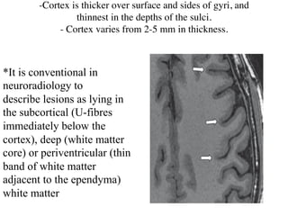
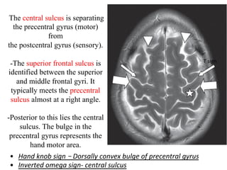
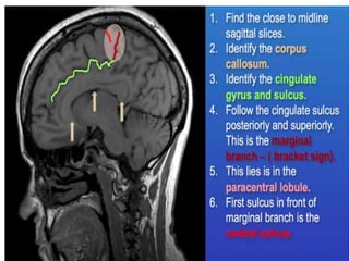
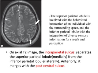
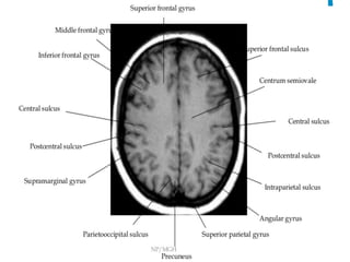
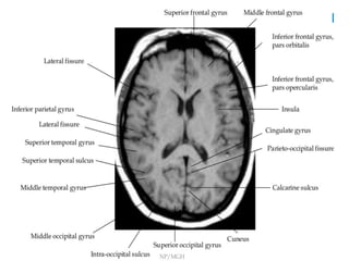
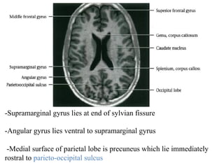
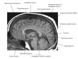
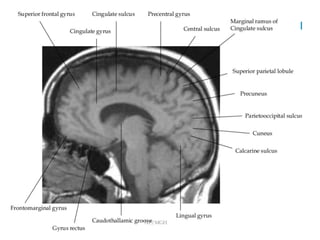
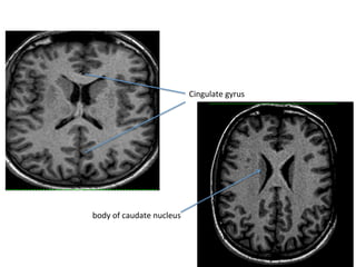
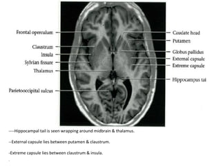
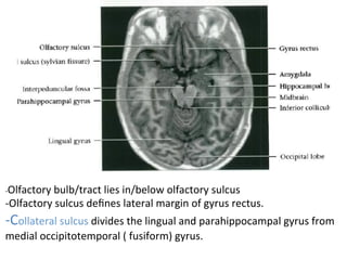
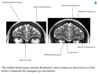
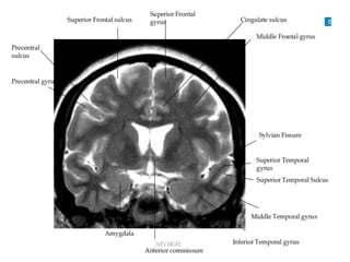
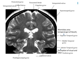
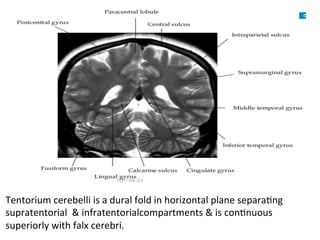
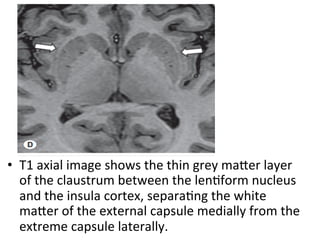
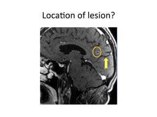
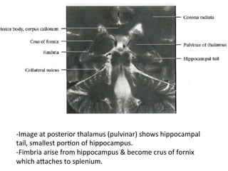
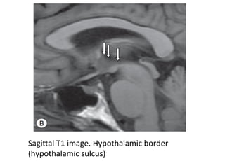
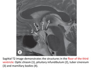
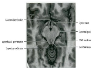
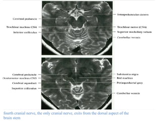
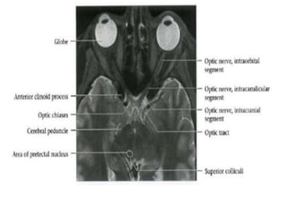
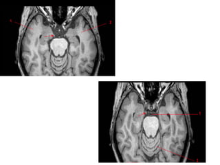


This document provides an overview of brain anatomy as seen on MRI. It describes key structures including sulci, gyri and other anatomical landmarks of the different lobes and regions of the brain. Key points covered include the location and functions of the precentral and postcentral gyri, central sulcus, sylvian fissure and other cortical structures. Subcortical structures like the hippocampus, amygdala and hypothalamus are also described. The document outlines anatomy from the cerebral cortex down through the brainstem.


![Radiological anatomy of_temporal_bone[1]](https://cdn.slidesharecdn.com/ss_thumbnails/radiologicalanatomyoftemporalbone1-171112100915-thumbnail.jpg?width=640&height=640&fit=bounds)


![Imaging in Neurovascular conflicts [Neurovascular compression syndrome ]](https://cdn.slidesharecdn.com/ss_thumbnails/cnv-141013092247-conversion-gate01-thumbnail.jpg?width=640&height=640&fit=bounds)


