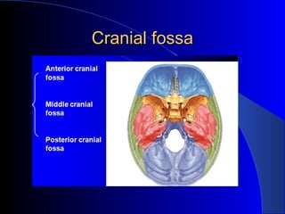
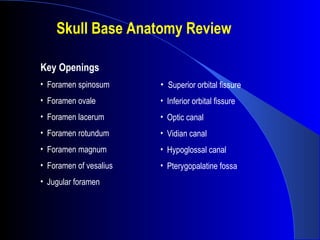
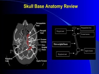
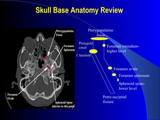
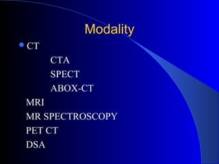
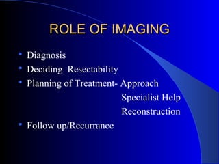
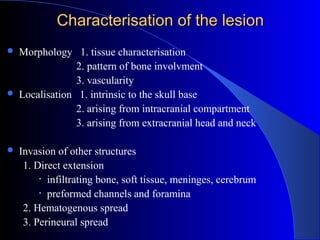
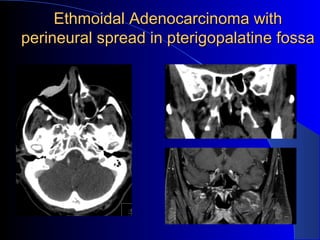
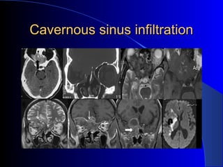
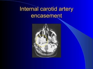
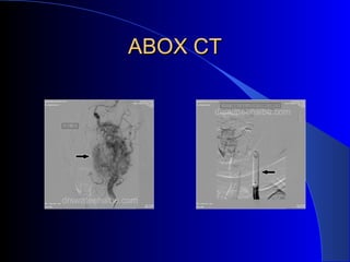
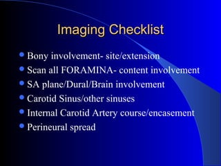
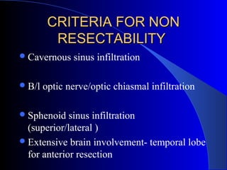
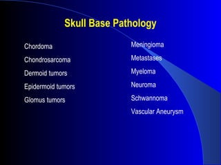
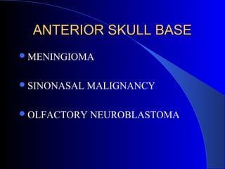
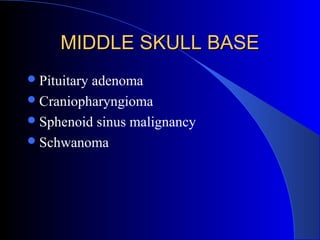
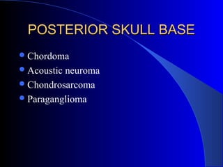
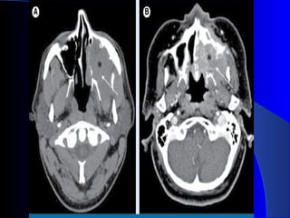
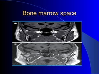
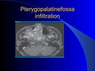
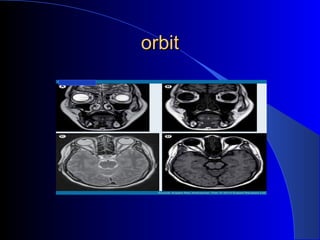
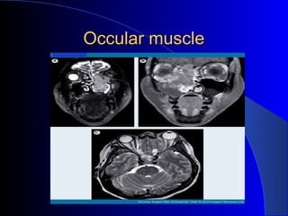
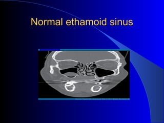
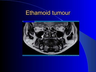
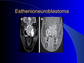
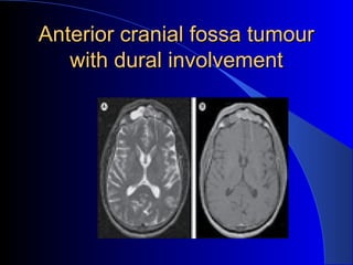
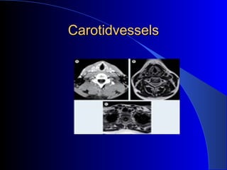
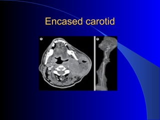
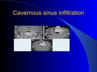
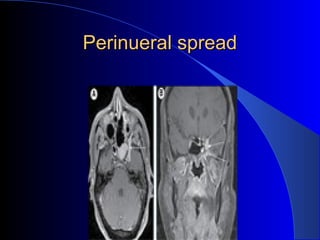
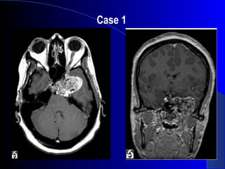
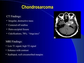
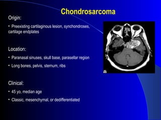
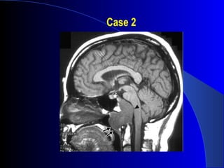
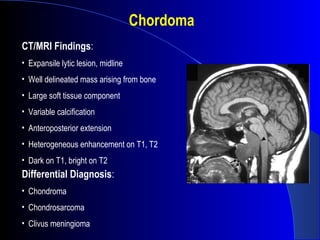
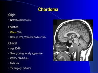
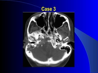
The document discusses imaging of the skull base. It begins with a review of skull base anatomy including key bones, fissures, sutures, and openings. It then covers the role of various imaging modalities like CT, MRI, and PET in diagnosing skull base lesions, determining resectability, treatment planning, and follow-up. Examples of imaging findings for conditions like sinonasal malignancy, perineural spread, and cavernous sinus infiltration are provided. Criteria for non-resectability are outlined. Common skull base pathologies are listed along with examples of three clinical cases.
Slides prepared and compiled by highly experienced ENT teacher, Dr. Krishna Koirala from Nepal , for teaching undergraduate and postgraduate ENT students in the field of otorhinolaryngology.
A clear and concise explanation of the basic concepts in the subject matter concerned.
He is the Head of department with a sound knowledge in the field of ENT to teach both undergraduate and postgraduate ENT students
- Video recording of this lecture in English language: https://youtu.be/lK81BzxMqdo
- Video recording of this lecture in Arabic language: https://youtu.be/Ve4P0COk9OI
- Link to download the book free: https://nephrotube.blogspot.com/p/nephrotube-nephrology-books.html
- Link to NephroTube website: www.NephroTube.com
- Link to NephroTube social media accounts: https://nephrotube.blogspot.com/p/join-nephrotube-on-social-media.html
Report Back from SGO 2024: What’s the Latest in Cervical Cancer?bkling
Are you curious about what’s new in cervical cancer research or unsure what the findings mean? Join Dr. Emily Ko, a gynecologic oncologist at Penn Medicine, to learn about the latest updates from the Society of Gynecologic Oncology (SGO) 2024 Annual Meeting on Women’s Cancer. Dr. Ko will discuss what the research presented at the conference means for you and answer your questions about the new developments.
Ozempic: Preoperative Management of Patients on GLP-1 Receptor Agonists Saeid Safari
Preoperative Management of Patients on GLP-1 Receptor Agonists like Ozempic and Semiglutide
ASA GUIDELINE
NYSORA Guideline
2 Case Reports of Gastric Ultrasound
Anti ulcer drugs and their Advance pharmacology ||
Anti-ulcer drugs are medications used to prevent and treat ulcers in the stomach and upper part of the small intestine (duodenal ulcers). These ulcers are often caused by an imbalance between stomach acid and the mucosal lining, which protects the stomach lining.
||Scope: Overview of various classes of anti-ulcer drugs, their mechanisms of action, indications, side effects, and clinical considerations.
MANAGEMENT OF ATRIOVENTRICULAR CONDUCTION BLOCK.pdfJim Jacob Roy
Cardiac conduction defects can occur due to various causes.
Atrioventricular conduction blocks ( AV blocks ) are classified into 3 types.
This document describes the acute management of AV block.
HOT NEW PRODUCT! BIG SALES FAST SHIPPING NOW FROM CHINA!! EU KU DB BK substit...GL Anaacs
Contact us if you are interested:
Email / Skype : kefaya1771@gmail.com
Threema: PXHY5PDH
New BATCH Ku !!! MUCH IN DEMAND FAST SALE EVERY BATCH HAPPY GOOD EFFECT BIG BATCH !
Contact me on Threema or skype to start big business!!
Hot-sale products:
NEW HOT EUTYLONE WHITE CRYSTAL!!
5cl-adba precursor (semi finished )
5cl-adba raw materials
ADBB precursor (semi finished )
ADBB raw materials
APVP powder
5fadb/4f-adb
Jwh018 / Jwh210
Eutylone crystal
Protonitazene (hydrochloride) CAS: 119276-01-6
Flubrotizolam CAS: 57801-95-3
Metonitazene CAS: 14680-51-4
Payment terms: Western Union,MoneyGram,Bitcoin or USDT.
Deliver Time: Usually 7-15days
Shipping method: FedEx, TNT, DHL,UPS etc.Our deliveries are 100% safe, fast, reliable and discreet.
Samples will be sent for your evaluation!If you are interested in, please contact me, let's talk details.
We specializes in exporting high quality Research chemical, medical intermediate, Pharmaceutical chemicals and so on. Products are exported to USA, Canada, France, Korea, Japan,Russia, Southeast Asia and other countries.
Title: Sense of Taste
Presenter: Dr. Faiza, Assistant Professor of Physiology
Qualifications:
MBBS (Best Graduate, AIMC Lahore)
FCPS Physiology
ICMT, CHPE, DHPE (STMU)
MPH (GC University, Faisalabad)
MBA (Virtual University of Pakistan)
Learning Objectives:
Describe the structure and function of taste buds.
Describe the relationship between the taste threshold and taste index of common substances.
Explain the chemical basis and signal transduction of taste perception for each type of primary taste sensation.
Recognize different abnormalities of taste perception and their causes.
Key Topics:
Significance of Taste Sensation:
Differentiation between pleasant and harmful food
Influence on behavior
Selection of food based on metabolic needs
Receptors of Taste:
Taste buds on the tongue
Influence of sense of smell, texture of food, and pain stimulation (e.g., by pepper)
Primary and Secondary Taste Sensations:
Primary taste sensations: Sweet, Sour, Salty, Bitter, Umami
Chemical basis and signal transduction mechanisms for each taste
Taste Threshold and Index:
Taste threshold values for Sweet (sucrose), Salty (NaCl), Sour (HCl), and Bitter (Quinine)
Taste index relationship: Inversely proportional to taste threshold
Taste Blindness:
Inability to taste certain substances, particularly thiourea compounds
Example: Phenylthiocarbamide
Structure and Function of Taste Buds:
Composition: Epithelial cells, Sustentacular/Supporting cells, Taste cells, Basal cells
Features: Taste pores, Taste hairs/microvilli, and Taste nerve fibers
Location of Taste Buds:
Found in papillae of the tongue (Fungiform, Circumvallate, Foliate)
Also present on the palate, tonsillar pillars, epiglottis, and proximal esophagus
Mechanism of Taste Stimulation:
Interaction of taste substances with receptors on microvilli
Signal transduction pathways for Umami, Sweet, Bitter, Sour, and Salty tastes
Taste Sensitivity and Adaptation:
Decrease in sensitivity with age
Rapid adaptation of taste sensation
Role of Saliva in Taste:
Dissolution of tastants to reach receptors
Washing away the stimulus
Taste Preferences and Aversions:
Mechanisms behind taste preference and aversion
Influence of receptors and neural pathways
Impact of Sensory Nerve Damage:
Degeneration of taste buds if the sensory nerve fiber is cut
Abnormalities of Taste Detection:
Conditions: Ageusia, Hypogeusia, Dysgeusia (parageusia)
Causes: Nerve damage, neurological disorders, infections, poor oral hygiene, adverse drug effects, deficiencies, aging, tobacco use, altered neurotransmitter levels
Neurotransmitters and Taste Threshold:
Effects of serotonin (5-HT) and norepinephrine (NE) on taste sensitivity
Supertasters:
25% of the population with heightened sensitivity to taste, especially bitterness
Increased number of fungiform papillae
TEST BANK for Operations Management, 14th Edition by William J. Stevenson, Ve...kevinkariuki227
TEST BANK for Operations Management, 14th Edition by William J. Stevenson, Verified Chapters 1 - 19, Complete Newest Version.pdf
Phone Us ❤85270-49040❤ #ℂall #gIRLS In Surat By Surat @ℂall @Girls Hotel With...
SKULL BASE IMAGING
1. IMAGING OF SKULL BASEIMAGING OF SKULL BASE
--Dr.A.Joseph Stalin(MCh PG)Dr.A.Joseph Stalin(MCh PG)
PROF .R.R UNIT
DEPT OF SURGICAL ONCOLOGY
GOVT ROYAPETTAH HOSPITAL
CHENNAI
9. ROLE OF IMAGINGROLE OF IMAGING
Diagnosis
Deciding Resectability
Planning of Treatment- Approach
Specialist Help
Reconstruction
Follow up/Recurrance
11. Characterisation of the lesionCharacterisation of the lesion
Morphology 1. tissue characterisation
2. pattern of bone involvment
3. vascularity
Localisation 1. intrinsic to the skull base
2. arising from intracranial compartment
3. arising from extracranial head and neck
Invasion of other structures
1. Direct extension
• infiltrating bone, soft tissue, meninges, cerebrum
• preformed channels and foramina
2. Hematogenous spread
3. Perineural spread
12. Agressive bone involvement patternAgressive bone involvement pattern
Osteolysis
Absent bone replaced by soft tissue
Thinned bone with soft tissue mass on
its both sides
Abnormal signal of the bone marrow
Calcifications within the soft tissue mass
13. Non-aggressive bone involvement patternNon-aggressive bone involvement pattern
Bone remodeling with bowing, thin or demineralized walls
Bone expansion with smooth contour or interrupted walls
Enlarged intramedullary cavity
Varying attenuation: ground-glass, radiolucent or sclerotic
14. INTRACRANIAL <> EXTRA CRANIALINTRACRANIAL <> EXTRA CRANIAL
Pharygeal mucosal space PMS Sinus Morgagni
Parapharyngeal space PPS Skull base
Carotid space CS Carotid canal
Jugular foramen
Mandibular space MS Foramen ovale
Retropharyngeal space RPS Basiocciput
15. Sinus frontalis Squamous CellSinus frontalis Squamous Cell
Cancer with intracranialCancer with intracranial
spreadspread
Nodular dural enhancing have
high specificity
Dural thickness > 5 mm
Coexistent leptomeningeal
enhancement
Hypointense leision
Brain parenchymal changes
16. Perineural spreadPerineural spread
Nerve enlargement and nerve enhancement
Obliteration of the fat in the foramina, fosse or fissures
Foraminal enlargement or destruction
Enhancing soft tissue in the cavernous sinus and Meckel cave
Neuropathic atrophy and fat replacement
Tumor growthTumor growth
Incresed permeability of endoneurial capillariesIncresed permeability of endoneurial capillaries
Rupture of the blood-nerve barrierRupture of the blood-nerve barrier
Contrast-enhancementContrast-enhancement
52. CONCLUSIONCONCLUSION
Thorough anatomical knowledge essential.
Both CT and MRI are needed.
Histological diagnosis not needed for
managing skull base tumours.
Main role of imaging is to plan the
recection .
Treatment options for skull base tumours –
resection +/_ radiotherapy
7 foramina
2 fissures
3 canals
All with fun stuff inside.
Hypoglossal artery = not always present.
Sometimes, can have a small emissary vein that runs through here that can protrude into the cerebellomedullary cistern and mimic a nerve sheath tumor.
2 parts:
Pars nervosa = anteromedial
Pars vascularis aka pars venosa = posterolateral
Separated by the jugular spur.
Pars nervosa: glossopharyngeal nerve,
inferior petrosal sinus- runs along petrooccipital fissure
Pars vascularis: vagus and spinal accessory nerve
Jugular bulb: confluence b/w sigmoid sinus and internal jugular vein.
Anterolateral to pars nervosa = petrous portion of the carotid artery.
F.S.:
V3 recurrent = mandibular branch.
Emissary veins in FS and FO connect cavernous sinus with pterygoid plexus of veins = path for nasaopharyngeal tumors.
Foramen of vesalius- inconstant. Anterior and medial to f. ovale. When it does occur, it contains (read slide)
F.L.:read slide
Vidian canal = aka pterygoid canal. Connects pterygopalatine fossa to foramen lacerum. Contains vidian artery (branch of maxillary artery) and nerve.
Vidian nerve = formed by merger of greater superficial petrosal nerve (branch of facial nerve) and deep petrosal nerve.
PPF = conduit for spread of tumor and infection
Communicates with inf orbital fissure, orbital apex,…
PPF - Sphenoplatine foramen = to nasal fossa
PPF – pterygomaxillary fissure = to masticator space
PPF- foramen rotundum = connection with Meckel’s cave, cavernous sinus
PPF- Vidian canal = to foramen lacerum
PPF- to greater/lesser palatine canals = to palate
PPF – foramen rotundum = connection with meckel’s cave, cav sinus, since we’ve mentioned it a few times now and b/c contains a lot of key elements:
Cav sinus- read slide.
V2- lateral wall of CS- then to foramen rotundum
V1- lateral wall of cav sinus- then to superior orbital fissure, along with CN III, IV, VI
Speaking of the superior orbital fissure…
Below SOF = IOF.
Optic canal = more superior to SOF.
Pitfalls calcified debris in fungal sinuitis and flow voids
associated with aggressive rapidly growing tumors , malignant neoplasms or infectious process( osteocorsoma, osteosarcoma and metastases)
High T2 signal and contrast enhancement meaning brain vasogen edema ; ddif linerar pattern of enhancemet rep reactiv dural changes of hyperemia, hypertrofia and inflammation
Adenoid cystic carcinoma, squamos cell carcinoma but even lymfoma, melanoma desmoid type basal cell ca rhabdomyosarcoma neurogenic tum juv angifibroma
Gd T1 and high resolusion T2 w ; Gd fs t1
etmoidal sinus cancer
Chordomas like the clivus
Chondrosarcomas like the cartilaginous endplates- petrooccipital suture
Glomus tumors- jugular bulb, middle ear, carotid body
Meningiomas- most common (20% of all brain tumors). But most = cerebral convexity, along and lateral to the falx.
10% clivus
20% sphenoid ridge
Schwannomas almost always develop from sensory nerves. Because the olfactory and optic nerves do not have a Schwann cell layer, they do not develop these tumors. The most common intracranial schwannoma = acoustic neuroma- it develops from the vestibular nerve. Bilateral acoustic neuromas = pathogomonic for NF2. (90% of the time, schwannomas are solitary).
The second most common intracranial schwannomas develop from the trigeminal nerve. Trigeminal schwannomas usually arise from the root or ganglion and occupy the middle fossa and, sometimes, the posterior fossa. Schwannomas of the other cranial nerves are rare.
T1 post contrast MR showing extraaxial lesion arising from the middle cranial fossa.
Heterogeneous enhancement.
Low signal areas = flow voids or calcs.
Coronal = involvement with skull base. Mass effect on temporal lobe.
Chondrosarcomas can occur anywhere in the skeletal system. Often = preexisting cartilaginous lesion like previously benign osteochondroma. In skull base = usually at cartilage endplates. E.g. petrooccipital suture.
Location: read slide
Clinical:
Most commonly, patients are diagnosed with chondrosarcomas during the third or fourth decade of life. Males are affected more often than females. Chondrosarcomas can be divided into classic, mesenchymal, and dedifferentiated tumors.
Mesenchymal, Dedifferentiated = high grade.
Classic low grade = like chordoma.
DDX: Chordoma- usually has marked bone destruction, midline (clivus)
Chondrosarcoma = significant soft tissue component, little bone destruction, arcs/nodules of calcification, eccentric locations- often centered in framen lacerum.
Sagittal T1-weighted MR image shows a large, hypointense soft-tissue mass that arises from the distal clivus with anterior extension into the nasopharynx (arrows) and extradural extension into the posterior fossa (arrowhead).
CT to assess degree of bone involvement. MRI to evaluate extension of tumor.
CT Findings:
The most characteristic appearance of intracranial chordoma is of a centrally located homogeneous soft tissue mass arising from the clivus and causing adjacent bone destruction.
Calcification is common but variable.
Areas of low attenuation within the soft tissue mass occasionally are found on CT, representing the myxoid and gelatinous material found on pathologic examination.
CT reliably demonstrates petrous apex involvement and lysis of the skull base foramina.
MRI Findings:
Mass originating from midline with extension primarily in the anteroposterior axis rather than laterally.
Well delineated.
Expands bone in the early stage = indicator that it arises from bone, not from adjacent structures.
Post gad = lobulated area, heterogeneous on T1 and T2 b/c of mucinous and gelatinous contents.
DDX: Chondroma- similar appearance but extend more laterally into sellar and cerebellopontine angles.
Clivus meningioma – homegeneous signal, dural attachment
Contrast enhanced T1 spin echo image. Chordoma of the upper part of the clivus with posterior extension into the pontine cistern.
Chordomas = benign tumor but has significant problems b/c of location, local invasion, recurrence.
Origin:
Notochord = early fetal axial skeleton. Gets surrounded by cartilage. Cartilage ossifies and notochord = squeezed out into intervertebral regions = nucleus pulposus of intervertebral disks.
Can get remnants then, along any position of the neural axis- turn into chordomas.
Location: read slide. Rare. &lt;0.2% of all intracranial tumors.
Clinical:read slide
CN deficits: HA, dysphagia, facial pain, facial paresis, visual loss, hearing loss, and ataxia.
CT to assess degree of bone involvement. MRI to evaluate extension of tumor.
CT Findings:
The most characteristic appearance of intracranial chordoma is of a centrally located homogeneous soft tissue mass arising from the clivus and causing adjacent bone destruction.
Calcification is common but variable.
Areas of low attenuation within the soft tissue mass occasionally are found on CT, representing the myxoid and gelatinous material found on pathologic examination.
CT reliably demonstrates petrous apex involvement and lysis of the skull base foramina.
MRI Findings:
Mass originating from midline with extension primarily in the anteroposterior axis rather than laterally.
Well delineated.
Expands bone in the early stage = indicator that it arises from bone, not from adjacent structures.
Post gad = lobulated area, heterogeneous on T1 and T2 b/c of mucinous and gelatinous contents.
CT imaging demonstrates the extent of bony destruction (white and black arrows) by the tumor. The normal jugular foramen on the left (arrow head) is shown for comparison.
Salt and pepper: multiple low signal intensity areas = flow voids in tumor.
When large- erode bone.
Glomus tumors arise from chemoreceptor cells.
These tumors are slow-growing hypervascular tumors that usually occur in the temporal bone.
Location: read slide- check other places for them b/c = multiple. E.g. Carotid body
Patients usually present with gradual hearing loss, unilateral pulsatile tinnitus, and lower cranial nerve palsies.
Approximately 1-3% of gangliogliomas produce catecholamines, so can get arrythmia, BP fluctuation.
May be locally invasive but rarely metastasize.