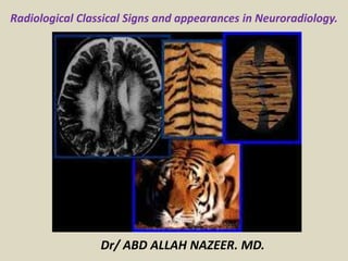
Presentation1.pptx, radiological classical signs and appearances in neuroradiology.
•Download as PPTX, PDF•
106 likes•8,331 views

This document describes several classical radiological signs seen on various imaging modalities like CT, MRI, ultrasound and X-ray. It provides images and descriptions of signs such as the "ice cream cone sign" seen on CT of the temporal bone, "CT reversal sign" seen in diffuse cerebral anoxia, "Mount Fuji sign" seen in tension pneumocephalus on CT, and "lemon sign" seen in spina bifida on ultrasound. Many other signs seen in different neurological conditions are also described along with example images, including "pancake brain sign", "molar tooth sign", "figure eight sign", and "tram track sign".

Recommended
More Related Content
What's hot
What's hot (20)
Viewers also liked
Viewers also liked (20)
Similar to Presentation1.pptx, radiological classical signs and appearances in neuroradiology.
Similar to Presentation1.pptx, radiological classical signs and appearances in neuroradiology. (20)
More from Abdellah Nazeer
More from Abdellah Nazeer (20)
Recently uploaded
Recently uploaded (20)
Presentation1.pptx, radiological classical signs and appearances in neuroradiology.
- 1. Radiological Classical Signs and appearances in Neuroradiology. Dr/ ABD ALLAH NAZEER. MD.
- 2. Ice-cream cone sign It reflects normal appearance of incudomalleolar joint formed by malleolar head and body of the incus on axial computed tomography (CT) sections. Anatomical identification of this anatomic structure is important in terms of ossicular luxation especially in trauma cases. The space between the ice-cream cone and the scutum is called Prussak’s space
- 3. High resolution, axial CT image demonstrating the “ice-cream sign” of the temporal bone (white arrow). The sign represents the typical appearance of the malleoincudal joint.
- 4. ICE CREAM CONE SIGN. Axial temporal bone CT. Picture and schematic drawing.
- 5. CT reversal sign The reversal sign is associated with diffuse anoxic- ischemic brain damage and almost always observed in children. This sign is characterized by a relative reversal of the attenuation between supra- and infratentorial structures. The grey-white matter distinction is lost and decreased, and there is a diffuse decrease in density in the cerebral grey and white matter. Thalami, brainstem, and cerebellum have a relatively increased density. It is closely related to child abuse, especially when accompanying intracranial bleeding.
- 6. CT reversal sign” is observed due to diffuse cerebral anoxia in non-contrasted CT examination.
- 7. Mount Fuji sign This sign is observed in bilateral subdural tension pneumocephalus. These air accumulations lead to compression in the frontal lobes and take a form of Mount Fuji on axial CT sections. It is most commonly seen after surgical decompression of chronic subdural hematoma. However, it may also be observed following a head trauma, otogenic infections, nitrous oxide anesthesia, and diving
- 8. ”Mount Fuji sign” due to tension pneumocephalus is observed in axial CT sections (parenchymal and bone window, white arrows)
- 9. Lemon sign The lemon sign is useful in identification of spina bifida and is commonly associated with hydrocephalus and Chiari II malformation. Loss of normal convex contour of the frontal bones in transverse fetal sonogram obtained at biparietal diameter. It has a high sensitivity and specificity in high-risk patients before the 24th gestational week. However, it is not specific for spina bifida and may be detected in encephalocele, Dandy-Walker malformation, thanatophoric dysplasia, cystic hygroma, corpus callosum agenesis, hydronephrosis, and umbilical vein varices
- 10. ”Lemon sign” is seen in the frontal bones in a fetus with myeloschisis, as detected in an obstetrical US performed at the 20th week of gestation (white arrows).
- 11. Axial sonogram of a fetal head demonstrating the lemon sign. Schematic drawing and picture.
- 12. Pancake brain sign This sign defines the appearance of abnormal brain tissue in cases with alobar holoprosencephaly. Holoprosencephaly is an anomaly caused by a prosencephalic division defect and characterized by varying degrees of fusion of cerebellar hemispheres, diencephalon, basal ganglia, and thalami. The pancake brain sign is formed by fusion of cerebral hemispheres associated with the presence of typical monoventricle at the center
- 13. ”Pancake brain appearance” formed by monoventricle cavity and cerebral hemispheric fusion is seen in T1-weighted MR image in a case with alobar holoprozencephaly.
- 14. Molar tooth sign Joubert syndrome is an autosomal recessive disorder characterized by abnormal eye movements, nystagmus, and difficulty in following mobile objects with eyes, apnea- tachypnea episodes, and motor retardation. Molar tooth sign represents abnormal antero- posterior orientation of superior cerebellar peduncles in a way similar to stems of a molar tooth on axial CT or magnetic resonance (MR) images. It is mainly observed in patients with Joubert syndrome.
- 15. Molar tooth sign” (star) at the level of pons and superior cerebellar peduncles coursing parallel to each other (white arrows) is seen in a T1-weighted MR section in a case with Joubert syndrome.
- 16. Joubert's syndrome - The 'Molar Tooth'
- 17. Figure eight sign Lissencephaly is a disorder caused by defective neuronal migration between the 8–14th gestational week and characterized by a lack of development of gyri and sulci. Lissencephaly is classified into two subgroups: complete (type 1 – agyria) or partial (type 2 – pachygyria). Type 1 lissencephaly is characterized by shallow sylvian fissures that are vertically oriented. In this type of lissencephaly, brain takes on an hour-glass or figure-8 appearance due to compression at the middle part by sylvian fissures on axial imaging.
- 18. An appearance similar to “figure eight” due to lissencephaly in the axial plane on CT examination.
- 20. Face of the giant panda sign This sign was first described by Hitoshi et al. in Wilson’s disease in 1991. It consists of high signal intensity in the tegmentum except for the red nucleus, preservation of signal intensity at the lateral portion of the pars reticulata of the substantia nigra, and hypointensity of the superior colliculus. The real pathology responsible for this appearance is the paramagnetic effect of the accumulation of heavy metals such as iron and copper in affected sites.
- 21. A “giant panda face” is observed in a T2-weighted axial MR image in a case with Wilson syndrome.
- 22. Radial band sign Radial bands are linear or curvilinear areas with an abnormal signal intensity extending from the periventricular region to the subcortical region, that are best observed on T2-weighted (T2W) and especially FLAIR MR images. It is believed that radial band sign is indicative of abnormal migration of dysplastic stem cells during the course of radial glial-neuronal unit in patients with tuberous sclerosis complex. Radial bands are hypo- /isointense on T1-weighted images and hyperintense on T2W and FLAIR images.
- 23. Hyperintense radial bands (black arrow) extending linearly at the level of the right cerebral hemisphere and a cortical tuber (short white arrow) located at the left parietal lobe in an axial FLAIR MR image in a case with tuberous sclerosis complex. In addition, MRI showing a subependymal nodule (thin black arrow).
- 24. String sign/Tigroid (Leopard skin) appearance This sign is characterized by multiple dark spots or stripes (spared perivascular white matter) of normal white matter intensity scattered within the bright demyelinated periventricular white matter on T2W images. Tigroid appearance of the white mater has been found in some cases with Pelizaeus-Merzbacher disease and metachromatic leukodystrophy. However, it has been recently reported that it may be observed in cases with lissencephaly accompanied by cerebellar hypoplasia.
- 25. STRIPE SIGN/TIGROID PATTERN. Linear hypointensities radiating from the ventricular margins within hyperintense periventricular white matter and the centrum semiovale on T2W MRI axial image. Schematic drawings and pictures.
- 26. A “tigroid appearance” is observed at periventricular white matter in axial T2- weighted MR sections in a 2-year-old girl with metachromatic leukodystrophy.
- 27. Open circle sign The open ring sign is a relatively specific sign for demyelination, helpful in distinguishing between ring enhancing lesions. It is observed in patients with multiple sclerosis. It is observed as a lesion showing contrast effect as a circle that incompletely encircles a demyelinated plaque. The lesion is a high-intensity one on T2W images and it may be difficult to distinguish from an abscess or astrocytoma in this form.
- 28. Post-contrast T1-weighted MR image showing an incomplete ring lesion enhancing in the right parietal region (black arrow).
- 29. Light bulb sign Diffusion-weighted (DW) MR imaging is the method that can delineate ischemic lesions in the brain at the earliest stage. With the help of this method, this lesion can be demonstrated after the onset of the event. The ischemic area shines like a light bulb at this stage (it appears darker on ADC images). This area forms the core of the infarcted region. The brightness diminishes by the 2nd–3rd month. In this way, acute and chronic infracts can be distinguished or acute lesions can be defined in patients with multiple lesions of varying age. The marked increase in DWI signal in areas of acute ischemia, relative to unaffected brain, is typically so striking that this finding has been referred to as the “light bulb sign” of acute stroke.
- 30. The b=1000 s/mm2 DWI showing an acute infarct as “light bulb” bright.
- 31. Keyhole sign The posterior fossa dimensions are normal in Dandy-Walker variants. There is a mild vermian hypoplasia and thus the vallecula becomes widened between the cerebellar hemispheres under the vermis. The fourth ventricle and cisterna magna communicate with each other through this wide vallecula. This appearance on axial CT and MR images is called “keyhole sign
- 32. Axial non-contrast CT image showing typical “key-hole” appearance of cisterna magna communicating with a dilated 4th ventricle (star).
- 33. Dawson finger It is detected on MR examination in multiple sclerosis. Demyelinating plaques are observed as focal signal areas on proton density and T2W MR images. These plaques are round or ovoid lesions limited particularly to the periventricular region. The appearance of periventricularly located ovoid lesions in the extended form along the ventricle is called Dawson finger.
- 34. Axial and parasagittal FLAIR MR images demonstrating multiple sclerosis plaques extending up through the corpus callosum (thin black arrows).
- 35. Cortical vein sign This was first described in MRI and also reported later on US and CT. It is used to differentiate extra-axial subarachnoid and subdural effusions from each other. On both CT and MRI, bridging veins extend from the cortical surface to the arachnoid. Appearance of bridging veins coursing in that manner in the extra-axial fluid is called a positive cortical vein sign and indicates that the fluid is located subarachnoidally. The fluid is located subdurally when these veins are invisible.
- 36. Post-contrast axial CT image showing the cortical veins (black arrows).
- 37. Caput medusa sign The most common vascular malformation in the bran is venous angiomas. They are most commonly observed in the frontal lobe and the posterior fossa. It has been suggested that they stem from a pause during brain development, i.e. when the arterial system completes its development but the venous system is not fully developed yet. The caput medusa sign, also known as a palm tree sign, refers to developmental venous anomalies of the brain, where a number of veins drain centrally towards a single drain vein. The appearance is reminiscent of Medusa, a gorgon of Greek mythology, who was encountered and defeated by Perseus. The sign is seen on both CT and MRI when contrast is administered
- 38. Contrast-enhanced T1-weighted axial MR image confirming converging tubular structures that represent a venous angioma (white arrow) in the medial aspect of the right cerebellar lobe.
- 39. Angel wing sign Chiari type II is the most common type of Chiari malformation. It is also known as Arnold-Chiari malformation. In 90% of cases there is also myelomeningocele, hydrocephalus, and corpus callosum agenesis. In these cases, prepontine migration of the cerebellum at the level of the middle cerebellar peduncle gives the brainstem an angel wing appearance on axial MR images
- 40. Axial T2-weighted MR image showing an “angel wing appearance” in the brainstem (black arrows).
- 41. Worm bag sign Arteriovenous malformations are space- occupying lesions formed by conglomerated large vessels. There may sometimes be a very small amount of brain tissue between the vessels in intracranial arteriovenous malformations. There is no brain tissue at all in some cases. Thus, such an appearance of large vessels resembles clustered worms and is called a worm bag sign
- 42. Sagittal T2-weigted MRI images showing a nidus of compact vessel with a typical appearance of “bag of black worms” in the left frontal region (white arrows).
- 43. Tectal beaking Chiari type II is the most common type of Chiari malformation. It is also known as Arnold-Chiari malformation. In 90% of cases there is also myelomeningocele, hydrocephalus, and corpus callosum agenesis. Variable degrees of fusion of the colliculi and tectum result in prominent beaking and inferior displacement of the tectal plate. In these cases, the appearance of the pointed tectum is called tectal beaking
- 44. Sagittal T1-weighted MRI demonstrating a small posterior fossa with a low-lying tentorial attachment posteriorly. The tectum is beaked (white arrow) and partial corpus callosum agenesis is present.
- 45. Double cortex appearance Because of the early arrest of neuronal migration, a symmetric circumferential band of heterotopic grey matter is separated from the overlying cortex by a thin band of white matter. On MRI, the brain appears to have a “double cortex” appearance. The condition is quite rare, found predominantly in females, and is occasionally associated with an X- linked dominant inheritance pattern.
- 46. Axial T2-weighted MR image showing four layers consisting of cortex, thin outer white matter, diffuse subcortical heterotopia, and inner white matter around the lateral ventricles, giving the appearance of a “double cortex”
- 47. Banana cerebellum sign The banana cerebellum sign is one of the many notable fruit-inspired signs, such as the “lemon sign”. In neural tube defects, folding of the cerebellum around the posterior brain stem due to inferior traction of the spinal cord causes the cerebellum to take the form of a banana. It has been reported that it may be present in 57% of fetuses with neural tube defect. In fetal hydrocephalus, a cerebellar deformation is observed in conjunction with ventriculomegaly and deletion of cisterna magna. In these cases, the cerebellum loses its normal central convexity and becomes compressed parallelly to the occipital bone, resembling a banana.
- 48. Transverse US image showing small posterior fossa and banana-shaped cerebellum (“banana sign”) (black arrows.
- 49. Viking helmet appearance The “Viking helmet” appearance refers to the lateral ventricles in the coronal projection in patients with dysgenesis of the corpus callosum. The cingulate gyrus is everted into narrowed and elongated frontal horns. Dysgenesis of the corpus callosum may be complete (agenesis) or partial and represents an “in utero” developmental anomaly.
- 50. Coronal view of MRI head of the patient demonstrating the lateral ventricles forming a “Viking helmet” appearance (white arrows) due to the absence of corpus callosum (black arrow).
- 51. The Tram-Track sign The tram-track sign is seen on skull radiographs as gyriform, curvilinear, parallel opacities that have the appearance of calcifications. A similar appearance can be seen on CTs. Sturge-Weber syndrome is a rare neurocutaneous syndrome that includes a facial port-wine stain and is associated with leptomeningeal angiomatosis. Weber demonstrated the characteristic gyriform intracranial calcifications. Calcifications are often gyriform and curvilinear and are most common in the parietal and occipital lobes. Calcifications can be more extensive but with frontal lobe and/or bilateral involvement. CT scans show calcifications in the areas of atrophy.
- 52. Lateral skull radiograph in a patient with Sturge-Weber syndrome showing parallel cortical calcifications (thin-white arrows). Contrast-enhanced axial T1-weighted MRI showing gyriform contrast enhancement in the right cerebral hemisphere (white arrows). There is brain atrophy on the right side. The cranial vault is asymmetric as secondary to brain atrophy.
- 53. Diamond-shaped fourth ventricle This appearance is seen in rhombencephalosynapsis. Rhombencephalosynapsis is a rare condition with most cases found in newborns and infants. Morphological findings are predominantly characterized by fusion of the cerebellar hemispheres and absence of the vermis, often accompanied by supratentorial anomalies. The size of the fourth ventricle is variable and in its axial plane it usually has a “keyhole or diamond shape”. This appearance is a result of dorsal and rostral convergence of the dentate nuclei, cerebellar peduncles and inferior colliculi
- 54. Axial T2-weighted MRI at the level of the posterior fossa showing antero-posterior elongation of the fourth ventricle giving it a “diamond shaped” appearance (arrows).
- 55. Bat wing 4th ventricle Bat wing 4th ventricle sign refers to the morphology of the fourth ventricle in the Joubert anomaly and related syndromes. The absence of the vermis with apposed cerebellar hemispheres give the fourth ventricle an appearance reminiscent of a bat with its wings outstretched. It is best demonstrated in axial imaging and could be easily missed in sagittal and coronal images
- 56. Axial T2-weighted MRI image at the level of the pontomedullary junction demonstrating the 4th ventricle that is shaped like a “bat wing” (arrow). In addition, axial T2-weighted MR image showing molar tooth sign (arrow).
- 57. Bat wing appearance of sylvian fissures Glutaric aciduria type 1 (GA-1) is an autosomal recessive inborn error of lysine, hydroxylysine and tryptophan metabolism that results from a deficiency of glutaryl-CoA dehydrogenase. The most striking finding on brain imaging is the presence of very wide CSF spaces anterior to the temporal lobes and within the sylvian fissures (giving a “bat wing” appearance). Widening of the sylvian fissures is a very characteristic finding in glutaric aciduria type I
- 58. Axial T1-weighted and T2-weighted images demonstrating widened sylvian fissures producing “bat-wings” appearance (arrows and stars).
- 59. Frog eye appearance Anencephaly is the most severe form of cranial neural tube defects (NTD) and is characterized by the absence of cortical tissue (although the brainstem and the cerebellum may be present) or cranial vault. Morphological spectrum within anencephaly ranges from holocrania (severest form) to merocrania (mildest form). Anencephaly may be radiologically detectable as early as at 11 weeks. A “frog eye” appearance may be seen in the coronal plane of US or MR images due to an absent cranial bone or brain, and bulging orbits
- 60. Fetal MR images demonstrating absent cranial bone/brain and bulging orbits (arrows). In addition, polyhydramnios is seen (star).
- 61. Boxcar ventricle sign Huntington’s disease is an autosomal dominant neurodegenerative disease, especially common in young adults. It has a course characterized by cognitive, behavioral, and muscle coordination disorders. In these cases, there may be an atrophy in basal ganglia, particularly in the caudate nucleus. Consequently, a widening may be seen in the frontal horns of the lateral ventricle. This particular appearance of frontal horns on multiplanar MR sections is called boxcar ventricle sign
- 62. Axial T2-weighted MR image showing bilateral atrophy of caudate nuclei and compensatory dilatation of lateral ventricles, a finding known as “boxcar ventricle” (black arrow).
- 63. "Cord sign" in cerebral venous thrombosis Cerebral venous thrombosis (CVT) is a rare entity, with variable clinical presentations. Seventy-five percent of the CVT occur in young women, between 20 and 40 years of age, with the superior sagittal sinus (SSS) being most frequently affected (62% of cases). Such increased incidence can be explained by pregnancy, puberty and use of oral contraceptives. The diagnosis can be achieved by means of CT (the most readily available), magnetic resonance imaging (MRI) (the method of choice) or by conventional angiography (CA) (the most invasive method). In 20% of cases, CT scans are normal. CVT findings can be classified in direct and indirect. The cord sign and the empty delta sign are direct signs of CVT. Indirect signs include: edema, infarction and hemorrhage. The cord sign is characterized as increased density of the sinuses or of the cortical or deep veins, originated from the thrombosed material inside the affected vessel. The cord sign is most frequently identified within two weeks after the first symptoms onset. With time, the thrombus becomes isodense and subsequently, hypodense.
- 65. "Empty delta sign" in venous sinuses thrombosis The empty delta sign may occur in cases of CVT, characteristically involving the SSS. On contrast-enhanced CT/MRI, the sign is characterized by a non-enhancing central triangular shaped area (the thrombus itself), limited by enhancing dura mater. Numerous factors may lead to CVT, as follows: inflammatory processes, infection, fibrosis of the venous sinuses walls, direct tumoral compression or/and extension, and hypercoagulable states. The empty delta sign is usually not identified at the first week (the material is isodense) as well as in chronic cases (more than two months), due to thrombus recanalization.
- 66. EMPTY DELTA SIGN. Note empty triangle on contrast-enhanced CT of the brain (thrombus in the dural sinus). Schematic drawing.
- 68. "Arrow sign" in ruptured middle cerebral artery aneurysm In ruptured aneurysms the pattern of distribution of subarachnoid hemorrhage can indicate its most likely location. In cases of bifurcation middle cerebral artery (MCA) aneurismal rupture the bleed may present the shape of an arrow, with the shaft and the tip representing blood in the horizontal segment of the Sylvian fissure and in the frontotemporal opercular area, respectively
- 70. "Dense artery sign" in acute middle cerebral artery infarction The dense MCA sign is one of the early signs of infarct. This is due an increase in density of its proximal segments, secondary to thrombosis. False-positive results may occur, particularly in cases of parietal calcification. It is important to observe that the distal branches of the MCAs rarely present parietal calcifications. Focal subarachnoid hemorrhage may simulate an abnormally dense MCA especially when located at the Sylvian fissure and constitute an additional cause for false positive results.
- 72. "Dot sign" in acute middle cerebral artery infarction The dot sign is one of the early signs of acute infarction and corresponds to a punctate hyperdensity in the Sylvian fissure. The signal represents thrombosis in the M2 and M3 segments of the MCA on plain CT scans. The presence of a thrombus/clot within the vessel alters and increases its density. The dot sign has a high specificity and high positive predictive value, but has low sensitivity.
- 74. "Hot nose sign" at brain death The hot nose sign can be seen in cases of brain death and it is defined by the presence of early and increased radiotracer activity in the nasopharyngeal region. It may also be seen as an intense blush (hyperemia) at CA examinations. The phenomenon is a result of a reduced blood flow in the internal carotid artery and increased flow in the external carotid branches. Such signal is not exclusive of brain death and may be found in different situations that lead to intracranial flow reduction in one or both internal carotid arteries
- 76. "Caput medusae sign" in developmental venous anomaly The caput medusae sign is indicative of developmental venous anomaly (DVA), and is identifiable at CA, CT and MRI. DVAs correspond to a network of dilated, abnormal medullary veins with radial distribution, converging into a dominant, calibrous transparenchymal vein, which may drain into a cortical vein, dural sinuses or into the deep venous system. DVAs are the most frequent intracranial vascular abnormalities, which are associated with cavernomas in around 30% of cases. Despite being considered incidental findings, in some cases these may lead to intracranial hemorrhage, thrombosis and venous infarction(11). Hemorrhages secondary to DVA are rarely found, with an annual risk of 0.7%
- 78. "Spoke wheel sign" in meningioma The spoke wheel sign refers to the typical angiographic appearance found in meningiomas. This sign corresponds to multiple small arteries radially distributed from a dominant feeding artery. Meningiomas are the most common primary intracranial tumors in adults. They are extra-axial, slow-growing, well-vascularized lesions with a benign behavior (grade I, according to the World Health Organization). Another remarkable and very common characteristic of meningiomas is the presence of a dural tail and, in 25% of cases, hyperostosis of the adjacent bone
- 80. "Onion skin sign" in Baló's concentric sclerosis The onion skin sign is considered pathognomonic for Baló's concentric sclerosis(14). According to the first reports on such disorder, most patients had an unfavorable history with progression either to death or disability. Recent cases however, have presented a less dramatic course. Baló's concentric sclerosis may occur as an isolated phenomenon or precede the development of multiple sclerosis. The lesions present a peculiar pattern of concentric lamellae of demyelination alternated with lamellae of myelinating or remyelinating white matter. Such lesions are most frequently found in the frontal lobes, but may be seen in the whole neuroaxis. Magnetic resonance imaging (MRI) is the best method for the disease diagnosis and follow-up. In spite of the high sensitivity of T2-weighted images to demonstrate demyelinating lesions, the concentric rings are better identified on T1-weighted images. The enhancement following contrast administration is variable and probably represents active areas of demyelination
- 82. "Eccentric target sign" in toxoplasmosis The eccentric or asymmetrical target sign is highly suggestive of central nervous system toxoplasmosis. The sign represents a ring enhancing abscess associated with an enhancing mural nodule. This finding is highly specific, but has low sensitivity, being found in approximately 30% of cases. The pathological correlation of such sign is not completely understood, but it is believed to represent internal folds and invaginations of the abscess walls
- 84. "Salt and pepper sign" in paraganglioma The appearance of salt and pepper is a highly sensitive and specific sign for head and neck paragangliomas. On T2weighted images, the salt- like appearance can be explained by the tumor matrix that appears hyperintense due to the presence of slow intratumor flow and hemorrhage and, on post-contrast T1-weighted images, by the presence of avid enhancement. The pepper-like appearance, can be explained in both on T1- and T2-weighted images by the presence of flow-voids of small vessels within these masses
- 85. SALT AND PEPPER SIGN. Axial MRI demonstrates the salt and pepper appearance due to the hypervascularity of this right mass (paraganglioma).
- 87. "Pancake brain sign" in alobar holoprosencephaly Pancake brain sign represents the appearance of the cerebral parenchyma in case of alobar holoprosencephaly. Holoprosencephaly is a malformation caused by a prosencephalic cleavage defect. Basically, holoprosencephalies are categorized into three major groups as follows: lobar holoprosencephaly, semilobar holoprosencephaly and alobar holoprosencephaly. Alobar holoprosencephaly is the most severe form of this malformation and presents a single ventricular cavity, fusion of frontal lobes, corpus callosum dysgenesis, alteration of the third ventricle, olfactory bulb and tracts, absence of interhemispheric fissure, besides fused thalami and basal ganglia.
- 89. "Hot cross bun sign" in C-type multiple systems atrophy The hot cross bun sign can be observed in multiple systems atrophy type C. Such sign is characterized by a cruciform pontine hyperintensity due to selective loss of neurons of the transverse pontocerebellar fibers, with preservation of the pontine tegmentum and of the fibers of the corticospinal tract. Multiple systems atrophy is a neurodegenerative disorder with varying degrees of involvement of the basal ganglia and the olivopontocerebellar complex.
- 90. The "face of the giant panda" sign in Wilson's disease The face of the giant panda pattern may be present in Wilson's disease. Such disease is characterized by hepatocellular degeneration caused by a genetic disorder of the copper metabolism with its consequential accumulation in tissues, particularly liver and brain. On MRI T2weighted sequences, one can observe hyperintensity in the pontine tegmentum, hypointensity of the periaqueductal gray matter and partially preserved signal in the red nuclei, in the lateral aspect of the substantia nigra pars reticulata and of the upper colliculus
- 92. "High heel foot print sign" in the skull base The high heel foot print sign is useful in the understanding of the intricate anatomy of the skull base and represent two relevant foramina. The anterior aspect of the high heel footprint represents the foramen ovale (FO), and the posterior aspect (the heel itself) the foramen spinosum (FS). The mandibular nerve, one of the three branches of the trigeminal nerve, is the main FO component(1). Also, the otic ganglion, the accessory meningeal artery, the lesser petrosal nerve and the emissary veins are found in this foramen. The middle meningeal artery is in the FS, and the absence of such artery is related to the persistent stapedial artery
- 94. "Dural tail sign" in meningiomas Dural tail corresponds to a thickened and abnormally enhancing segment of dura mater adjacent to a lesion whose shape is similar to a tail. Dural tail signs, which had been described as highly specific for meningiomas, can also be seen in other pathologies such as extra- and intra-axial tumors. It may correspond to isolated vascular changes, tumor invasion, adjacent non-continuous tumor growth, and tumor like micronodules. The dural tail sign is poorly specific for meningiomas, but presents good sensitivity, ranging from 50% and 80%
- 96. DURAL TAIL. Coronal T1-WI MR shows enhancement of the dura mater in continuity with a mass. Meningioma (arrows).
- 98. "Martini glass sign" in persistent hyperplastic primary vitreous Persistent hyperplastic primary vitreous (PHPV) is characterized by the presence of congenital embryonic remnants of hyaline vessels. The primary vitreous is supplied by the embryonal hyaloid circulation, which regresses at birth. In the posterior form of PHPV (the most common) a connective fibrovascular tissue is seen attached to the lens, connecting laterally to abnormally elongated ciliary process. At MRI a retrolental soft tissue and vascular mass is observed in association with a central, low-signal linear image corresponding to the remnant hyaloid vasculature that connects the crystalline lens to the optic nerve head, resembling the image of a martini glass. Associatedly, the vitreous may present high signal intensity because of hemorrhage, besides the presence of a small ocular globe
- 100. "Tram-track sign" in optic nerve sheath meningioma Optic nerve sheath meningiomas correspond to approximately two thirds of the primary tumors in the optic nerve-sheath complex, and are most frequently found in women between their third and fifth decade of life. The tram-track sign is better visualized in the axial plane of enhanced CT or MRI, and corresponds to a central linear hypodensity/hypointensity (optic nerve) delimitated by the contrast uptake of the optic nerve sheath at each of the sides affected by the meningioma itself. The tram-track sign is extremely useful in the differentiation between optic nerve sheath meningiomas and optic nerve gliomas. The optic nerve may be thickened and infiltrated by the glioma, but its sheath generally does not demonstrate contrast uptake. The tram-track pattern, in spite of being a characteristic sign, is not specific of optic nerve sheath meningiomas, and may occur in orbit pseudotumors, perioptic neuritis, sarcoidosis, leukemia and lymphoma.
- 101. OPTIC NERVE TRAM-TRACK SIGN. Contrast-enhanced CT scan and MRI images demonstrate tram-track sign (two enhancing areas of tumor separated from each other by the negative defect of the optic nerve)in two different cases of optic nerve sheath meningioma.
- 103. "Boxcar ventricle sign" in Huntington's disease The boxcar ventricle sign represents the prominent aspect of the lateral ventricles observed in the coronal plane in cases of Huntington's disease, secondary to atrophy of the basal nuclei, particularly the caudate nuclei. Huntington's disease is an autosomal dominant neurodegenerative disease, which affects particularly young adult individuals. Huntington's disease causes muscles discoordination and cognitive and behavioral alterations. The finding of ventricular dilatation, as well as basal ganglia atrophy, is very sensitive, but poorly specific
- 105. "Empty orbit sign" in neurofibromatosis type 1 Neurofibromatosis type 1 is an autosomal dominant disease with variable presentation, with cerebral and spinal changes seen in one third of the patients. Among the possible alterations, café- au-lait spots, Lisch nodules, plexiform fibromas and optic nerve gliomas are highlighted. The empty orbit sign represents the appearance of the orbit on plain films of the skull and on CT scan because of the lack of the innominate line due to dysplasia of the greater wing of the sphenoid, shortening of the lateral wall of the orbit and flattening of the orbital angle.
- 107. "En coup de sabre" sign in localized scleroderma Localized scleroderma is characterized by the presence of sclerotic lesions on the skin and subcutaneous tissues. This is different from systemic sclerosis because of the absence of significant systemic involvement; and generally presents a better prog-nosis(29). Localized scleroderma invariably affects the head, presenting as a linear, usually frontoparietal lesion (scleroderma "en coup de sabre“), with progressive facial hemiatrophy or Parry-Romberg syndrome where the atrophy extends beyond the skin to involve the subcutaneous cellular tissue, muscles and bones. Abnormal MRI findings are observed in 90% of cases and include hyperintensity on T2weighted images of the corpus callosum, subcortical regions, deep gray matter and brainstem; and most of times are ipsilateral. Focal atrophy that is the main dermatological finding may also be observed in the cerebral parenchyma.
- 109. Medusa head sign The medusa head sign is seen in a developmental venous anomaly (DVA), where multiple tributaries arranged in a radial fashion drain into a larger vein. This sign is best seen on gadolinium-enhanced T1W images. DVAs are usually located in the juxtacortical and periventricular regions and are commonly seen in the frontal and parietal lobes and in the brachium pontis. DVA is considered a non pathologic variation of venous drainage and, by itself, is usually not of any clinical significance. However, it can occur in association with a cavernoma ; it is seen in approximately 25–30% of cavernomas
- 110. Medusa head sign. Postcontrast axial T1W MRI image of the brain (A) shows a developmental venous anomaly (arrow), with multiple, small, radiating veins forming a ‘Medusa head’ in the left cerebellar hemisphere. Postcontrast axial T1W MRI image of the supratentorial brain (B) shows a large developmental venous anomaly with multiple radiating veins (arrows) draining into it. Postcontrast axial T1W image of the brain (C) shows a large developmental venous anomaly and a round hyperintense lesion with a dark rim (arrow), suggestive of a cavernoma, anterior to it
- 111. MEDUSA HEAD SIGN. MR shows venous malformation. Multiple tributaries arranged in a radial fashion drain into a larger vein (arrows).
- 112. Contrast-enhanced T1-weighted axial MR image confirming converging tubular structures that represent a venous angioma in medial aspect of right cerebellar lobe.
- 113. Moya moya appearance Moya moya is a Japanese term that means ‘puff of smoke.’ It represents the angiographic appearance of basal telangiectasias, which consist of dilated collateral branches of the lenticulostriate and thalamostriate arteries. It is usually seen in the anterior circulation in association with internal carotid artery stenosis. When the moya moya appearance is seen along with idiopathic occlusion of the internal carotid arteries it is called moya moya disease; when the occlusion is secondary to some other disease it is called moya moya syndrome. Causes of moya moya syndrome include NF1, sickle cell disease, bacterial meningitis, polyarteritis nodosa, radiation therapy, tuberculosis, and atherosclerosis. Histopathology of occluded arteries in moya moya disease shows endothelial hyperplasia and fibrosis without inflammatory reaction. Children with moya moya usually have ischemia or infarction, while adults with moya moya usually have hemorrhage. The treatment of moya moya includes anticoagulation, hypertransfusion, encephalo-duro-arterio-synangiosis (EDAS), anastomosis of the superficial temporal artery with the intracranial arteries, and sympathectomy or cervical ganglionectomy.
- 114. Moya moya appearance. Lateral anterior oblique view (A) of an internal carotid artery (thick short arrow) angiogram shows multiple, small, tortuous collateral vessels in the distribution of the middle cerebral artery (arrows), suggestive of the moya moya (puff of smoke) appearance. Axial view of the MRI angiogram (B) shows complete occlusion of the middle cerebral arteries bilaterally. Arrows indicate the internal carotid arteries
- 115. Moyamoya angiographic pattern. Schematic drawing and picture.
- 116. Eye-of-the-tiger sign This sign represents marked low signal intensity of the globus palladi on T2W MRI images. This low signal surrounds a central, small hyperintense area, producing the eye-of-the-tiger appearance. The sign is seen in what was once known as Hallervorden-Spatz (HS) syndrome but is now called neurodegeneration with brain iron accumulation (NBIA) or pantothenate kinase II (PANC2)-associated neurodegeneration. The marked low signal intensity of the globus palladi is a result of excessive iron accumulation and the central high signal is attributed to gliosis, increased water content, and neuronal loss with disintegration, vacuolization, and cavitation of the neuropil. Iron levels in blood and CSF are normal. The HS syndrome is a neurodegenerative disorder associated with extrapyramidal dysfunction and dementia. It is a neuroaxonal dystrophy, with the pathologic triad of iron deposition, axonal spheroids, and gliosis in the globus pallidi. MRI is important for differentiating HS syndrome from infantile axonal dystrophy, which does not show iron deposition. Mutation of the gene for pantothenate kinase 2 is the cause for the syndrome. The sign can be seen in other extra- pyramidal Parkinsonian disorders such as cortical-basal ganglionic degeneration, Steele-Richardson-Olszewski syndrome, and early-onset levodopa-responsive Parkinsonism.
- 117. EYE-OF-THE-TIGER SIGN. Axial T2-WI MR shows low signal surrounding a central, small hyperintense area, producing the eye-of-the-tiger appearance in the globus pallidus bilaterally (arrows). Hallervorden-Spatz disease.
- 118. The eye-of-the-tiger sign. Axial T2W MRI image of the brain shows hypointensity of the globus palladi (arrows). There is relative hyperintensity of the central part, giving the globus palladi the appearance of the eyes of a tiger. This appearance is seen in Hallervorden-Spatz syndrome
- 119. Tau sign The tau sign represents the appearance of the pre-sellar internal carotid artery (ICA) when a persistent trigeminal artery (PTA) originates from it, on a T1W sagittal MRI image . The configuration of the flow void in the presellar segment of the ICA with the PTA arising from it, resembles the Greek letter ‘τ’ (tau). The sign is suggestive of a PTA. The PTA arises from the ICA as it exits the carotid canal and enters the cavernous sinus. It joins the distal third of the basilar artery between the origins of the anterior, inferior, and superior cerebellar arteries. A PTA can be of two types: 1) the artery may supply the entire vertebrobasilar system distal to the anastomosis or 2) the anastomosis may mainly supply the superior cerebellar arteries bilaterally. PTA can be associated with aneurysms, arteriovenous malformations, moya moya disease, and other persistent carotid-vertebrobasilar anastomosis. Other persistent arteries that are responsible for communications between the carotid and vertebrobasilar systems are persistent hypoglossal and otic arteries.
- 120. Tau sign. Sagittal T1-W image of the brain shows (A) flow voids of the internal carotid artery (ICA) in the precavernous segment (thick short arrow), in the cavernous segment (medium-sized arrow), and a persistent trigeminal artery (thin long arrow). Together, these flow voids form the Greek letter ‘τ’ (tau). Sagittal view of the MRI angiogram shows the persistent trigeminal artery (arrow) arising from the ICA and joining the basilar artery in its mid segment
- 121. Harlequin appearance Harlequin appearance of the orbit represents the elevation of the superolateral angle of the orbit along with a flat frontal bone on a plain radiograph. It is seen in coronal craniosynostosis, where the anteroposterior growth of the skull is limited. There is also relative increase in the transverse diameter of the skull, which is called brachycephaly. The orbit is shallow, the lesser wing of the sphenoid is elevated, and the greater wing is expanded. The innominate line (superior border of the greater wing of the sphenoid) appears as a dense ridge. The sign can be unilateral or bilateral.
- 122. Harlequin appearance. Frontal view of the skull (A) in a child with Apert syndrome shows elevated superolateral angles of both orbits giving the appearance of a ‘harlequin mask.’ Frontal (B) and lateral (C) surface-shaded display 3D CT views of the skull show the harlequin appearance of the orbits. The sagittal suture (arrow in B) and the lambdoid suture (black arrows in C) are wide open. The coronal suture (thin arrows in C) is fused, which is suggestive of coronal craniosynostosis
- 123. Skull AP radiograph shows HARLEQUIN APPEARANCE. Schematic drawing and picture of a mask.
- 124. Central pontine myelinolysis (CPM) is a non-inflammatory demyelinating disease of the white matter tracts traversing the pons. Since the peripheral pontine fibers are typically spared it is referred to as central pontine myelinolysis. The predominant involvement of the transverse pontine fibers and relative sparing of the descending corticospinal tracts results in a characteristic "Owls- eye" appearance on axial T2- weighted MR imaging. If the areas of demyelination coalesce, axial T2- weighted MR images resemble "Face of the Piglet". This sign was first reported by Wagner et al. The pons with its characteristic appearance resembles the snout, while the internal carotid arteries and the fourth ventricle constitute the eyes and mouth of the piglet respectively. Corresponding T1-weighted MR images may show this characteristic pattern of signal alteration in the basal pons as resembling the face of a monkey - also referred to as the "Monkey sign" of CPM
- 128. "Butterfly Glioma" refers to a high grade astrocytoma, usually a glioblastoma multiforme, which crosses the midline via the corpus callosum. The term butterfly refers to the symmetric wing like extensions across the midline. Mostly butterfly gliomas occur in the frontal lobes crossing via the genu of the corpus callosum, but posterior butterfly lesions (crossing through the splenium) are also encountered. The differential diagnoses include primary cerebral lymphoma and tumefactive demyelination .
- 129. The "Target sign" or "Bull's eye" is a favorite radiologic descriptor of everything from bowel intussusception to cerebral tuberculoma. CT target sign, although uncommon, is considered nearly pathognomonic for cerebral tuberculoma when it is seen. The three zone T2- weighted target (bull's eye) sign at MR imaging has not been described in any other condition other than Cerebral Toxoplasmosis [9]. The T2- weighted target or bull's eye sign is characterized by a hypointense core, an intermediate hyperintense region, and a peripheral hypointense rim.
- 130. "Batwings dilatation" of sylvian fissures and wide CSF spaces anterior to temporal lobes in a child should alert the radiologist of the possibility of Glutaric acidemia type-I. Glutaric acidemia type-I is a rare autosomal recessive disorder caused by the deficiency of a mitochondrial enzyme glutaryl- CoA dehydrogenase. This enzyme deficiency is responsible for the improper breakdown of the amino acids resulting in an elevated plasma and urine level of glutaric acid. Glutaric acid accumulation in the body is responsible for neurotoxicity in the basal ganglia and fronto- temporal cortex.
- 131. "Steer-horn" lateral ventricles refer to the abnormal shape of the frontal horns of the lateral ventricle on coronal MR images in patients with corpus callosum agenesis. The lack of supporting deep white matter fibers and associated redirection of longitudinal callosal fibers (Probst bundles), result in alteration in the configuration of the ventricles, with the frontal horns taking on a "steer or bull's horn" appearance in the coronal plane
- 132. Tectal "Beaking" refers to the triangular ("beaked") appearance of the tectum in Arnold Chiari type-II malformation (Fig. 32, 33). The tectum is abnormal in virtually all patients with Chiari -II malformation. The quadrigeminal plate is partially or completely fused. The midbrain is elongated caudally and posteriorly to overlie the midline cerebellum and pons. Variable degrees of fusion of the colliculi and tectum result in a triangular ("beaked") tectum.
- 133. The "Zebra" sign refers to the streaky pattern of hemorrhage along the cerebellar folia in patients with remote cerebellar hemorrhage (RCH). Remote cerebellar hemorrhage or cerebellar hemorrhage distant from the site of surgery is a rare, usually benign, complication that most often occurs after supratentorial craniotomy.
- 134. "Rodent facies" refers to the oro-facial manifestations of thalassemia major which include abnormally prominent maxillary and cheek bones and a depressed nasal bridge. Erythroid hyperplasia and extramedullary hematopoiesis in thalassemic patients results in hypertrophy of osseous structures and a consequent prominence of the malar eminences. Proliferation of marrow within the frontal and facial bones impedes pneumatization of the paranasal sinuses. Radiographs and CT examination show dilatation of marrow spaces with coarse osseous trabeculations. A generalized loss of bone density accompanies thinning of the cortex of maxillary and mandibular bones with obliterated paranasal sinuses.
- 135. "Penguin" sign on brain MRI is an interesting radiological sign seen in patients with progressive supranuclear palsy (PSP). It refers to atrophy of the midbrain tegmentum, with a relatively preserved pons on mid-sagittal T1-weighted images. The "penguin" sign can be helpful in distinguishing progressive supranuclear palsy from multisystem atrophy and Parkinson disease. Patients with Parkinson disease, multisystem atrophy & corticobasal degeneration have no midbrain atrophy & hence do not show this sign. The penguin sign is useful for establishing the diagnosis of PSP; and is reported to have a sensitivity of nearly 100%.
- 136. Normally the upper border of the midbrain is convex. The atrophy of the midbrain in PSP results in a concave upper border of the midbrain with the typical 'humming bird sign' (figure).
- 137. Multi System Atrophy (MSA) MSA is also one of the atypical parkinsonian syndromes. MSA is a rare neurological disorder characterized by a combination of parkinsonism, cerebellar and pyramidal signs, and autonomic dysfunction.
- 138. Metachromatic leukodystrophy (MLD) is an autosomal recessive disorder caused by a deficiency of the lysosomal enzyme arylsulfatase. The decreased activity of arylsulfatase enzyme accounts for failure of myelin breakdown and reutilization, thus resulting in dysmyelination. At T2- weighted MR imaging, MLD manifests as symmetric confluent areas of high signal intensity in the periventricular white matter typically sparing the subcortical U fibers during the early stages. The sparing of the perivascular white matter within the periventricular white matter and centrum semiovale is responsible for the characteristic "Tigroid" and "Leopard skin" pattern.
- 139. A "Tiger-striped" cerebellar foliar pattern that consists of alternating bands on T1- and T2-weighted images is considered almost pathognomonic of Lhermitte-Duclos disease. The maintenance of the overall cerebellar architecture in spite of the thickened, and hyperplastic folia is responsible for this characteristic imaging appearance . Lhermitte- Duclos disease (LDD) (also known as dysplastic gangliocytoma).
- 140. Up to 50 percent of tumefactive demyelinating lesions demonstrate abnormal contrast enhancement, often in the form of ring enhancement. Commonly the enhancement pattern is in the form of an "open ring (Horse- shoe enhancement)", with the incomplete portion of the ring facing the cerebral cortex. The enhancing segment of the ring is thought to represent the zone of active demyelination accordingly favouring the white matter side of the lesion. The nonenhancing central part represents a more chronic phase of demyelination.
- 141. Thank You.