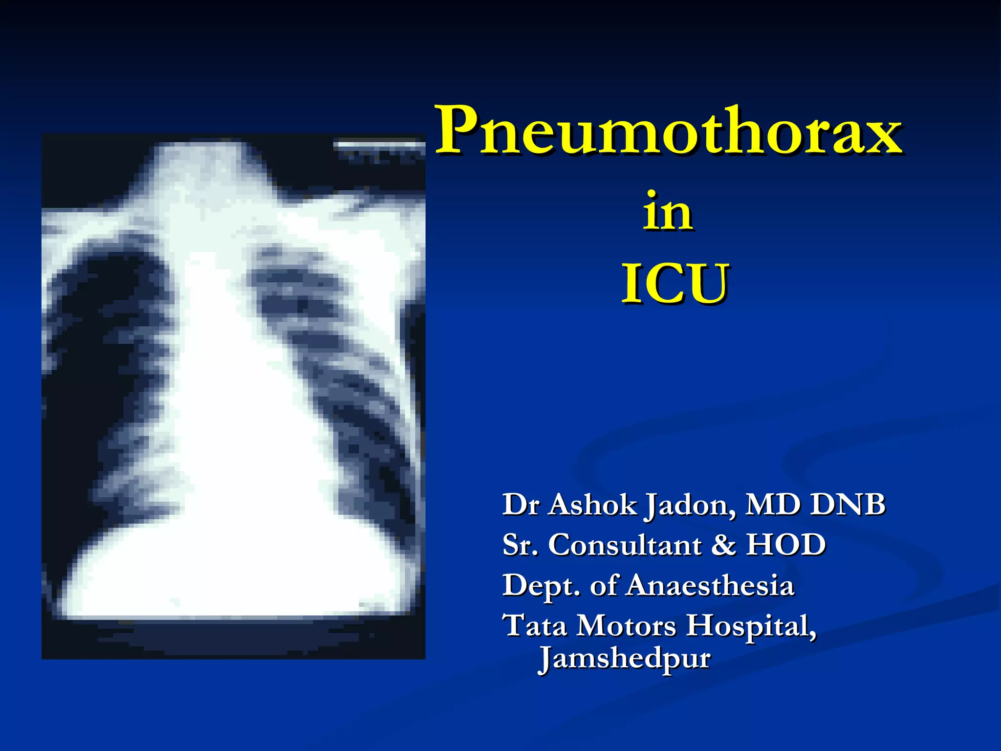
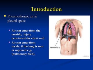
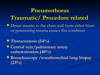
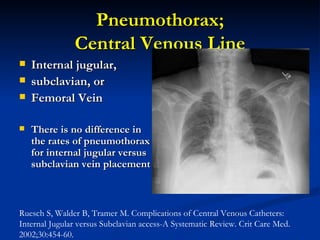
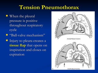
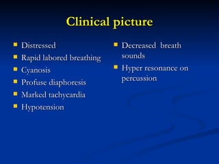
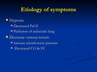
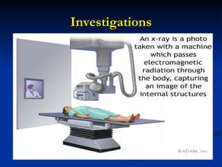
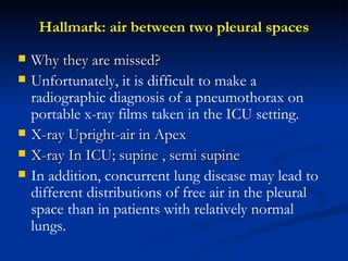
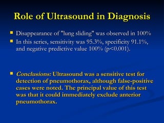
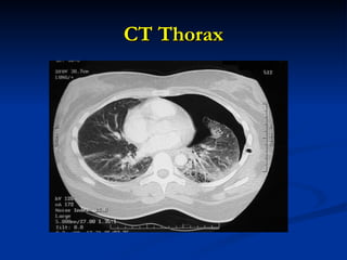
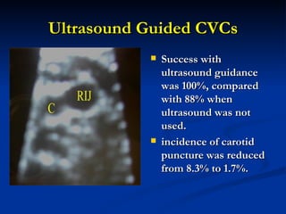
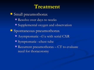
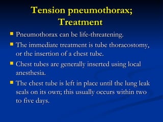
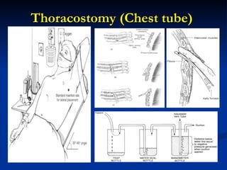
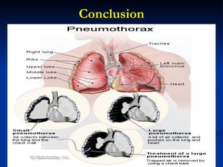
Pneumothorax   in  ICU Dr Ashok Jadon, MD DNB Sr. Consultant & HOD  Dept. of Anaesthesia Tata Motors Hospital, Jamshedpur
Introduction Pneumothorax; air in pleural space Air can enter from the outside;  injury penetrated the chest wall  Air can enter from inside, if the lung is torn or ruptured e.g. (pulmonary bleb).
Pneumothorax is the most common serious pleural complication in the ICU  Pneumothorax may be difficult to diagnose  when their locations are atypical,  when the patient has underlying cardiopulmonary disease  altered mental status
ICU pts : High Risk Group  Serious systemic disease  Hemodynamically unstable; Invasive Procedures Ventilator/ Resuscitations Postoperative Patients; shifted from another invasive environment  Trauma; admitted to ICU Penetrating Injury of Chest/ Abdomen # Rib Resuscitation Central Line
De Lassence et al  Anesthesiology. 2006 Jan;104(1):5-13.   Incidence 1.4% on day 5 and 3.0% on day 30.  Risk factors History of adult immunodeficiency syndrome Diagnosis of acute respiratory distress syndrome  Cardiogenic pulmonary edema at admission  Central vein or pulmonary artery catheter insertion  Use of inotropic agents during the first 24 h Pneumothorax in the intensive care unit: incidence & risk factors,
Close  Associations  for high incidence Disease; ARDS Ventilation; Incidence (4 to 15%). Procedures Thoracentesis,  Central venous catheter placement, Bronchoscopy Pericardiocentesis  Tracheostomy
Types/ Etiology Spontaneous Primary Secondary Iatrogenic / Traumatic Open/ Close Tension Pneumothorax
Spontaneous Pneumothorax  This refers to a condition in which the lung collapses with no apparent injury or trauma Pulmonary blebs COPD; Emphysematous Bullae AIDS/ Lung Tumor Infective or Infiltrative Lung Disease  Cigarette smokers & Recreational drug users are at greater risk for spontaneous pneumothorax.
How Mechanical Ventilation Responsible for Pneumothorax ?
Biotrauma Barotrauma and Volutrauma Atelectrauma
Barotrauma and Volutrauma Ventilator-induced lung injury by high levels of mechanical stress and strain that occur when  high airway pressures ( Barotrauma) and  high volumes  (Volutrauma) are delivered .  This stress and strain can disrupt the pulmonary fibroelastic skeleton and trigger a secondary inflammatory response.
Atelectrauma Moderate degrees of stress and strain related to the cyclic opening and closing of parts of the lung may directly induce the release of inflammatory mediators and noxious proteinases.
 
Relationship between ventilatory settings and barotrauma in the ARDS   incidence of barotrauma 0% to 49%,  High incidence correlated strongly  P(plat), above 35 cm H2O,  Compliance below 30 ml/cm H2O  Aspiration Pneumonia In a prospective study 38 percent of patients developed pneumothorax and pneumo-mediastinum.  Mohamed Boussarsar  Intensive Care Med. 2002 ;28 (4):406-13.
Pneumothorax Traumatic/ Procedure related  Direct trauma to the chest wall from either blunt or penetrating trauma causes this condition Thoracentesis (54%) Central vein/pulmonary artery catheterization (40%) Bronchoscopy /transbronchial lung biopsy (23%)
Pneumothorax; Central Venous Line Internal jugular,  subclavian, or  Femoral Vein There is no difference in the rates of pneumothorax for internal jugular versus subclavian vein placement   Ruesch S, Walder B, Tramer M. Complications of Central Venous Catheters: Internal Jugular versus Subclavian access-A Systematic Review. Crit Care Med. 2002;30:454-60.
Pneumothorax after insertion of central venous catheters in the intensive care unit: association with month of year and week of month Highest in July and August and in the first week of the month (beginning of intensive care unit (ICU) rotation).  The rate of PTX after insertion of CVCs is greatest in the last week of the month(2.7%) than during the first, second or third weeks (1.7%, 1.8% and 1.4%, respectively).  Najib T Ayas- Quality and Safety in Health Care  2007; 16 :252-255
Symptoms Sharp, stabbing chest pain that worsens on breathing or with deep inspiration. Pain often radiates to the shoulder and or back  A dry, hacking cough may occur because of irritation of the diaphragm.
Tension Pneumothorax When the pleural pressure is positive throughout respiratory cycle  “ Ball-valve mechanism” Injury to pleura creates a  tissue flap  that opens on inspiration and closes on expiration
Epidemiology Kolef reviewed 464 ICU patients 28 (6%) developed pneumothorax 9 patients missed the initial diagnosis  3 (33%) developed tension pneumothorax In diagnosed 19 patients only 1 (5%) develop pneumothorax  Tocino & coworkers  Missed pneumothorax 34/112 (30%) 16/34 patients developed tension pneumothorax
Clinical picture Distressed Rapid labored breathing Cyanosis Profuse diaphoresis Marked tachycardia Hypotension Decreased  breath sounds Hyper resonance on percussion
Etiology of symptoms Hypoxia Decreased PaO2 Perfusion of atelectatic lung Decrease venous return  increase intrathoracic pressure Decreased CO & SV
Investigations A A A A A
Hallmark: air between two pleural spaces Why they are missed?  Unfortunately, it is difficult to make a radiographic diagnosis of a pneumothorax on portable x-ray films taken in the ICU setting. X-ray Upright-air in Apex X-ray In ICU; supine , semi supine  In addition, concurrent lung disease may lead to different distributions of free air in the pleural space than in patients with relatively normal lungs.
Distribution of air
Always look for…. Subtle radiographic signs of pneumothorax Relative hyperlucency over the upper abdominal quadrants Deep costophrenic angle (the deep sulcus sign)
Role of Ultrasound in Diagnosis Disappearance of &quot;lung sliding&quot; was observed in 100%  In this series, sensitivity was 95.3%, specificity 91.1%, and negative predictive value 100% (p<0.001). Conclusions:  Ultrasound was a sensitive test for detection of pneumothorax, although false-positive cases were noted. The principal value of this test was that it could immediately exclude anterior pneumothorax.
CT Thorax
Pneumothorax Prevention during CVCs Remove patient from ventilator before advancing the needle.  Choose the right side rather than left,  Avoid multiple attempts when possible Check post procedure x-ray,
Ultrasound Guided CVCs Success with ultrasound guidance was 100%, compared with 88% when ultrasound was not used.  incidence of carotid puncture was reduced from 8.3% to 1.7%.
Treatment Small pneumothorax Resolve over days to weeks Supplemental oxygen and observation Spontaneous pneumothorax Asymptomatic –f/u with serial CXR Symptomatic –chest tube Recurrent pneumothorax – CT to evaluate need for thoracotomy
Tension pneumothorax; Treatment  Pneumothorax can be life-threatening.  The immediate treatment is tube thoracostomy, or the insertion of a chest tube.  Chest tubes are generally inserted using local anesthesia.  The chest tube is left in place until the lung leak seals on its own; this usually occurs within two to five days.
Thoracostomy (Chest tube)
Pulmonary blebs Pulmonary blebs can be resected, preventing future pneumothorax.  Thoracoscopic surgical procedure.  A stapling device is inserted into the chest during, and the segment of lung with blebs is stapled across and then removed
complications of air of pleural parenchyma Sub-pleural air cysts ;  Secondary infections in the cysts, Pseudomonas sepsis & death.  Systemic Gas Embolism It is possible for extra-alveolar air to enter the systemic circulation if there is a bronchovenous communication and an adequate pressure gradient.
Prognosis Paucity of clinical data describing the prognostic factors associated with patient outcomes
Prognosis Seven patients (12%) had to undergo external suction and pleurodesis for persistent air leaks.  Five patients still had air leaks, and the chest tube was not removed during their ICU stay.  The mean duration of ICU stay was 24 ± 19 days (median, 17.5 days).  The mean duration of chest tube drainage was 10 ± 11 days (median, 6 days).  The mortality rate for patients with pneumothorax was 68%.
Prognosis contd.. Patients with procedure-related pneumothorax had a lower risk of mortality.  Patients who had tension pneumothorax and concurrent septic shock had a higher risk of mortality.  pneumothorax due to barotrauma, tension pneumothorax, and concurrent septic shock were significantly and independently associated with death.
Conclusion
 
Twist in Tale..
CXR with recurrent right-sided pneumothorax, despite thoracostomy tube in place CT scan with giant bullae and anterior pneumothorax after insertion of a second thoracostomy tube
Message Put Chest Drain; Be Happy But, Be Observant and Do Follow -Up  Not only Till Patient Go Home  Later on too.
Thank You

pneumothorax in ICU

  • 1.
    Pneumothorax in ICU Dr Ashok Jadon, MD DNB Sr. Consultant & HOD Dept. of Anaesthesia Tata Motors Hospital, Jamshedpur
  • 2.
    Introduction Pneumothorax; airin pleural space Air can enter from the outside; injury penetrated the chest wall Air can enter from inside, if the lung is torn or ruptured e.g. (pulmonary bleb).
  • 3.
    Pneumothorax is themost common serious pleural complication in the ICU Pneumothorax may be difficult to diagnose when their locations are atypical, when the patient has underlying cardiopulmonary disease altered mental status
  • 4.
    ICU pts :High Risk Group Serious systemic disease Hemodynamically unstable; Invasive Procedures Ventilator/ Resuscitations Postoperative Patients; shifted from another invasive environment Trauma; admitted to ICU Penetrating Injury of Chest/ Abdomen # Rib Resuscitation Central Line
  • 5.
    De Lassence etal Anesthesiology. 2006 Jan;104(1):5-13. Incidence 1.4% on day 5 and 3.0% on day 30. Risk factors History of adult immunodeficiency syndrome Diagnosis of acute respiratory distress syndrome Cardiogenic pulmonary edema at admission Central vein or pulmonary artery catheter insertion Use of inotropic agents during the first 24 h Pneumothorax in the intensive care unit: incidence & risk factors,
  • 6.
    Close Associations for high incidence Disease; ARDS Ventilation; Incidence (4 to 15%). Procedures Thoracentesis, Central venous catheter placement, Bronchoscopy Pericardiocentesis Tracheostomy
  • 7.
    Types/ Etiology SpontaneousPrimary Secondary Iatrogenic / Traumatic Open/ Close Tension Pneumothorax
  • 8.
    Spontaneous Pneumothorax This refers to a condition in which the lung collapses with no apparent injury or trauma Pulmonary blebs COPD; Emphysematous Bullae AIDS/ Lung Tumor Infective or Infiltrative Lung Disease Cigarette smokers & Recreational drug users are at greater risk for spontaneous pneumothorax.
  • 9.
    How Mechanical VentilationResponsible for Pneumothorax ?
  • 10.
    Biotrauma Barotrauma andVolutrauma Atelectrauma
  • 11.
    Barotrauma and VolutraumaVentilator-induced lung injury by high levels of mechanical stress and strain that occur when high airway pressures ( Barotrauma) and high volumes (Volutrauma) are delivered . This stress and strain can disrupt the pulmonary fibroelastic skeleton and trigger a secondary inflammatory response.
  • 12.
    Atelectrauma Moderate degreesof stress and strain related to the cyclic opening and closing of parts of the lung may directly induce the release of inflammatory mediators and noxious proteinases.
  • 13.
  • 14.
    Relationship between ventilatorysettings and barotrauma in the ARDS incidence of barotrauma 0% to 49%, High incidence correlated strongly P(plat), above 35 cm H2O, Compliance below 30 ml/cm H2O Aspiration Pneumonia In a prospective study 38 percent of patients developed pneumothorax and pneumo-mediastinum. Mohamed Boussarsar Intensive Care Med. 2002 ;28 (4):406-13.
  • 15.
    Pneumothorax Traumatic/ Procedurerelated Direct trauma to the chest wall from either blunt or penetrating trauma causes this condition Thoracentesis (54%) Central vein/pulmonary artery catheterization (40%) Bronchoscopy /transbronchial lung biopsy (23%)
  • 16.
    Pneumothorax; Central VenousLine Internal jugular, subclavian, or Femoral Vein There is no difference in the rates of pneumothorax for internal jugular versus subclavian vein placement Ruesch S, Walder B, Tramer M. Complications of Central Venous Catheters: Internal Jugular versus Subclavian access-A Systematic Review. Crit Care Med. 2002;30:454-60.
  • 17.
    Pneumothorax after insertionof central venous catheters in the intensive care unit: association with month of year and week of month Highest in July and August and in the first week of the month (beginning of intensive care unit (ICU) rotation). The rate of PTX after insertion of CVCs is greatest in the last week of the month(2.7%) than during the first, second or third weeks (1.7%, 1.8% and 1.4%, respectively). Najib T Ayas- Quality and Safety in Health Care 2007; 16 :252-255
  • 18.
    Symptoms Sharp, stabbingchest pain that worsens on breathing or with deep inspiration. Pain often radiates to the shoulder and or back A dry, hacking cough may occur because of irritation of the diaphragm.
  • 19.
    Tension Pneumothorax Whenthe pleural pressure is positive throughout respiratory cycle “ Ball-valve mechanism” Injury to pleura creates a tissue flap that opens on inspiration and closes on expiration
  • 20.
    Epidemiology Kolef reviewed464 ICU patients 28 (6%) developed pneumothorax 9 patients missed the initial diagnosis 3 (33%) developed tension pneumothorax In diagnosed 19 patients only 1 (5%) develop pneumothorax Tocino & coworkers Missed pneumothorax 34/112 (30%) 16/34 patients developed tension pneumothorax
  • 21.
    Clinical picture DistressedRapid labored breathing Cyanosis Profuse diaphoresis Marked tachycardia Hypotension Decreased breath sounds Hyper resonance on percussion
  • 22.
    Etiology of symptomsHypoxia Decreased PaO2 Perfusion of atelectatic lung Decrease venous return increase intrathoracic pressure Decreased CO & SV
  • 23.
  • 24.
    Hallmark: air betweentwo pleural spaces Why they are missed? Unfortunately, it is difficult to make a radiographic diagnosis of a pneumothorax on portable x-ray films taken in the ICU setting. X-ray Upright-air in Apex X-ray In ICU; supine , semi supine In addition, concurrent lung disease may lead to different distributions of free air in the pleural space than in patients with relatively normal lungs.
  • 25.
  • 26.
    Always look for….Subtle radiographic signs of pneumothorax Relative hyperlucency over the upper abdominal quadrants Deep costophrenic angle (the deep sulcus sign)
  • 27.
    Role of Ultrasoundin Diagnosis Disappearance of &quot;lung sliding&quot; was observed in 100% In this series, sensitivity was 95.3%, specificity 91.1%, and negative predictive value 100% (p<0.001). Conclusions: Ultrasound was a sensitive test for detection of pneumothorax, although false-positive cases were noted. The principal value of this test was that it could immediately exclude anterior pneumothorax.
  • 28.
  • 29.
    Pneumothorax Prevention duringCVCs Remove patient from ventilator before advancing the needle. Choose the right side rather than left, Avoid multiple attempts when possible Check post procedure x-ray,
  • 30.
    Ultrasound Guided CVCsSuccess with ultrasound guidance was 100%, compared with 88% when ultrasound was not used. incidence of carotid puncture was reduced from 8.3% to 1.7%.
  • 31.
    Treatment Small pneumothoraxResolve over days to weeks Supplemental oxygen and observation Spontaneous pneumothorax Asymptomatic –f/u with serial CXR Symptomatic –chest tube Recurrent pneumothorax – CT to evaluate need for thoracotomy
  • 32.
    Tension pneumothorax; Treatment Pneumothorax can be life-threatening. The immediate treatment is tube thoracostomy, or the insertion of a chest tube. Chest tubes are generally inserted using local anesthesia. The chest tube is left in place until the lung leak seals on its own; this usually occurs within two to five days.
  • 33.
  • 34.
    Pulmonary blebs Pulmonaryblebs can be resected, preventing future pneumothorax. Thoracoscopic surgical procedure. A stapling device is inserted into the chest during, and the segment of lung with blebs is stapled across and then removed
  • 35.
    complications of airof pleural parenchyma Sub-pleural air cysts ; Secondary infections in the cysts, Pseudomonas sepsis & death. Systemic Gas Embolism It is possible for extra-alveolar air to enter the systemic circulation if there is a bronchovenous communication and an adequate pressure gradient.
  • 36.
    Prognosis Paucity ofclinical data describing the prognostic factors associated with patient outcomes
  • 37.
    Prognosis Seven patients(12%) had to undergo external suction and pleurodesis for persistent air leaks. Five patients still had air leaks, and the chest tube was not removed during their ICU stay. The mean duration of ICU stay was 24 ± 19 days (median, 17.5 days). The mean duration of chest tube drainage was 10 ± 11 days (median, 6 days). The mortality rate for patients with pneumothorax was 68%.
  • 38.
    Prognosis contd.. Patientswith procedure-related pneumothorax had a lower risk of mortality. Patients who had tension pneumothorax and concurrent septic shock had a higher risk of mortality. pneumothorax due to barotrauma, tension pneumothorax, and concurrent septic shock were significantly and independently associated with death.
  • 39.
  • 40.
  • 41.
  • 42.
    CXR with recurrentright-sided pneumothorax, despite thoracostomy tube in place CT scan with giant bullae and anterior pneumothorax after insertion of a second thoracostomy tube
  • 43.
    Message Put ChestDrain; Be Happy But, Be Observant and Do Follow -Up Not only Till Patient Go Home Later on too.
  • 44.