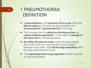
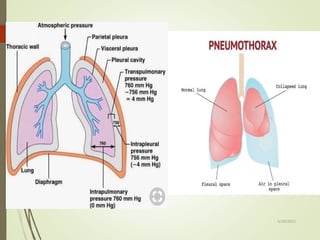
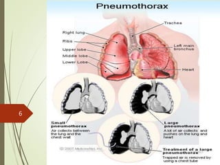
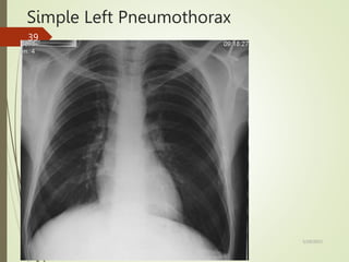
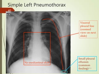
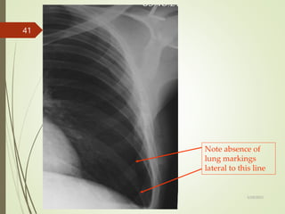
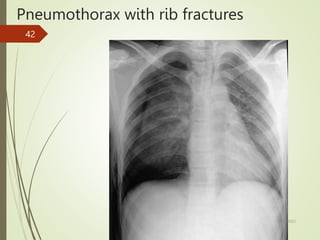
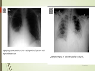
SURGERY I : CHEST CONDITION.
GROUP 4-DCM MARCH 2023
ASSIGNMENT
1 ABUBAKAR
2 FAHAD ISMAIL
3 MISHI SOZA
4 YUSSUF BILE
5/29/2023
1
CONTENT TO BE COVERED
1 PNEUMOTHORAX
2 HAEMOTHORAX
3 EMPYEMA THORACIC
4 SURGICAL EMPHYSEMA
5 CARDIAC TAMPONADE
5/29/2023
2
objectives
 Definition
 Epidemiology/Incidence
 Pathophysiology
 Classification
 Risk factors/Causes
 Clinical presentation
 Investigation
 Management
 Complication
5/29/2023
3
1 PNEUMOTHORAX
DEFINITION
 A pneumothorax is the presence of air or gas within the
pleural cavity i.e. the potential spaces between the
visceral pleural & parietal pleural of the lungs.
 This is usually from the defect on the lung surface e.g.
rapture bullae(Large blister ) or through the damage of
the chest wall e.g. following trauma.
 Air within the pleural cavity causes the physiological
pleural seal to be lost ,meaning the normal negative
pressure in this space , that aid the lung expanding within
the chest wall movement is lost.
 This impedes(prevent) lung expansion & leads to partial
or total lung collapse.
5/29/2023
4
Cont..
5/29/2023
5
5/29/2023
6
EPIDEMIOLOGY/INCIDENCE
 Annual incidence of pneumothorax is around 9% per
100,000
 Primary pneumothoraces occur most commonly in tall thin
men aged between 20-40
 They are less common in women- consider the possibility of
underlying lung disease e.g. LAM, Catamental
pneumothorax
 Cigarette or cannabis smoking is a major risk factor for
pneumothorax increasing the risk by factor of 22 in men &
9 in women
5/29/2023
7
Cont..
 The mechanism is unclear ; a smoking induced influx of
inflammatory cells may both break down elastic lung fibers
(causing bulla formation ) & cause small airway obstruction
(increasing alveolar pressure & the likelihood of interstitial
air leak)
 More common in patient with Marfans syndrome &
homocystinuria
 May rarely be farmilial
5/29/2023
8
PATHOPHYSIOLOGY
 As air enters the pleural space which normally have a
negative pressure , the elastic recoil in the lung tissue
causing either a partial or full lung collapse.
NORMAL PHYSIOLOGY
 Pleural space has a negative pressure
 Chest wall expand ►surface tension between parietal &
visceral pleural expands the lungs.
 Lung tissue has an elastic recoil► innate tendency to
collapse inward.
5/29/2023
9
Traumatic pneumothorax
 Closed pneumothorax : blunt trauma →lung damage →air
flow from the lung into the pleural spaces.
 Open pneumothorax : penetrating trauma to the chest
wall→ pathway for air directly into pleural spaces.
 Close & open pneumothorax : In closed pneumothorax air
travel in & out of the pleural spaces from the lungs .
 However in an open pneumothorax a defect in the chest
wall allows air to move in & out of the pleural spaces.
5/29/2023
10
Cont..
5/29/2023
11
Iatrogenic pneumothorax
 Induced in a patient by the treatment or comment of a
physician
 Lung surgery
 Central venous catheter insertion
 Thoracentesis -removal of fluid around the lung
 Mechanical ventilation
 Esophageal procedure
5/29/2023
12
Spontaneous pneumothorax
 Ruptured bleb→ air flow from the lungs into the pleural
spaces→ positive pleural pressure→ compressed lung.
 Lung collapse until an equilibrium is achieved or the rupture
seals
 Vital capacity & ↓ partial pressure of oxygen
 Primary/idiopathic
Rapture apical subpleural bleb or bullae
 Secondary
 Chronic obstruction-COPD account for 50%
5/29/2023
13
Cont..
 Bronchiectasis
 Lung malignancies
 Lung infection- pneumocystic jirovecii, TB, bacterial
pneumonia,
 Genetic disease-cystic fibrosis, marfans syndrome ,
homocystinuria
 Cystic lung disorders – lymphangioleimyomatosis, diffuse
Langerhans cell histiocytosis, lymphocytic interstitial
pneumonia, thoracic endometriosis (catemanial
pneumothorax)
 Smoking is a mojor risk for primary & secondary
5/29/2023
14
Tension pneumothorax
 Life threatening & can develop from any type of a
pneumothorax.
 Air enters the pleural space through a one way mechanism
→air cannot escape.
 Air accumulate in the pleural space with each inspiratory
phase→ ↑ pleural space pressure → shifting of
mediastinum→ compression of the contralateral lung →
hypoxia.
 Eventually compress of the vena cava & atria →↓ venous
return to the heart & ↓ cardiac function .
 Leads to rapid cardiopulmonary collapse.
5/29/2023
15
Cont..
5/29/2023
16
Cont..
 Tension pneumothorax:
 Spontaneous & traumatic pneumothorax can develop into a
tension pneumothorax if the defect that allow air into the
pleural space becomes one way valve (air enters during
inspiration but cannot escape during exhalation which
causes rising pressure in the pleural cavity , shifting the
mediastinum to the contralateral side
5/29/2023
17
Cont..
 Simple
 Mediastinum remains central
 Clinical condition stable
 Can wait for CXR to confirm diagnosis
 Tension
 Progressive build up of air in the pleural space, causing
a shift of the heart and mediastinal structures away
from side of pneumothorax
 Clinical condition unstable
 Do not wait for CXR to confirm diagnosis
5/29/2023
18
CLASSIFICATION
5/29/2023
19
Cont.…
5/29/2023
20
RISK FACTORS/CAUSES
 Sex-men are at high risk
 Smoking
 Age
 Genetics
 Lung disease
 Mechanical ventilation
 History of pneumothorax
5/29/2023
21
CLINICAL PRESENTATION
 Shortness of breath of varying degree depending on the
size of the pneumothorax & patients factors e.g. lung
disease.
 Sudden onset chest pain, often pleuritic in nature , small
spontaneous pneumothorax can be asymptomatic
particularly in younger patients.
 O/E there will be:
Hyperresonance on percussion
Reduced or absent breath sound on auscultation
Reduced chest expansion
Decrease in tactile fremitus
5/29/2023
22
Cont..
 In cases of tension pneumothorax:
Patients will be hypoxic
Tachycardiac
Hypotensive
Potential distended neck vein
Tracheal deviation away from the affected side
Cyanosis
Tachypnea-abnormal rapid breathing
 Cardiovascular-jugular venous distension
-Pulsus paradoxus-↓ stroke volume
5/29/2023
23
Cont..
MNEMONIC
 Signs & symptoms of tension pneumothorax is summarized
with P-THORAX
 P-Pleuritis pain
 T-Tracheal deviation
 H-Hyper- resonance
 O-Onset sudden chest pain
 R-Reduced breath sound
 A-Absent fremitus
 X-X-ray showing collapes
5/29/2023
24
INVESTIGATION
 Initial investigation should run alongside this(expect for
cases of tension pneumothorax when urgent needle
decompression is required in 2 or 3rd ICS
 Tension pneumothorax is a clinical diagnosis &
management should not wait for imaging confirmation
1 Plain chest radiograph(CXR)
 The size of pneumothorax is determined by measuring
interpleural distance at the level of hilum.
 Should be performed in upright position (when possible)
5/29/2023
25
Cont..
General findings
 White visceral pleural lining defining lung & pleural air
 Bronchovascular markings are not visible beyond pleural edge
 Deep sulcus
 Ipsilateral hemidiaphragm elevation
Tension pneumothorax
 Potential mediastinum shift
 Trachea deviation
 Ipsilateral hemidiaphragm flattening
 Ribs are spread a part
5/29/2023
26
Cont..
2 Routine blood –FBC
-CPR
-U & Es & clotting
 Arterial blood gas (ABG)
 Electrocardiogram (ECG)
3 CT imaging
 determine underlying cause in context of trauma &
concurrent injuries
 Findings –air in the space , can evaluate the location ,
pleural pathology & lung disease
5/29/2023
27
Cont..
5/29/2023
28
Cont..
4 Ultrasound
 Presence of a lung point –boundary between the lung &
pneumothorax
 Lung sliding will be absent at the location of pneumothorax
5/29/2023
29
MANAGEMENT
 Management is determined by both size or type of the
pneumothorax & patient factor.
 As a minimal ensure all patient have sufficient analgesia &
started on oxygen if required.
 For patient with chest drain inserted ensure it is attached
to underwater seal.
INITIAL MANAGEMENT
 Primary spontaneous pneumothorax those that are small
(<2) & asymptomatic patients should be admitted for
observation.
5/29/2023
30
Cont..
 Symptomatic or large primary pneumothoraces needle
decompression should be attempted placed in 2nd or
3rd intercostal spaces at the midclavicular line if no
improvement chest drain via seldinger technique to be
placed
 5th ICS space in the anterior or midaxillary line in SAFTEY
TRIAGE is another option-followed by chest tube
placement.
 Small spontaneous pneumothorax will required
admission for observation with a low threshold for
attempting needle decompression ,
 Those that are large & symptomatic required chest drain
via seldinger technique to be placed.
5/29/2023
31
Cont..
 Traumatic pneumothoraces will normally require surgical
chest drain insertion or otherwise admitting for observation
if small & asymptomatic.
 Importantly there is no role in needle decompression in
traumatic non-tensioning pneumothoraces.
 For traumatic tension pneumothoraces either needle
decompression (in 5th intercostal space mid-axillary line)
or finger thoracostomy is required prior to chest drain
insertion.
5/29/2023
32
Cont..
FURTHER INVESTIGATION
 Considered in those with persistence air leak or failure of lung
re-expansion.
 Spontaneous cases medical pleurodesis is often trailed
resulting in partial obliteration of the pleural space through
introducing irritant agent aiming to prevent recurrences
,alternatively Heimlich valve can be trailed a one way valve
attached to a chest tube & enable evacuation of air that is not
under tension.
 Those failing these intervention or in traumatic cases should ne
considered surgical intervention which includes video
assisted thoracoscopic surgery (VATS) for pleurectomy +/-
pleural abrasion or open thoracostomy& pleurectomy .
5/29/2023
33
COMPLICATIONS
 Hypoxemic respiratory failure-low level of oxygen
 Respiratory or cardiac arrest-heart suddenly stop pumping blood
 Hemopneumothorax-combination of pneumothorax &
hemothorax.
 Bronchopulmonary fistula-abnormal communication btwn
bronchial tree & pleural cavity
 Pulmonary edema –following lung re-expansion
 Empyema-collection of pus in the pleural cavity
 Pneumomediastinum-presence of air in mediastinum
 Pneumopericardium-presence of air in pericardium
 Pneumoperitoneum-presence of air in peritoneal cavity
 Pyopneumothorax-accumulation of gas & pus in pleural cavity
5/29/2023
34
Cont..
COMPLICATIONS OF SURGICAL PROCEDURE
 Failure to cure the problem
 Acute respiratory distress or failure
 Infection of the pleural spaces
 Cutaneous or systemic infection
 Persistent air leak
 Re-expansion pulmonary
 Pain at the site of chest tube insertion
 Prolonged tube drainage & hospital stay
5/29/2023
35
DIFFERENTIAL DIAGNOSIS
 Pulmonary embolism(PE)
 Hemothorax
 Pleural effusion
 Myocardial infarction
 Pericarditis
5/29/2023
36
R
•Right lung more translucent than left
•Faint line just visible (zoomed view to follow)
5/29/2023
37
•Pencil-thin white line
running parallel to chest
wall
•No lung markings lateral
to the line
Blade of right scapula
Right pneumothorax
5/29/2023
38
Simple Left Pneumothorax
5/29/2023
39
Simple Left Pneumothorax
No mediastinal shift
Small pleural
effusion
(common
finding)
Visceral
pleural line
(zoomed
view on next
slide)
5/29/2023
40
Note absence of
lung markings
lateral to this line
5/29/2023
41
Pneumothorax with rib fractures
5/29/2023
42
Tension right pneumothorax
5/29/2023
43
Tension right pneumothorax
Mediastinal shift to
left 5/29/2023
44
HEMOTHORAX
 Hemothorax is the accumulation of blood in the intrapleural
spaces.
 Bleeding is usually from intercostal artery in lacerated chest
wall or from underlying contused lung, heart or great vessel.
 Massive hemothorax is bleeding of more than 1500ml into
pleural cavity
5/29/2023
45
EPIDEMIOLOGY
 Hemothorax can be associated with a single rib fracture.
 Approximately 150,000 deaths occurs from trauma each
year.
 Approximately 3times this number of individuals are
permanently disabled because of trauma.
 Chest injuries occurs in approximately 60% of multiple
trauma cases.
5/29/2023
46
PATHOPHYSIOLOGY
 Accumulation of blood in the pleural space caused by
bleeding from; penetration or blunt lung injury, chest wall
vessels or intercostal vessels.
 Hemothorax is manifested by;
 >hemodynamic response-hypovolemic shock rapid
bleeding.
 >respiratory response-slow bleeding.
 Blood that enters the pleural cavity is exposed to the
motion of the diaphragm, the lungs, and other intrathoracic
structures.
5/29/2023
47
CLASSIFICATIONS
TRAUMATIC HEMOTHORAX
 Occurs due to penetration injury of the lungs, heart, great
vessels, or chest wall
non Traumatic hemothorax
 Malignancy pleural diseases(sarcoma, angiosarcoma)
 Bleeding disorders(hemophilia, thrombocytopenia, rupture of
thoracic aorta)
 Necrotizing infection
 Pulmonary embolism with infarction
Iatrogenic hemothorax
 Causes;
Central venous catheterization
5/29/2023
48
Cont..
 Injury during trans lumber aortography
 Thoracocentesis
 Pleural biopsy
 Trans brachial biopsy
5/29/2023
49
CAUSES OF HEMOTHORAX
 Traumatic
 Infection/infestation
 Congenital
 Degenerative
 Neoplastic(benign/malignant)
 Rib fractures associated with pneumothorax
 Iatrogenic
 Tuberculosis
 Non pulmonary intrathoracic vascular pathology(e.g.
Pancreatic pseudocyst, splenic artery aneurysm)
5/29/2023
50
Clinical presentation
 Restlessness
 Hypovolemic shock
 Tachypnea
 Dyspnea
 Cyanosis
 Diminished breath sounds on affected side
 Hypo resonance(dullness on percussion) on affected side.
 Chest wall deformity
 Crepitus upon palpation over fractured ribs
5/29/2023
51
investigations
 Imaging studies
X-ray
USG(ultrasound sonography test)
CT chest
Angiography
MRI(Magnetic resonance test)
Thoracoscopy
Nuclear scan
5/29/2023
52
management
 ABC of resuscitation
 Large bow cannular &begin IV fluids-crystolliods
 Vital check up including SPO2
 Intercostal drainage tube thoracostomy
 Large bore tube in 5th spacing between mid and posterior axillary
lines
 Can be done before x-ray
 Draining of blood from chest cavity
 Thoracostomy(indicated when total chest tube output exceeds 1500ml
within 24hrs)
 Video assisted thoracoscopic surgery(VATS)
 Shock care due to blood loss
5/29/2023
53
5/29/2023
54
5/29/2023
55
complication
 Clot retention
 Pleural infection
 Pleural effusion
 Fibrothorax
 Collapsed lung
 Respiratory failure
 Hemorrhagic shock
5/29/2023
56
EMPHYEMA
DEFINITION
 Accumulation of pus in pleural cavity.
 Emphyema come from Greek word Empyein which means
pus-producing suppurates
 Also called Pyothorax
 Its also a secondary disease to other underlyind diseases
5/29/2023
57
EPIDEMIOLOGY
Etiology
Local cause
Chest causes; thoracic wall abscess
Penetrating wound
Oesteomyelitis of ribs
Pleural cause Pneumothorax
Hemothorax
Pulmonary cause Lung abscess
Bronchitis and Pneumonia
5/29/2023
58
Cont..
Latrojenic causes Pasthoractomy
Esophageal perforation
Systemic cause Septicaemia
Bacteriology ; Strep. Pneumoniae,H.influenza and Staph. Aureus
STAGES IN EMPYEMA FORMATION
Stage 1 Exudative stage
Stage 2 Fibrino purulent stage
Stage 3 Organizing stage
5/29/2023
59
STAGE 1 EXUDATIVE PHASE
This is purely on inflammatory process in which there is
increase in permeability of small blood vessesls leading to
exudation of fluid in the pleural cavity.
The fluid is very thin with low cellular content.
Approximately in 7 days
STAGE 2 FIBRINO PURULENT STAGE
This is whereby there is fibrin clot and fibrin membranes in the
pleural cavity leading to fluid loculation
From day 7 to 21 days
5/29/2023
60
CONTI…….
STAGE 3 ORGANIZING STAGE
Proliferation of fibroblasts on the pleural surface,which forms
covering preventing adequate lung expansion.
There is also scarring of pleural membranes with possible
inability of the lung to expand
Takes about 4 to 6 weeks
5/29/2023
61
TREATMENT &
 Treatment of the underlying cause of precipitating factor
 Mild cases –observation is appropriate
 Patient with discomfort –give high oxygen concertation
 Use of empiric broad spectrum antibiotic
 Extensive phase -2cm intravascular incision bilaterally can
reduce further subcutaneous expansion
 In severe cases ICT on one side or both sides placement
5/29/2023
62
DIFFERENTIAL DIAGNOSIS
 Esophageal rapture
 Pneumothorax
 Tracheal /lower/diaphragm perforation
5/29/2023
63
5/29/2023
64
5/29/2023
65
5/29/2023
66
AETIOLOGY
 Trauma to the chest
 TB
 Uremia
 Idiopathic pericarditis
 Infectious disease
 Anticoagulation
 hyperthyroidism
 Connective tissue disease
 Post pericardiotomy syndrome
 Malignant disease
5/29/2023
67
CLINICAL FEATURES /SIGNS
 Hypotension/shock(rapid weak pulse)
 Grossly distended neck vein (raised JVP)
 Elevated central venous pressure
 Severe distress
 faint heart sound
 Penetrating injury in the proximity to the heart
 The classic findings /a hallmark signs of beck triad
1Hypotension
 2 Distended neck vein
 3 Faint heart sound
5/29/2023
68
BECKS TRAID
 Collection of three clinical signs associated with pericardial
tamponade which is due to excessive accumulation of fluid
within the pericardial sac
5/29/2023
69
SYMPTOMS
1 Sharp pain in the chest –pain may radiate to the nearby parts
of the body like abdomen ,arm ,neck & shoulder
2 trouble breathing /breathing rapidly
3 fainting ,dizziness/light headache
4 changes in skin color
5 heart palpitation
6 fast pulse
7 Altered mental status /confusion
8 decreased urine output
5/29/2023
70
DIAGNOSI/INVESTIGATION
 History taking
 Physical examination
 Echocardiogram
 Chest x-ray
 Computerized tomography
 Heart catheterization
 Ultrasound
5/29/2023
71
MANAGEMENT
 Removal of the fluid around the heart
 Its done through pericardiocentesis –the procedure use a
needle that is inserted into the chest until in enters the
pericardial sac & the fluid is aspirated
 Surgery
5/29/2023
72
COMPLICATION
 Shock
 Heart failure
 Death
PERICARDIOCENTESIS & SURGERY COMPLICATION
 Bleeding
 Injury to the heart chambers
 Heart attack
 Infections
 Injury to nearby organs
5/29/2023
73
DIFFERENTIAL DIAGNOSIS
 Cardiogenic shock
 Constrictive pericarditis
 Pneumothorax
 Pulmonary embolism
5/29/2023
74

CHEST INJURY GROUP FOUR.pptx

  • 1.
    SURGERY I :CHEST CONDITION. GROUP 4-DCM MARCH 2023 ASSIGNMENT 1 ABUBAKAR 2 FAHAD ISMAIL 3 MISHI SOZA 4 YUSSUF BILE 5/29/2023 1
  • 2.
    CONTENT TO BECOVERED 1 PNEUMOTHORAX 2 HAEMOTHORAX 3 EMPYEMA THORACIC 4 SURGICAL EMPHYSEMA 5 CARDIAC TAMPONADE 5/29/2023 2
  • 3.
    objectives  Definition  Epidemiology/Incidence Pathophysiology  Classification  Risk factors/Causes  Clinical presentation  Investigation  Management  Complication 5/29/2023 3
  • 4.
    1 PNEUMOTHORAX DEFINITION  Apneumothorax is the presence of air or gas within the pleural cavity i.e. the potential spaces between the visceral pleural & parietal pleural of the lungs.  This is usually from the defect on the lung surface e.g. rapture bullae(Large blister ) or through the damage of the chest wall e.g. following trauma.  Air within the pleural cavity causes the physiological pleural seal to be lost ,meaning the normal negative pressure in this space , that aid the lung expanding within the chest wall movement is lost.  This impedes(prevent) lung expansion & leads to partial or total lung collapse. 5/29/2023 4
  • 5.
  • 6.
  • 7.
    EPIDEMIOLOGY/INCIDENCE  Annual incidenceof pneumothorax is around 9% per 100,000  Primary pneumothoraces occur most commonly in tall thin men aged between 20-40  They are less common in women- consider the possibility of underlying lung disease e.g. LAM, Catamental pneumothorax  Cigarette or cannabis smoking is a major risk factor for pneumothorax increasing the risk by factor of 22 in men & 9 in women 5/29/2023 7
  • 8.
    Cont..  The mechanismis unclear ; a smoking induced influx of inflammatory cells may both break down elastic lung fibers (causing bulla formation ) & cause small airway obstruction (increasing alveolar pressure & the likelihood of interstitial air leak)  More common in patient with Marfans syndrome & homocystinuria  May rarely be farmilial 5/29/2023 8
  • 9.
    PATHOPHYSIOLOGY  As airenters the pleural space which normally have a negative pressure , the elastic recoil in the lung tissue causing either a partial or full lung collapse. NORMAL PHYSIOLOGY  Pleural space has a negative pressure  Chest wall expand ►surface tension between parietal & visceral pleural expands the lungs.  Lung tissue has an elastic recoil► innate tendency to collapse inward. 5/29/2023 9
  • 10.
    Traumatic pneumothorax  Closedpneumothorax : blunt trauma →lung damage →air flow from the lung into the pleural spaces.  Open pneumothorax : penetrating trauma to the chest wall→ pathway for air directly into pleural spaces.  Close & open pneumothorax : In closed pneumothorax air travel in & out of the pleural spaces from the lungs .  However in an open pneumothorax a defect in the chest wall allows air to move in & out of the pleural spaces. 5/29/2023 10
  • 11.
  • 12.
    Iatrogenic pneumothorax  Inducedin a patient by the treatment or comment of a physician  Lung surgery  Central venous catheter insertion  Thoracentesis -removal of fluid around the lung  Mechanical ventilation  Esophageal procedure 5/29/2023 12
  • 13.
    Spontaneous pneumothorax  Rupturedbleb→ air flow from the lungs into the pleural spaces→ positive pleural pressure→ compressed lung.  Lung collapse until an equilibrium is achieved or the rupture seals  Vital capacity & ↓ partial pressure of oxygen  Primary/idiopathic Rapture apical subpleural bleb or bullae  Secondary  Chronic obstruction-COPD account for 50% 5/29/2023 13
  • 14.
    Cont..  Bronchiectasis  Lungmalignancies  Lung infection- pneumocystic jirovecii, TB, bacterial pneumonia,  Genetic disease-cystic fibrosis, marfans syndrome , homocystinuria  Cystic lung disorders – lymphangioleimyomatosis, diffuse Langerhans cell histiocytosis, lymphocytic interstitial pneumonia, thoracic endometriosis (catemanial pneumothorax)  Smoking is a mojor risk for primary & secondary 5/29/2023 14
  • 15.
    Tension pneumothorax  Lifethreatening & can develop from any type of a pneumothorax.  Air enters the pleural space through a one way mechanism →air cannot escape.  Air accumulate in the pleural space with each inspiratory phase→ ↑ pleural space pressure → shifting of mediastinum→ compression of the contralateral lung → hypoxia.  Eventually compress of the vena cava & atria →↓ venous return to the heart & ↓ cardiac function .  Leads to rapid cardiopulmonary collapse. 5/29/2023 15
  • 16.
  • 17.
    Cont..  Tension pneumothorax: Spontaneous & traumatic pneumothorax can develop into a tension pneumothorax if the defect that allow air into the pleural space becomes one way valve (air enters during inspiration but cannot escape during exhalation which causes rising pressure in the pleural cavity , shifting the mediastinum to the contralateral side 5/29/2023 17
  • 18.
    Cont..  Simple  Mediastinumremains central  Clinical condition stable  Can wait for CXR to confirm diagnosis  Tension  Progressive build up of air in the pleural space, causing a shift of the heart and mediastinal structures away from side of pneumothorax  Clinical condition unstable  Do not wait for CXR to confirm diagnosis 5/29/2023 18
  • 19.
  • 20.
  • 21.
    RISK FACTORS/CAUSES  Sex-menare at high risk  Smoking  Age  Genetics  Lung disease  Mechanical ventilation  History of pneumothorax 5/29/2023 21
  • 22.
    CLINICAL PRESENTATION  Shortnessof breath of varying degree depending on the size of the pneumothorax & patients factors e.g. lung disease.  Sudden onset chest pain, often pleuritic in nature , small spontaneous pneumothorax can be asymptomatic particularly in younger patients.  O/E there will be: Hyperresonance on percussion Reduced or absent breath sound on auscultation Reduced chest expansion Decrease in tactile fremitus 5/29/2023 22
  • 23.
    Cont..  In casesof tension pneumothorax: Patients will be hypoxic Tachycardiac Hypotensive Potential distended neck vein Tracheal deviation away from the affected side Cyanosis Tachypnea-abnormal rapid breathing  Cardiovascular-jugular venous distension -Pulsus paradoxus-↓ stroke volume 5/29/2023 23
  • 24.
    Cont.. MNEMONIC  Signs &symptoms of tension pneumothorax is summarized with P-THORAX  P-Pleuritis pain  T-Tracheal deviation  H-Hyper- resonance  O-Onset sudden chest pain  R-Reduced breath sound  A-Absent fremitus  X-X-ray showing collapes 5/29/2023 24
  • 25.
    INVESTIGATION  Initial investigationshould run alongside this(expect for cases of tension pneumothorax when urgent needle decompression is required in 2 or 3rd ICS  Tension pneumothorax is a clinical diagnosis & management should not wait for imaging confirmation 1 Plain chest radiograph(CXR)  The size of pneumothorax is determined by measuring interpleural distance at the level of hilum.  Should be performed in upright position (when possible) 5/29/2023 25
  • 26.
    Cont.. General findings  Whitevisceral pleural lining defining lung & pleural air  Bronchovascular markings are not visible beyond pleural edge  Deep sulcus  Ipsilateral hemidiaphragm elevation Tension pneumothorax  Potential mediastinum shift  Trachea deviation  Ipsilateral hemidiaphragm flattening  Ribs are spread a part 5/29/2023 26
  • 27.
    Cont.. 2 Routine blood–FBC -CPR -U & Es & clotting  Arterial blood gas (ABG)  Electrocardiogram (ECG) 3 CT imaging  determine underlying cause in context of trauma & concurrent injuries  Findings –air in the space , can evaluate the location , pleural pathology & lung disease 5/29/2023 27
  • 28.
  • 29.
    Cont.. 4 Ultrasound  Presenceof a lung point –boundary between the lung & pneumothorax  Lung sliding will be absent at the location of pneumothorax 5/29/2023 29
  • 30.
    MANAGEMENT  Management isdetermined by both size or type of the pneumothorax & patient factor.  As a minimal ensure all patient have sufficient analgesia & started on oxygen if required.  For patient with chest drain inserted ensure it is attached to underwater seal. INITIAL MANAGEMENT  Primary spontaneous pneumothorax those that are small (<2) & asymptomatic patients should be admitted for observation. 5/29/2023 30
  • 31.
    Cont..  Symptomatic orlarge primary pneumothoraces needle decompression should be attempted placed in 2nd or 3rd intercostal spaces at the midclavicular line if no improvement chest drain via seldinger technique to be placed  5th ICS space in the anterior or midaxillary line in SAFTEY TRIAGE is another option-followed by chest tube placement.  Small spontaneous pneumothorax will required admission for observation with a low threshold for attempting needle decompression ,  Those that are large & symptomatic required chest drain via seldinger technique to be placed. 5/29/2023 31
  • 32.
    Cont..  Traumatic pneumothoraceswill normally require surgical chest drain insertion or otherwise admitting for observation if small & asymptomatic.  Importantly there is no role in needle decompression in traumatic non-tensioning pneumothoraces.  For traumatic tension pneumothoraces either needle decompression (in 5th intercostal space mid-axillary line) or finger thoracostomy is required prior to chest drain insertion. 5/29/2023 32
  • 33.
    Cont.. FURTHER INVESTIGATION  Consideredin those with persistence air leak or failure of lung re-expansion.  Spontaneous cases medical pleurodesis is often trailed resulting in partial obliteration of the pleural space through introducing irritant agent aiming to prevent recurrences ,alternatively Heimlich valve can be trailed a one way valve attached to a chest tube & enable evacuation of air that is not under tension.  Those failing these intervention or in traumatic cases should ne considered surgical intervention which includes video assisted thoracoscopic surgery (VATS) for pleurectomy +/- pleural abrasion or open thoracostomy& pleurectomy . 5/29/2023 33
  • 34.
    COMPLICATIONS  Hypoxemic respiratoryfailure-low level of oxygen  Respiratory or cardiac arrest-heart suddenly stop pumping blood  Hemopneumothorax-combination of pneumothorax & hemothorax.  Bronchopulmonary fistula-abnormal communication btwn bronchial tree & pleural cavity  Pulmonary edema –following lung re-expansion  Empyema-collection of pus in the pleural cavity  Pneumomediastinum-presence of air in mediastinum  Pneumopericardium-presence of air in pericardium  Pneumoperitoneum-presence of air in peritoneal cavity  Pyopneumothorax-accumulation of gas & pus in pleural cavity 5/29/2023 34
  • 35.
    Cont.. COMPLICATIONS OF SURGICALPROCEDURE  Failure to cure the problem  Acute respiratory distress or failure  Infection of the pleural spaces  Cutaneous or systemic infection  Persistent air leak  Re-expansion pulmonary  Pain at the site of chest tube insertion  Prolonged tube drainage & hospital stay 5/29/2023 35
  • 36.
    DIFFERENTIAL DIAGNOSIS  Pulmonaryembolism(PE)  Hemothorax  Pleural effusion  Myocardial infarction  Pericarditis 5/29/2023 36
  • 37.
    R •Right lung moretranslucent than left •Faint line just visible (zoomed view to follow) 5/29/2023 37
  • 38.
    •Pencil-thin white line runningparallel to chest wall •No lung markings lateral to the line Blade of right scapula Right pneumothorax 5/29/2023 38
  • 39.
  • 40.
    Simple Left Pneumothorax Nomediastinal shift Small pleural effusion (common finding) Visceral pleural line (zoomed view on next slide) 5/29/2023 40
  • 41.
    Note absence of lungmarkings lateral to this line 5/29/2023 41
  • 42.
    Pneumothorax with ribfractures 5/29/2023 42
  • 43.
  • 44.
    Tension right pneumothorax Mediastinalshift to left 5/29/2023 44
  • 45.
    HEMOTHORAX  Hemothorax isthe accumulation of blood in the intrapleural spaces.  Bleeding is usually from intercostal artery in lacerated chest wall or from underlying contused lung, heart or great vessel.  Massive hemothorax is bleeding of more than 1500ml into pleural cavity 5/29/2023 45
  • 46.
    EPIDEMIOLOGY  Hemothorax canbe associated with a single rib fracture.  Approximately 150,000 deaths occurs from trauma each year.  Approximately 3times this number of individuals are permanently disabled because of trauma.  Chest injuries occurs in approximately 60% of multiple trauma cases. 5/29/2023 46
  • 47.
    PATHOPHYSIOLOGY  Accumulation ofblood in the pleural space caused by bleeding from; penetration or blunt lung injury, chest wall vessels or intercostal vessels.  Hemothorax is manifested by;  >hemodynamic response-hypovolemic shock rapid bleeding.  >respiratory response-slow bleeding.  Blood that enters the pleural cavity is exposed to the motion of the diaphragm, the lungs, and other intrathoracic structures. 5/29/2023 47
  • 48.
    CLASSIFICATIONS TRAUMATIC HEMOTHORAX  Occursdue to penetration injury of the lungs, heart, great vessels, or chest wall non Traumatic hemothorax  Malignancy pleural diseases(sarcoma, angiosarcoma)  Bleeding disorders(hemophilia, thrombocytopenia, rupture of thoracic aorta)  Necrotizing infection  Pulmonary embolism with infarction Iatrogenic hemothorax  Causes; Central venous catheterization 5/29/2023 48
  • 49.
    Cont..  Injury duringtrans lumber aortography  Thoracocentesis  Pleural biopsy  Trans brachial biopsy 5/29/2023 49
  • 50.
    CAUSES OF HEMOTHORAX Traumatic  Infection/infestation  Congenital  Degenerative  Neoplastic(benign/malignant)  Rib fractures associated with pneumothorax  Iatrogenic  Tuberculosis  Non pulmonary intrathoracic vascular pathology(e.g. Pancreatic pseudocyst, splenic artery aneurysm) 5/29/2023 50
  • 51.
    Clinical presentation  Restlessness Hypovolemic shock  Tachypnea  Dyspnea  Cyanosis  Diminished breath sounds on affected side  Hypo resonance(dullness on percussion) on affected side.  Chest wall deformity  Crepitus upon palpation over fractured ribs 5/29/2023 51
  • 52.
    investigations  Imaging studies X-ray USG(ultrasoundsonography test) CT chest Angiography MRI(Magnetic resonance test) Thoracoscopy Nuclear scan 5/29/2023 52
  • 53.
    management  ABC ofresuscitation  Large bow cannular &begin IV fluids-crystolliods  Vital check up including SPO2  Intercostal drainage tube thoracostomy  Large bore tube in 5th spacing between mid and posterior axillary lines  Can be done before x-ray  Draining of blood from chest cavity  Thoracostomy(indicated when total chest tube output exceeds 1500ml within 24hrs)  Video assisted thoracoscopic surgery(VATS)  Shock care due to blood loss 5/29/2023 53
  • 54.
  • 55.
  • 56.
    complication  Clot retention Pleural infection  Pleural effusion  Fibrothorax  Collapsed lung  Respiratory failure  Hemorrhagic shock 5/29/2023 56
  • 57.
    EMPHYEMA DEFINITION  Accumulation ofpus in pleural cavity.  Emphyema come from Greek word Empyein which means pus-producing suppurates  Also called Pyothorax  Its also a secondary disease to other underlyind diseases 5/29/2023 57
  • 58.
    EPIDEMIOLOGY Etiology Local cause Chest causes;thoracic wall abscess Penetrating wound Oesteomyelitis of ribs Pleural cause Pneumothorax Hemothorax Pulmonary cause Lung abscess Bronchitis and Pneumonia 5/29/2023 58
  • 59.
    Cont.. Latrojenic causes Pasthoractomy Esophagealperforation Systemic cause Septicaemia Bacteriology ; Strep. Pneumoniae,H.influenza and Staph. Aureus STAGES IN EMPYEMA FORMATION Stage 1 Exudative stage Stage 2 Fibrino purulent stage Stage 3 Organizing stage 5/29/2023 59
  • 60.
    STAGE 1 EXUDATIVEPHASE This is purely on inflammatory process in which there is increase in permeability of small blood vessesls leading to exudation of fluid in the pleural cavity. The fluid is very thin with low cellular content. Approximately in 7 days STAGE 2 FIBRINO PURULENT STAGE This is whereby there is fibrin clot and fibrin membranes in the pleural cavity leading to fluid loculation From day 7 to 21 days 5/29/2023 60
  • 61.
    CONTI……. STAGE 3 ORGANIZINGSTAGE Proliferation of fibroblasts on the pleural surface,which forms covering preventing adequate lung expansion. There is also scarring of pleural membranes with possible inability of the lung to expand Takes about 4 to 6 weeks 5/29/2023 61
  • 62.
    TREATMENT &  Treatmentof the underlying cause of precipitating factor  Mild cases –observation is appropriate  Patient with discomfort –give high oxygen concertation  Use of empiric broad spectrum antibiotic  Extensive phase -2cm intravascular incision bilaterally can reduce further subcutaneous expansion  In severe cases ICT on one side or both sides placement 5/29/2023 62
  • 63.
    DIFFERENTIAL DIAGNOSIS  Esophagealrapture  Pneumothorax  Tracheal /lower/diaphragm perforation 5/29/2023 63
  • 64.
  • 65.
  • 66.
  • 67.
    AETIOLOGY  Trauma tothe chest  TB  Uremia  Idiopathic pericarditis  Infectious disease  Anticoagulation  hyperthyroidism  Connective tissue disease  Post pericardiotomy syndrome  Malignant disease 5/29/2023 67
  • 68.
    CLINICAL FEATURES /SIGNS Hypotension/shock(rapid weak pulse)  Grossly distended neck vein (raised JVP)  Elevated central venous pressure  Severe distress  faint heart sound  Penetrating injury in the proximity to the heart  The classic findings /a hallmark signs of beck triad 1Hypotension  2 Distended neck vein  3 Faint heart sound 5/29/2023 68
  • 69.
    BECKS TRAID  Collectionof three clinical signs associated with pericardial tamponade which is due to excessive accumulation of fluid within the pericardial sac 5/29/2023 69
  • 70.
    SYMPTOMS 1 Sharp painin the chest –pain may radiate to the nearby parts of the body like abdomen ,arm ,neck & shoulder 2 trouble breathing /breathing rapidly 3 fainting ,dizziness/light headache 4 changes in skin color 5 heart palpitation 6 fast pulse 7 Altered mental status /confusion 8 decreased urine output 5/29/2023 70
  • 71.
    DIAGNOSI/INVESTIGATION  History taking Physical examination  Echocardiogram  Chest x-ray  Computerized tomography  Heart catheterization  Ultrasound 5/29/2023 71
  • 72.
    MANAGEMENT  Removal ofthe fluid around the heart  Its done through pericardiocentesis –the procedure use a needle that is inserted into the chest until in enters the pericardial sac & the fluid is aspirated  Surgery 5/29/2023 72
  • 73.
    COMPLICATION  Shock  Heartfailure  Death PERICARDIOCENTESIS & SURGERY COMPLICATION  Bleeding  Injury to the heart chambers  Heart attack  Infections  Injury to nearby organs 5/29/2023 73
  • 74.
    DIFFERENTIAL DIAGNOSIS  Cardiogenicshock  Constrictive pericarditis  Pneumothorax  Pulmonary embolism 5/29/2023 74