Microorganisms require nutrients for growth and metabolism. There are two categories of essential nutrients: macro-nutrients which are needed in large amounts to maintain cell structure and metabolism, and micro-nutrients which are needed in trace amounts to help enzyme function and maintain protein structure. Microorganisms obtain carbon, nitrogen, and other macro-nutrients from both inorganic and organic sources, while micro-nutrients like metals serve as catalysts in enzymes. Microorganisms are also classified based on their energy and electron sources as phototrophs or chemotrophs, and lithotrophs or organotrophs.
In this presentation we can see.
What is microbial nutrition and what kind of nutrients take by the microbes, types of nutrients and how microbes uptake nutrients and classification of microorganisms on the basis of nutrition. And Growth factors for microbial growth .What is passive diffusion ,active transport and phagocytosis,
The physical factors affects the growth of microorganism.
1) Temperature
Temperature is the most important factor that influences the rate of enzyme catalysed reactions and rate of growth.
For every organisms there is an optimum temperature for growth and minimum temperature for inhibiting the growth.
Most extreme the microbes need liquid water to grow.(330C).
some algae and fungi grow at 55-60 degreeC.
Prokaryotes are grow at 100 degreeC.
Based on temperature the microorganisms are classified into two 4.
A fimbria (Latin for 'fringe', plural fimbriae), also referred to as an "attachment pilus" by some scientists, is an appendage that can be found on many Gram-negative and some Gram-positive bacteria, that is thinner and shorter than a flagellum. This appendage ranges from 3–10 nanometers in diameter and can be up to several micrometers long. Fimbriae are used by bacteria to adhere to one another and to adhere to animal cells and some inanimate objects. A bacterium can have as many as 1,000 fimbriae. Fimbriae are only visible with the use of an electron microscope. They may be straight or flexible.
A pilus (Latin for 'hair'; plural: pili) is a hair-like appendage found on the surface of many bacteria and archaea.[1] The terms pilus and fimbria (Latin for 'fringe'; plural: fimbriae) can be used interchangeably, although some researchers reserve the term pilus for the appendage required for bacterial conjugation. All pili in the latter sense are primarily composed of pilin proteins, which are oligomeric.
FOLLOW US ON YOUTUBE # BIOTECH SIMPLIFIED #
This presentation gives the bird's eye view of bacterial nutrition along with some other issues required to understand bacterial diversity as far as nutrition is concerned.
dr. ihsan alsaimary microbial nutrition and nutritional requirementsdr.Ihsan alsaimary
prof . dr. ihsan edan alsaimary
department of microbiology - college of medicine - university of basrah - basrah -IRAQ
ihsanalsaimary@gmail.com
00964 7801410838
Ppt on microbial nutrition. what are different nutrient required by microorganism, with a special focus on yeast for those who are dealing with alcoholic fermentation. nutritional classification of microorganism also given
microbial nutrition and nutritional requirements dr. ihsan alsaimarydr.Ihsan alsaimary
prof . dr. ihsan edan alsaimary
department of microbiology - college of medicine - university of basrah - basrah -IRAQ
ihsanalsaimary@gmail.com
00964 7801410838
Nutrition of Bacteria: Bacteria primarily rely on autotrophic and heterotrophic nourishment. Heterotrophic bacteria rely on the food produced by other species, whereas phototrophic bacteria synthesize their own food using a variety of colors. The host cell provides the nutrients and other necessities for parasitic microorganisms. To learn more about bacterial nutrition and the specific form of bacterial feeding, see this article.
It contain more information about Amino acids and their structure. Then , contain both physical and chemical properties. Next Classification of amino acids based on nutritional requirements, based on metabolic fate, Position of NH2 group, etc.,
Transmission electron microscopy (TEM)- by sivasangari Shanmugam. Transmission electron microscopy (TEM) is a technique used to observe the features of very small specimens.
SEM is a type of electron microscope designed for directly studying the surfaces of solid objects, that utilizes a beam of focused electron of relatively low energy as an electron probe that is scanned in a regular manner over the specimen.
Electron microscopy by SIVASANGARI SHANMUGAM.
Electron microscopy is a technique for obtaining high-resolution images of biological and non-biological specimens.
Phase contrast microscopy by sivasangari shanmugam
Phase-contrast microscopy, first described by Dutch physicist Frits Zernike in 1934.
It can be utilized to produce high-contrast images of transparent specimens, such as living cells (usually in culture), microorganisms, thin tissue slices, fibers, latex dispersions, glass fragments, and subcellular particles (including nuclei and other organelles).
BRIGHT FIELD MICROSCOPY by SIVASANGARI SHANMUGAM
bRIGHT FIELD MICROSCOPY is also called a compound microscope. The name bright - field is derived from the fact that the specimen is dark and contrasted by the surrounding bright viewing field.
DARK FIELD MICROSCOPY by SIVASANGARI SHANMUGAM
Dark-field microscopy is ideally used to illuminate unstained samples causing them to appear brightly lit against a dark background.
This type of microscope contains a special condenser that scatters light and causes it to reflect off the specimen at an angle
LIGHT MICROSCOPY by SIVASANGARI SHANMUGAM
The optical microscope, The functions of a light microscope is based on its ability to focus a beam of light through, which is very small and transparent, to produce an image.
Earliest Galaxies in the JADES Origins Field: Luminosity Function and Cosmic ...Sérgio Sacani
We characterize the earliest galaxy population in the JADES Origins Field (JOF), the deepest
imaging field observed with JWST. We make use of the ancillary Hubble optical images (5 filters
spanning 0.4−0.9µm) and novel JWST images with 14 filters spanning 0.8−5µm, including 7 mediumband filters, and reaching total exposure times of up to 46 hours per filter. We combine all our data
at > 2.3µm to construct an ultradeep image, reaching as deep as ≈ 31.4 AB mag in the stack and
30.3-31.0 AB mag (5σ, r = 0.1” circular aperture) in individual filters. We measure photometric
redshifts and use robust selection criteria to identify a sample of eight galaxy candidates at redshifts
z = 11.5 − 15. These objects show compact half-light radii of R1/2 ∼ 50 − 200pc, stellar masses of
M⋆ ∼ 107−108M⊙, and star-formation rates of SFR ∼ 0.1−1 M⊙ yr−1
. Our search finds no candidates
at 15 < z < 20, placing upper limits at these redshifts. We develop a forward modeling approach to
infer the properties of the evolving luminosity function without binning in redshift or luminosity that
marginalizes over the photometric redshift uncertainty of our candidate galaxies and incorporates the
impact of non-detections. We find a z = 12 luminosity function in good agreement with prior results,
and that the luminosity function normalization and UV luminosity density decline by a factor of ∼ 2.5
from z = 12 to z = 14. We discuss the possible implications of our results in the context of theoretical
models for evolution of the dark matter halo mass function.
Professional air quality monitoring systems provide immediate, on-site data for analysis, compliance, and decision-making.
Monitor common gases, weather parameters, particulates.
Slide 1: Title Slide
Extrachromosomal Inheritance
Slide 2: Introduction to Extrachromosomal Inheritance
Definition: Extrachromosomal inheritance refers to the transmission of genetic material that is not found within the nucleus.
Key Components: Involves genes located in mitochondria, chloroplasts, and plasmids.
Slide 3: Mitochondrial Inheritance
Mitochondria: Organelles responsible for energy production.
Mitochondrial DNA (mtDNA): Circular DNA molecule found in mitochondria.
Inheritance Pattern: Maternally inherited, meaning it is passed from mothers to all their offspring.
Diseases: Examples include Leber’s hereditary optic neuropathy (LHON) and mitochondrial myopathy.
Slide 4: Chloroplast Inheritance
Chloroplasts: Organelles responsible for photosynthesis in plants.
Chloroplast DNA (cpDNA): Circular DNA molecule found in chloroplasts.
Inheritance Pattern: Often maternally inherited in most plants, but can vary in some species.
Examples: Variegation in plants, where leaf color patterns are determined by chloroplast DNA.
Slide 5: Plasmid Inheritance
Plasmids: Small, circular DNA molecules found in bacteria and some eukaryotes.
Features: Can carry antibiotic resistance genes and can be transferred between cells through processes like conjugation.
Significance: Important in biotechnology for gene cloning and genetic engineering.
Slide 6: Mechanisms of Extrachromosomal Inheritance
Non-Mendelian Patterns: Do not follow Mendel’s laws of inheritance.
Cytoplasmic Segregation: During cell division, organelles like mitochondria and chloroplasts are randomly distributed to daughter cells.
Heteroplasmy: Presence of more than one type of organellar genome within a cell, leading to variation in expression.
Slide 7: Examples of Extrachromosomal Inheritance
Four O’clock Plant (Mirabilis jalapa): Shows variegated leaves due to different cpDNA in leaf cells.
Petite Mutants in Yeast: Result from mutations in mitochondrial DNA affecting respiration.
Slide 8: Importance of Extrachromosomal Inheritance
Evolution: Provides insight into the evolution of eukaryotic cells.
Medicine: Understanding mitochondrial inheritance helps in diagnosing and treating mitochondrial diseases.
Agriculture: Chloroplast inheritance can be used in plant breeding and genetic modification.
Slide 9: Recent Research and Advances
Gene Editing: Techniques like CRISPR-Cas9 are being used to edit mitochondrial and chloroplast DNA.
Therapies: Development of mitochondrial replacement therapy (MRT) for preventing mitochondrial diseases.
Slide 10: Conclusion
Summary: Extrachromosomal inheritance involves the transmission of genetic material outside the nucleus and plays a crucial role in genetics, medicine, and biotechnology.
Future Directions: Continued research and technological advancements hold promise for new treatments and applications.
Slide 11: Questions and Discussion
Invite Audience: Open the floor for any questions or further discussion on the topic.
The increased availability of biomedical data, particularly in the public domain, offers the opportunity to better understand human health and to develop effective therapeutics for a wide range of unmet medical needs. However, data scientists remain stymied by the fact that data remain hard to find and to productively reuse because data and their metadata i) are wholly inaccessible, ii) are in non-standard or incompatible representations, iii) do not conform to community standards, and iv) have unclear or highly restricted terms and conditions that preclude legitimate reuse. These limitations require a rethink on data can be made machine and AI-ready - the key motivation behind the FAIR Guiding Principles. Concurrently, while recent efforts have explored the use of deep learning to fuse disparate data into predictive models for a wide range of biomedical applications, these models often fail even when the correct answer is already known, and fail to explain individual predictions in terms that data scientists can appreciate. These limitations suggest that new methods to produce practical artificial intelligence are still needed.
In this talk, I will discuss our work in (1) building an integrative knowledge infrastructure to prepare FAIR and "AI-ready" data and services along with (2) neurosymbolic AI methods to improve the quality of predictions and to generate plausible explanations. Attention is given to standards, platforms, and methods to wrangle knowledge into simple, but effective semantic and latent representations, and to make these available into standards-compliant and discoverable interfaces that can be used in model building, validation, and explanation. Our work, and those of others in the field, creates a baseline for building trustworthy and easy to deploy AI models in biomedicine.
Bio
Dr. Michel Dumontier is the Distinguished Professor of Data Science at Maastricht University, founder and executive director of the Institute of Data Science, and co-founder of the FAIR (Findable, Accessible, Interoperable and Reusable) data principles. His research explores socio-technological approaches for responsible discovery science, which includes collaborative multi-modal knowledge graphs, privacy-preserving distributed data mining, and AI methods for drug discovery and personalized medicine. His work is supported through the Dutch National Research Agenda, the Netherlands Organisation for Scientific Research, Horizon Europe, the European Open Science Cloud, the US National Institutes of Health, and a Marie-Curie Innovative Training Network. He is the editor-in-chief for the journal Data Science and is internationally recognized for his contributions in bioinformatics, biomedical informatics, and semantic technologies including ontologies and linked data.
THE IMPORTANCE OF MARTIAN ATMOSPHERE SAMPLE RETURN.Sérgio Sacani
The return of a sample of near-surface atmosphere from Mars would facilitate answers to several first-order science questions surrounding the formation and evolution of the planet. One of the important aspects of terrestrial planet formation in general is the role that primary atmospheres played in influencing the chemistry and structure of the planets and their antecedents. Studies of the martian atmosphere can be used to investigate the role of a primary atmosphere in its history. Atmosphere samples would also inform our understanding of the near-surface chemistry of the planet, and ultimately the prospects for life. High-precision isotopic analyses of constituent gases are needed to address these questions, requiring that the analyses are made on returned samples rather than in situ.
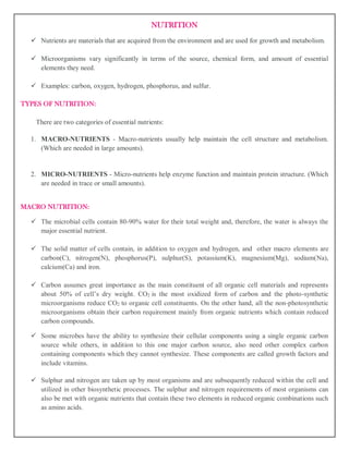
1. NUTRITION
Nutrients are materials that are acquired from the environment and are used for growth and metabolism.
Microorganisms vary significantly in terms of the source, chemical form, and amount of essential
elements they need.
Examples: carbon, oxygen, hydrogen, phosphorus, and sulfur.
TYPES OF NUTRITION:
There are two categories of essential nutrients:
1. MACRO-NUTRIENTS - Macro-nutrients usually help maintain the cell structure and metabolism.
(Which are needed in large amounts).
2. MICRO-NUTRIENTS - Micro-nutrients help enzyme function and maintain protein structure. (Which
are needed in trace or small amounts).
MACRO NUTRITION:
The microbial cells contain 80-90% water for their total weight and, therefore, the water is always the
major essential nutrient.
The solid matter of cells contain, in addition to oxygen and hydrogen, and other macro elements are
carbon(C), nitrogen(N), phosphorus(P), sulphur(S), potassium(K), magnesium(Mg), sodium(Na),
calcium(Ca) and iron.
Carbon assumes great importance as the main constituent of all organic cell materials and represents
about 50% of cell’s dry weight. CO2 is the most oxidized form of carbon and the photo-synthetic
microorganisms reduce CO2 to organic cell constituents. On the other hand, all the non-photosynthetic
microorganisms obtain their carbon requirement mainly from organic nutrients which contain reduced
carbon compounds.
Some microbes have the ability to synthesize their cellular components using a single organic carbon
source while others, in addition to this one major carbon source, also need other complex carbon
containing components which they cannot synthesize. These components are called growth factors and
include vitamins.
Sulphur and nitrogen are taken up by most organisms and are subsequently reduced within the cell and
utilized in other biosynthetic processes. The sulphur and nitrogen requirements of most organisms can
also be met with organic nutrients that contain these two elements in reduced organic combinations such
as amino acids.
2. A few microorganisms are capable of reducing elemental nitrogen to ammonia and this process of
nitrogen assimilation is known as biological nitrogen fixation.
Most of the microorganisms need molecular oxygen for respiration. In these, the oxygen serves as
terminal electron acceptor, and such organisms are referred to as ‘obligate aerobes’.
MICRO NUTRITION:
The microorganisms, in general do not use only macro or major elements but also others like cobalt,
copper, manganese, molybdenum, nickel, selenium, tungsten, vanadium and zinc which are required in
residual fraction by nearly all microorganisms.
These elements are often referred to as minor or micro nutrients or trace elements. The micronutrients or
trace elements are nevertheless just as critical to cell function as are the macronutrients.
They are metals playing the role of cell’s catalysts and many of them are play a structural role in various
enzymes.
Some microorganisms, however, need additional specific mineral nutrients, for example, diatoms and
some microalgae require silica, supplied as silicate, to impregnate their cell walls.
CLASSIFICATION MICROORGANISM BASED ON NUTRITION UTILIZATIONS:
1. Based on energy source
I. Phototrophs
II. Chemotrophs
2. Based on electron source
I. Lithotrophs
II. Organotrophs
PHOTOTROPHS:
The organisms which can utilize light as an energy source are known as phototrophs. These bacteria
gain energy from light.
CHEMOTROPHS:
These bacteria gain energy from chemical compounds. They cannot carry out photosynthesis.
LITHOTROPHS:
Some organisms can use reduced organic compounds as electron donors and are termed as Lithotrophs.
They can be Chemolithotrophs and Photolithotrophs
3. Photo-lithotrops: These bacteria gain energy from light and use reduced inorganic compounds
such as H2S as a source of electrons.
eg: Chromatium okeinii.
Chemo-lithotrophs: These bacteria gain energy from reduced inorganic compounds such as
NH3 as a source of electron
eg; Nitrosomonas.
ORGANOTROPHS:
Some organisms can use organic compounds as electron donors and are termed as organotrophs.
Some can be Chemoorganotrophs and Photoorganotrophs.
Photo-organotrophs: These bacteria gain energy from light an d use organic compounds such
as Succinate as a source of electrons.
eg; Rhodospirillum.
Chemo-organotrophs: These bacteria gain energy from organic compounds such as glucose
and ammino acids as a source of electrons.
eg; Pseudomonas pseudoflora.
Some bacteria can live ether chemo-lithotrophs or chemo-
organotrophs like Pseudomonas pseudoflora as they can use either glucose or H2S as electron source.